Ob Gyn Questions and answers, study set. 100% coverage. Rated A+
Document Content and Description Below
Ob Gyn Questions and answers, study set. 100% coverage. Rated A+ Stages of Labor Normal labor progress is slower before 6 cm than after 6 cm, and this should be considered when making a diagno... sis of a protraction disorder. Most common cause of protraction / arrest -Hypocontractile Uterine Activity other reasons: -Cephalopelvic Disproportion this diagnosis is often based upon observation of protracted or arrested labor during the active phase. In this setting, it is often due to fetal malposition (eg, extended or asynclitic fetal head) or malpresentation (mentum posterior, brow) rather than a true disparity between fetal size and maternal pelvic dimensions Occiput posterior (OP) position — Persistent OP position is associated with longer duration of first and second stages of labor, as well as a higher risk of arrest of descent requiring operative delivery Neuraxial anesthesia (epidural, spinal) Obesity - ✔✔*First Stage* (avg 10-12hrs null, 6-8 multi) : onset of contraction to complete cervical dilation -Latent: gradual cervical change -Active: Rapid cervical change (>1 cm dilation/hr) *Protraction*: Friedman considered the minimum rate of acceptable cervical dilation during the active phase of the first stage of labor to be 1.2 cm/hour for nulliparous patients and 1.5 cm/hour for multiparous patients. A slower rate of cervical dilation was diagnostic of protracted labor. *Arrest of labor *is diagnosed at cervical dilation ≥6 cm dilation in a patient with ruptured membranes and: ●No cervical change for ≥4 hours despite adequate contractions ●No cervical change for ≥6 hours with inadequate contractions Tx: start Oxytocin and evaluate cervical change after four hours of adequate uterine contractions with oxytocin or six hours of inadequate uterine contractions with oxytocin prior to making a diagnosis of labor arrest and performing a cesarean delivery ( under 200 MVUs) *Second Stage* : cervical dilation to fetus expulsion -Passive: from dilation to pushing -Active: pushing to expulsion a second stage longer than two hours in nulliparas (three hours when regional analgesia is used), and longer than one hour in multiparas (two hours when regional analgesia is used) ●Nulliparous women: No progress (descent, rotation) after ≥4 hours with epidural anesthesia and ≥3 hours without epidural anesthesia ●Multiparous women: No progress (descent, rotation) after ≥3 hours with epidural anesthesia and ≥2 hours without epidural anesthesia *Third Stage* Expulsion of fetus to expulsion of placenta Nonreassuring Fetal Status - ✔✔Repetitive Late Decels Bradycardias Loss of Variability Tx: O2, turned onto left side to decrease IVC compression and increase uterine perfusion, DC oxytocin until reassured if prolonged decel due to hypertonus (single contraction over 2 min) or Tachysystole (greater than 5 contractions in 10min peroid) the patient can be given a dose of terbutaline to help relax the uterus. Preterm Labor - ✔✔*Risk Factors * No partner Low socioeconomic status Anxiety Depression Life events (divorce, separation, death) Abdominal surgery during pregnancy Occupational issues (upright posture, use of industrial machines, physical exertion, mental or environmental stress related to work or working conditions) Multiple gestation Polyhydramnios Uterine anomaly, including diethylstilbestrol-induced changes in uterus and leiomyomas Preterm premature rupture of membranes History of second trimester abortion History of cervical surgery Premature cervical dilatation or effacement (short cervical length) Sexually transmitted infections Systemic infection, pyelonephritis, appendicitis, pneumonia Bacteriuria Periodontal disease Placenta previa Placental abruption Vaginal bleeding, especially in more than one trimester Previous preterm delivery Substance abuse Smoking Maternal age (<18 or >40) African-American race Poor nutrition and low body mass index Inadequate prenatal care Anemia (hemoglobin <10 g/dL) Excessive uterine contractility Low level of educational achievement Maternal first degree family history of spontaneous preterm birth, especially if the pregnant woman herself was born preterm Fetal anomaly Fetal growth restriction Environmental factors (eg, heat, air pollution) Placental Abruption RF History of Abruption (also in sisters) Abdominal Trauma Cocaine or other drugs Polyhydroaminos Chronic HTN Pre-eclampsia, Eclampsia, GA HTN PROM Chorioamnionitis Previous Ischemic Placental disease (FGR, SFGA) Maternal Age Parity Smoking during preg Male infant Uterine abnormalities, cocaine use, and smoking are additional less common causes of abruption. Uterine anomalies (eg, bicornuate uterus), uterine synechiae, and leiomyoma are mechanically and biologically unstable sites for placental implantation; abruption at these sites may be due to inadequate decidualization and/or shear. Suboptimal trophoblastic implantation may also explain the increased risk of abruption among women with a prior cesarean - ✔✔Intense contractions, dark flowing vaginal bleed, preterm labor = Abruption (Nbme q) abrupt onset of vaginal bleeding, mild to moderate abdominal and/or back pain, and uterine contractions. Back pain is prominent when the placenta is on the posterior wall of the uterus. The uterus is often firm, and may be rigid and tender. Contractions are usually high frequency and low amplitude, but a contraction pattern typical of labor is also possible and labor may proceed rapidly. Imaging — Identification of a retroplacental hematoma is the classic ultrasound finding of placental abruption Dx: Clinical Tx: unstable at any gestational age (eg, significant coagulopathy, hypotension, and/or ongoing major blood loss), or the fetal heart rate tracing is nonreassuring at any gestational age, or the gestational age is ≥36 weeks, we suggest *expeditious delivery via c/s* Uterine Rupture RF grand multiparity, advancing maternal age, dystocia resulting in protracted labor, macrosomia, multiple gestation, and abnormal placentation (eg, placenta accreta, increta, or percreta) inherent or acquired weakness of the myometrium, disorders of the collagen matrix (Ehlers-Danlos type IV) [9-12], and abnormal architecture of the uterine cavity (bicornuate uteri, uterus didelphys, "blind uterine horns") Rupture of an unscarred uterus has also been associated with trauma (eg, traffic accident, domestic violence, gunshot wound) and obstetric maneuvers (eg, internal version and breech extraction, instrumental delivery, manual removal of the placenta). -higher incidence in TOLAC - ✔✔Uterine rupture is most common in women with a prior hysterotomy. Signs of uterine rupture may include the sudden onset fetal heart rate (FHR) abnormalities (FETAL BRADYCARDIA) , vaginal bleeding, constant abdominal pain, cessation of uterine contractions, recession of the presenting part (loss of station), and maternal hypotension and tachycardia Postpartum, uterine rupture is characterized by pain and persistent vaginal bleeding despite use of uterotonic agents. Tx = definitive is hysterectomy Subchorionic Hematoma - ✔✔believed to result from partial detachment of the chorionic membranes from the uterine wall, in contrast to abruption, which is due to detachment of the placenta from the uterine wall Patients are asymptomatic or experience light vaginal bleeding. In contrast to abruption, abdominal pain is typically absent, a minority of patients experience cramping or contractions, and the diagnosis is usually made before rather than after 20 weeks of gestation ultrasound findings of a hypoechoic or anechoic crescent-shape area behind the fetal membranes, which may also elevate the edge of the placenta Placenta Previa RF ●Previous placenta previa ●Previous cesarean delivery ●Multiple gestation ●Multiparity ●Advanced maternal age ●Infertility treatment ●Previous abortion ●Previous intrauterine surgical procedure ●Maternal smoking ●Maternal cocaine use ●Male fetus ●Non-white race Prelabor cesarean delivery may increase previa risk in a subsequent delivery, more than previous intrapartum cesarean or vaginal delivery - ✔✔refers to the presence of placental tissue that extends over or lies proximate to the internal cervical os. Sequelae include the potential for severe bleeding and preterm birth, as well as the need for cesarean delivery. Placenta previa should be suspected in any woman beyond 20 weeks of gestation who presents with painless vaginal bleeding. For women who have not had a second trimester ultrasound examination, antepartum bleeding after 20 weeks of gestation should prompt sonographic determination of placental location before digital vaginal examination is performed because palpation of the placenta can cause severe hemorrhage. Dx: TA -US --> TV-US f the placental edge covers the internal os, the placenta is labeled a previa . If the placental edge is <2 cm from, but not covering, the internal os, the placenta is labeled as low-lying Tx: *asymptomatic placenta previa*, we monitor placental position with ultrasound examination as an outpatient and counsel these patients to avoid excess physical activity and to call their provider promptly if bleeding or labor occurs. We perform cesarean delivery at 36 to 37 weeks *Women with active bleeding* are hospitalized for close maternal and fetal monitoring and supportive care. Indications for emergency cesarean delivery include refractory life threatening maternal hemorrhage, nonreassuring fetal status, and significant vaginal bleeding after 34 weeks of gestation. *Give Anti-D Ig for symptomatic Rh - moms* *Csection at 36-37 weeks but if placenta edge is more than 10mm from internal os you may attempt vaginal delivery* Placenta Accreta RF risk increases w/ previous C-sections Placenta Previa history of uterine surgery (eg, myomectomy entering the uterine cavity, hysteroscopic removal of intrauterine adhesions, cornual resection of ectopic pregnancy, dilatation and curettage, endometrial ablation), cesarean scar pregnancy, maternal age greater than 35 years history of pelvic irradiation - ✔✔Placenta accreta refers to an abnormality of placental implantation in which the anchoring placental villi attach to myometrium rather than decidua, resulting in a morbidly adherent placenta. Placenta increta (chorionic villi penetrate into the myometrium) and placenta percreta (chorionic villi penetrate through the myometrium to the uterine serosa or adjacent organs) are related, but more severe, abnormalities of placental implantation The first clinical manifestation of placenta accreta is usually profuse, life-threatening hemorrhage that occurs at the time of attempted manual placental separation. Part, or all, of the placenta remains strongly attached to the uterine cavity, and no plane of separation can be developed Dx: 2nd/3rd trimester US - loss of placental homogeneity (mulitple sonolucent spaces = venous lakes in mymoetrium) or loss / thinning of normal hypoechoic area behind placenta w/ retroplacental myometrium thickness less than 1mm or loss of bladder line w/ uterine serosa (bulging of placenta into the posterior wall of bladder) -1st trimester suspicious w/ gestational sac in lower uterine segment (instead of fundus) near scar Summary: US showing multiple vascular lacunae within the placenta, loss or disruption of the retroplacental clear space and bladder line) Tx: Betamethason at 34 weeks deliver within 48 hrs. Place balloon catheters into the internal iliac arters prior to hysterectomy leaving the placenta undisturbed in situ Bleeding During Pregnancy Uterine size larger than expected for dates suggests a multiple gestation, possibly with miscarriage of one of the multiples, gestational trophoblastic disease (molar pregnancy), or other uterine pathology (fibroids can cause an irregularly enlarged uterus) - ✔✔1st trimester -ectopic preg -miscarriage (MCC) -implantation of normal pregnancy -polyps, ectriopion, infection/inflammation, trophoblastic disease Steps: Stabalize (+CBC) --> HPI, Physicial + Speculum +TV- US 2nd and 3rd trimester ●Bloody show associated with cervical insufficiency or labor (by definition, labor occurs after 20 weeks) ●Miscarriage (by definition, miscarriage occurs before 20 weeks) ●Placenta previa ●Abruptio placenta ●Uterine rupture ●Vasa previa Bleeding before 20 weeks The presence of only light, intermittent, painless bleeding suggests bloody show from cervical insufficiency, a small marginal placental separation, or a cervical or vaginal lesion (eg, polyp, infection, cancer). Heavier bleeding, particularly when associated with pain, is more consistent with impending miscarriage or a larger placental separation (ie, abruption) Bleeding After 20 weeks ●Placenta previa (20 percent) ●Abruptio placenta (30 percent) ●Uterine rupture (rare) ●Vasa previa (rare) In contrast to bleeding in the first half of pregnancy, digital examination of the cervix should be avoided in women presenting with bleeding in the second half of pregnancy until placenta previa has been excluded. Primary Dysmenorrhea RF: age <30 years, body mass index <20 kg/m2, smoking, menarche before age 12, longer menstrual cycles/duration of bleeding, irregular or heavy menstrual flow, and history of sexual assault Pathogenesis: Prostaglandins released from endometrial sloughing at the beginning of menses play a major role in inducing contractions. These contractions are nonrhythmic or incoordinate, occur at high frequency (more than 4 or 5 per 10 minutes), often begin from an elevated basal tone (more than 10 mmHg), and result in high intrauterine pressures (frequently more than 150 to 180 mmHg, sometimes exceeding 400 mmHg) [15]. When uterine pressure exceeds arterial pressure, uterine ischemia develops and anaerobic metabolites accumulate, which stimulate type C pain neurons resulting in dysmenorrhea. Tx: Nonsteroidal antiinflammatory agents and hormonal contraceptives are the mainstays of therapy ( NSAIDS before APAP ie Ibuprofen or mefenamic acid ) then both estrogen-progestin and progestin-only methods are effective. -if refractory for 3m w/ both we try a laparoscopy - ✔✔Primary dysmenorrhea refers to the presence of recurrent, crampy, lower abdominal pain that occurs during menses in the absence of demonstrable disease that could account for these symptoms. Nausea, diarrhea, fatigue, headache, and a general sense of malaise often accompany the pain The majority of women with primary dysmenorrhea do not have any risk factors for the disorder. The pain starts one to two days before or with the onset of menstrual bleeding and then gradually diminishes over 12 to 72 hours. It is recurrent, occurring in most, if not all, menstrual cycles. The pain is usually crampy and intermittently intense, but may be a continuous dull ache. It is usually confined to the lower abdomen and suprapubic area. Although the pain is usually strongest in the midline, some women also have severe back and/or thigh pain. Nonmidline pain, especially if unilateral, suggests a uterine anomaly or alternative diagnosis *Secondary Dysmenorrhea* Important findings on pelvic examination may include: •Imperforate hymen (hematometrocolpos) •Vulvar, anal, and/or vaginal trauma (sexual assault) •Vaginal discharge (PID) •Vaginal bleeding (ectopic pregnancy, acute abruption, spontaneous abortion, normal labor) •Cervical motion tenderness (PID) •Uterine tenderness (PID, acute abruption, dysmenorrhea, uterine rupture) •Adnexal tenderness (PID, ectopic pregnancy, ovarian torsion) •Adnexal mass (ovarian mass [cyst, abscess, or tumor], ectopic pregnancy, PID, endometriosis) •Tenderness of the posterior cul-de-sac, rectovaginal septum, or uterosacral ligaments on rectovaginal examination (endometriosis) - ✔✔Secondary dysmenorrhea has the same clinical features, but occurs in women with a disorder that could account for their symptoms, such as endometriosis, adenomyosis, or uterine fibroids. -onset after 25 -AUB -nonmidline pain -absence of N/V/D backpain, dizziness, headache during menses -dysparenia or dyschezia -progression of symptoms *Pelvic inflammatory disease* is most common in women 15 to 25 years of age. It is characterized by lower abdominal pain, which is usually BILATERAL and ranges from mild to severe. The onset of pain is often during or shortly after menses, and may worsen during coitus or with jarring movement. •Purulent endocervical discharge •Acute cervical motion and adnexal tenderness Women with *endometriosis* typically report pelvic pain that is both related to menses and occurs at times other than menses. They may have premenstrual spotting, dyspareunia, dyschezia, poor relief of symptoms with NSAIDs, progressively worsening symptoms, and inability to attend work or school during menses. •Uterosacral ligament abnormalities, such as nodularity, thickening, or focal tenderness •Lateral displacement of the cervix due to asymmetric involvement of one uterosacral ligament by endometriosis, causing it to shorten (figure 2) • Retroverted uterus •Cervical stenosis •Adnexal enlargement from an endometrioma Women with *adenomyosis* typically present with dysmenorrhea after age 35 years •bulky globular mildly tender uterus *Fibroids* are rare in adolescents but are common after 35yr • enlarged, irregularly-shaped, nontender uterus, which may also be palpable abdominally. Ruptured ovarian cyst - Sudden onset of lateralizing pain, especially in association with exercise or sexual intercourse Premenstrual Dysphoric Disorder - ✔✔5 or more of following the week prior to mense declining w/in a few days after the onset of menses w/ 1 of them being a core symptom Core: Affective Liability, Irritability, markedly depressed mood, marked anxiety Non-core: anhedonia, dc concentration, lethargy, appetite changes, sleep changes, overwhelmed feeling, physical sx (breast tenderness, bloating, muscle jx pt, HA) fxal life difficulties -exclude other psych paths first MUST OCCUR FOR AT LEAST 2 CYCLES Tx: SSRIs -Fluoxetine -{Paroxetine - Citalopram - Sertraline Gestational Landmarks on US - ✔✔Gestational sac - 4.5 to 5 weeks Yolk sac - 5 weeks Cardiac activity - 5.5 to 6 weeks Measurable crown-rump length - 6 weeks Endometriosis Endometrioma The classical example is of an unilocular cyst with acoustic enhancement with diffuse homogeneous ground-glass echoes as a result of the haemorrhagic debris it also is a common cause of an elevated CA125 level in the premenopausal patient with an adnexal mass - ✔✔Pelvic examination findings consistent with endometriosis include: posterior vaginal fornix tenderness; palpable tender nodules in the posterior cul-de-sac, uterosacral ligaments, or rectovaginal septum; lateral displacement of the cervix; fixation of adnexa or uterus in a retroverted position; and/or a tender adnexal mass Tx: NSAIDS + Low dose cyclical E+P or P only Adenomyosis - ✔✔ Impreforate Hymen - ✔✔ Ovarian Torsion RF ovarian mass 5cm or greater reproductive age pregnancy Torsion is more likely to occur with benign cysts or neoplasms rather than malignant lesions, possibly because malignant masses are more likely to be fixed in place. Ovulation induction Prior Torsion Tx: Laparoscopy For most premenopausal patients with ovarian torsion, we recommend detorsion and ovarian conservation rather than salpingo-oophorectomy. Ovarian cystectomy is often performed if a benign mass is present. Patients with an ovarian mass that is suspicious for malignancy require salpingo-oophorectomy. Ovaries that are hemorrhagic and/or edematous are most likely viable and, thus, oophorectomy should be reserved for necrotic/gelatinous/dead tissue (black and atrophic). Salpingo-oophorectomy is also reasonable for postmenopausal women. -prevent recurrance w/ high dose oral contraceptions if cysts - ✔✔acute onset of moderate to severe pelvic pain, often with nausea and possibly vomiting, in a woman with an adnexal mass WAXING AND WANING "waves" of n/v low grade fever Dx: U/S -edematous enlarged ovary compared to contralateral - Multiple small peripheral follicles ("string of pearls") - similiar to PCOS appearance -Abnormal ovarian location - The normal location of the ovaries is lateral to the uterus, but in torsion, they may be located anterior to the uterus -Decreased or absent Doppler flow within the ovary or Whirlpool sign (concentric hypoechoic stripes) A definitive diagnosis of ovarian torsion is made by direct visualization of a rotated ovary at the time of surgical evaluation. The decision to proceed with surgery is based upon a clinical diagnosis, which is often based upon the presence of acute pelvic pain and an adnexal mass with a sonographic appearance consistent with torsion Note: The embryological ovary migrates from the level of the 10th thoracic vertebrae and descends to the true pelvis by puberty. Thus, in early life, the ovary is an abdominal organ and more susceptible to torsion. Tubo-ovarian Abscess - ✔✔characteristic sonographic appearance of an abscess is a complex, multilocular mass Ruptured ovarian cyst - ✔✔Rupture of an ovarian cyst is often accompanied by sonographic evidence of hematoperitoneum or free fluid in the pelvis Cervical Cancer RF Early onset of sexual activity Multiple sexual partners A high-risk sexual partner (eg, a partner with multiple sexual partners or known HPV infection) History of sexually transmitted infections (eg, Chlamydia trachomatis, genital herpes) History of vulvar or vaginal squamous intraepithelial neoplasia or cancer Immunosuppression Early age at first birth (younger than 20 years old) and increasing parity (3 or more full term births) are also associated with an increased risk of cervical cancer; these are also likely due to exposure to HPV through sexual intercourse Low socioeconomic status - ✔✔pelvic ultrasound reveals a fundal placenta and a fetus measuring 18 weeks with normal cardiac activity. Vaginal examination reveals a 3-centimeter lesion arising off the posterior lip of the cervix. It easily bleeds with palpation and is hard in consistency Endometrial Cancer Increasing age Women 50- to 70-years-old have a 1.4 percent risk of endometrial cancer Unopposed estrogen therapy Tamoxifen therapy Early menarche Late menopause (after age 55) Nulliparity Polycystic ovary syndrome (chronic anovulation) Obesity Diabetes mellitus Estrogen-secreting tumor Lynch syndrome (hereditary nonpolyposis colorectal cancer) Cowden syndrome Family history of endometrial, ovarian, breast, or colon cancer - ✔✔ Vulvular cancer - ✔✔ Breast Cancer - ✔✔ Ovarian Cancer - ✔✔ Adnexal Masses If premenopausal In the absence of pain or intraperitoneal bleeding, observation for a time period between two weeks and three months and possibly therapy with oral contraceptive pills is appropriate. The oral contraceptive pills will keep a new cyst from forming so as to decrease confusion at the follow-up ultrasound, but do not help the current cyst regress According to aafp if premenopausal 1st test = pregnancy test then US if -internal echos, septae, exrecrescenes, papillations or over 10 cm then surg - if less than 10cm do trial of NSAIDs and serial tv- us 4 - 6 weeks if postmenopausal any mass over 10cm is surgical if under do ultrasound for malignant clues if negative check CA 125 if over 35 units/mL its surgical if not...serial ultrasound every 4-6 w ●High risk - Solid component that is not hyperechoic and is often nodular or papillary Septations, if present, that are thick (>2 to 3 mm) Color or power Doppler demonstration of flow in the solid component Presence of ascites (any peritoneal fluid in postpostmenopausal women and more than a small amount of peritoneal fluid in premenopausal women is abnormal) Peritoneal masses, enlarged nodes, or matted bowel (may be difficult to detect) ●Intermediate risk - Not anechoic and/or unilocular, but no features of malignancy (eg, a mass with thin septations or low level echoes) ●Low risk - Anechoic unilocular fluid filled cysts with thin walls - ✔✔Simple cysts less than 2 cm in diameter are considered physiologic. Larger and complex cysts are more likely to be nonphysiologic. asymptomatic simple cysts <6 cm on ultrasound examination can be observed with or without administration of oral contraceptive pills. A solid ovarian mass in childhood is always considered malignant until proven otherwise by histological examination. ●Alpha-fetoprotein (AFP) is an oncofetal antigen that is a glycoprotein. It is produced by endodermal sinus tumors, mixed germ cell tumors, and immature teratomas. ●Lactate dehydrogenase (LDH) is elevated with dysgerminomas ●CA-125 is a marker for epithelial ovarian cancer that is highly sensitive, but not very specific, since it is elevated with many intraperitoneal processes (eg, endometriosis, pelvic inflammatory disease, pregnancy, Crohn's disease) ●Human chorionic gonadotropin (hCG) is produced by trophoblastic cells and thus will be elevated with pregnancy, hydatidiform moles, placental site tumors, nongestational choriocarcinoma, and embryonal ovarian carcinomas ●Carcinoembryonic antigen (CEA) can be produced by epithelial or germ cell tumors ●Inhibin and mullerian inhibiting substance (MIS) concentrations are elevated in children with granulosa-theca cell tumors ●Thrombocytosis has been associated with ovarian malignancies in girls and adolescents [52]. Because it is readily available, the platelet count is useful in the emergency evaluation of a torsed ovary with suspicion for malignancy. Theca Lutein Cyst: luteinized follicle cysts that form as a result of overstimulation from high hCG levels or extreme sensitivity to hCG. Bilateral multiseptated cystic adnexal masses in a woman with gestational trophoblastic disease, multiple gestation, ovarian hyperstimulation, or a pregnancy complicated by fetal hydrops are likely to represent theca lutein cysts, rather than malignancy. Most are asymptomatic, but maternal virilization, hyperemesis gravidarum, preeclampsia, or thyroid dysfunction may occur. The cysts gradually resolve weeks to months after the source of hCG is eliminate Corpus luteum of pregnancy - An early intrauterine pregnancy is always associated with a corpus luteum cyst, which is typically less than 2.5 cm in diameter. However, the corpus luteum may occasionally become enlarged and painful due to hemorrhage. Luteoma - Luteoma is a nonneoplastic ovarian change associated with pregnancy that can simulate a neoplasm on clinical, gross, or microscopic examination [10]. Luteomas involute spontaneously after delivery or are adequately treated by a conservative surgical approach. The diagnosis should be suspected in the presence of a solid adnexal mass and maternal hirsutism or virilization Hydrops Fetalis - ✔✔ Spontaneous Abortion - ✔✔ UTIs - ✔✔ Breast Abscess / Mastitis DIFFERENTIAL DIAGNOSIS ●Plugged duct - A plugged duct is a localized area of milk stasis within the milk duct that causes distention of mammary tissue. Symptoms include a palpable lump with tenderness. A plugged duct may be distinguished from mastitis and breast abscess by the absence of systemic findings. Severe engorgement - Engorgement occurs due to interstitial edema with onset of lactation or at other times with accumulation of excess milk. Mastitis may be distinguished from severe engorgement in that engorgement is bilateral, with generalized involvement [1]. Severe engorgement is not typically associated with systemic symptoms of fever and myalgias. ●Galactocele - A galactocele (also known as a milk retention cyst) is a cystic collection of fluid that is usually caused by an obstructed milk duct. Galactoceles present as soft cystic masses; they are not tender and are not associated with systemic manifestations. Ultrasonography may demonstrate a simple milk cyst or a complex mass. The diagnosis can be made on the basis of the clinical history and needle aspiration, which yields a milky substance. (See "Common problems of breastfeeding and weaning", section on 'Galactoceles'.) ●Inflammatory breast cancer - Inflammatory breast cancer (IBC) should be considered if a breast infection does not resolve with appropriate treatment. Clinical manifestations include skin thickening due to edema, erythema, and peau d'orange appearance (picture 1). It is often associated with axillary lymphadenopathy. The diagnosis is established via biopsy. - ✔✔Patients with *primary breast abscess* present with localized, painful inflammation of the breast associated with fever and malaise, along with a FLUCTUANT, tender, PALPABLE MASS *Lactational mastitis * typically presents as a firm, red, tender, swollen area of one breast associated with fever >38.3ºC in a nursing mother. Systemic complaints may include myalgia, chills, malaise, and flu-like symptoms. In the early stages, the presentation can be subtle with few clinical signs; patients with advanced infection may present with a large area of breast swelling with overlying skin erythema. Reactive lymphadenopathy may be associated with axillary pain and swelling. Most primary breast abscesses are caused by Staphylococcus aureus. Methicillin-resistant S. aureus (MRSA) is becoming an increasingly important pathogen in cases of lactational and nonlactational mastitis Tx: Needle aspiration is an appropriate initial approach for abscess drainage when the overlying skin is intact . The use of ultrasound guidance ensures complete drainage and facilitates aspiration of loculated areas as well as collections that may not be appreciated on physical exam The patient should be reexamined every two to three days and the cavity imaged with ultrasound and aspiration repeated in a similar manner until there is no further fluid visible in the abscess cavity or the fluid aspirated is serous In the setting of nonsevere infection in the absence of risk factors for methicillin-resistant S. aureus (MRSA), outpatient therapy may be initiated with *dicloxacillin or cephalexin, pending culture results if allergic = clindamycin* -if MRSA risk factors (diabetic) use TMP-SMX or clindamycin Women should be encouraged to continue breastfeeding following breast infection, even in the setting of incision and drainage. If there is difficulty with breastfeeding because the incision interferes with nursing on the affected breast, the infant cannot relieve breast fullness, or there is concern about spreading infection to the baby, hand expression or breast pumping can be effective for maintaining the milk supply until breastfeeding can resume Fat Necrosis of Breast - ✔✔ Lichen Planus Lichen planus is a chronic dermatologic disorder involving the hair-bearing skin and scalp, nails, oral mucous membranes and vulva. This disease manifests as inflammatory mucocutaneous eruptions characterized by remissions and flares. The exact etiology is unknown, but is thought to be multifactorial. Vulvar symptoms include irritation, burning, pruritus, contact bleeding, pain and dyspareunia. Clinical findings vary with a lacy, reticulated pattern of the labia and perineum, with or without scarring and erosions as well. With progressive adhesion formation and loss of normal architecture, the vagina can become obliterated. Patients may also experience oral lesions, alopecia and extragenital rashes. Treatment is challenging, since no single agent is universally effective and consists of multiple supportive therapies and topical superpotent corticosteroids. - ✔✔52-year-old nulliparous woman presents with long-standing vulvar and vaginal pain and burning. DYSPARUNIA, painful sitting and clothing. Gums bleed. Products on genitals hurt. Her physical examination is remarkable for inflamed gingiva and a whitish reticular skin change on her buccal mucosa. A fine papular rash is present around her wrists bilaterally. Pelvic examination reveals white plaques with intervening red erosions on the labia minora as shown in below picture. A speculum cannot be inserted into her vagina because of extensive adhesions. Vulvar vestibulitis - ✔✔a constellation of symptoms and findings limited to the vulvar vestibule, which include severe pain on vestibular touch or attempted vaginal entry, tenderness to pressure and erythema of various degrees abrupt onset and are described as a sharp, burning and rawness sensation. Women may experience pain with tampon insertion, biking or wearing tight pants, and avoid intercourse because of marked introital dyspareunia. Vestibular findings include exquisite tenderness to light touch of variable intensity with or without focal or diffuse erythematous macules. - Treatment includes use of tricyclic antidepressants to block sympathetic afferent pain loops, pelvic floor rehabilitation, biofeedback, and topical anesthetics. Surgery with vestibulectomy is recommended for patients who do not respond to standard therapies and are unable to tolerate intercourse. Lichen Simplex Chronicus 0-year-old G1P1 woman presents with a history of chronic vulvar pruritus. The itching is so severe that she scratches constantly and is unable to sleep at night. She reports no significant vaginal discharge or dyspareunia. She does not take antibiotics. Her medical history is unremarkable. Pelvic examination reveals normal external genitalia with marked lichenification (increased skin markings) and diffuse vulvar edema and erythema as shown in picture below. Saline microscopy is negative. Potassium hydroxide testing is negative. Vaginal pH is 4.0. The vaginal mucosa is normal - ✔✔common vulvar non-neoplastic disorder, results from chronic scratching and rubbing, which damages the skin and leads to loss of its protective barrier. Over time, a perpetual itch-scratch-itch cycle develops, and the result is susceptibility to infection, ease of irritation and more itching. Symptoms consist of severe vulvar pruritus, which can be worse at night. Clinical findings include thick, lichenified, enlarged and rugose labia, with or without edema. The skin changes can be localized or generalized. Diagnosis is based on clinical history and findings, as well as vulvar biopsy. Treatment involves a short-course of high-potency topical corticosteroids and antihistamines to control pruritus. Mucopurulent Cervicitis - ✔✔Ceftriaxone and azithromycin Lichen Sclerosis -steps (when to biopsy) -tx -diff increased risk of squamous cell cancer of the vulva -proper hygiene AVOID (substitute with) -pantyhose (wear stockings), synthetic underwear (wear cotton or none), jeans or other tight pants (wear loose pants skirts and dresses), swimsuits thongs leotards (wear loose cotton), pantyliners (wear tampons or cotton pas), scented soaps or shampoos (use fragrance free pH neutral soap), bubble baths (tub baths in morning and night w/o additives), scented detergents (unscented), washcloths (fingers), douches, powders, sprays, dyed toilet paper, high dryers (patting) - ✔✔benign, chronic, progressive dermatologic condition characterized by marked inflammation, epithelial thinning, and distinctive dermal changes accompanied by pruritus and pain. -classic appearance is thin, white, wrinkled skin localized to the labia minora and/or labia majora -areas of epithelial hyperplasia from chronic rubbing are often seen. Fissuring is frequently present perianally, in the intralabial folds, or around the clitoris. At the end-stages, the vulva is pallid and featureless due to midline fusion -Vulvar pruritus is a common symptom of vulvar lichen sclerosus and can be so intense that it interferes with sleep. Other symptoms include pruritus ani, painful defecation, rectal bleeding, dyspareunia, and dysuria. Dx: Clinical + Biopsy Histology Tx: Educate + Topical Corticosteroids (clobetasol or halobetasol) daily for 6 - 12 w then taper (if thickened plaques may inject corticosteroids) 64-year-old G2P2 woman 12m of vulvar pruritus. No discharge. No OTC med helps. Dysparunia. Hx of HTN and allergic rhinitis. loss of the labia minora with resorption of the clitoris (phimosis). The vulvar skin appears thin and pale and involves the perianal area as in the picture below. No ulcerations are present. The vagina is mildly atrophic, but appears uninvolved. Which of the following is the most likely diagnosis in this patient? Lichen sclerosus is a chronic inflammatory skin condition that most commonly affects Caucasian premenarchal girls and postmenopausal women. The exact etiology is unknown, but is most likely multifactorial. Patients typically present with extreme vulvar pruritus and may also present with vulvar burning, pain and introital dyspareunia. Early skin changes include polygonal ivory papules involving the vulva and perianal areas, waxy sheen on the labia minora and clitoris, and hypopigmentation. The vagina is not involved. More advanced skin changes may include fissures and erosions due to a chronic itch-scratch-itch cycle, mucosal edema and surface vascular changes. Ultimately, scarring with loss of normal architecture, such as introital stenosis and resorption of the clitoris (phimosis) and labia minora, may occur. Treatment involves use of high-potency topical steroids. There is less than a 5% risk of developing squamous cell cancer within a field of lichen sclerosus. Women G3P2 w/ Type 2 diabetes at 38 weeks, the cervix is 2cm dialated, membranes are intact, fundal height 42 cm, 4hrs after admission the cervix is completely dialated and the vertix is occiput anterior at -1 station. Over next hr contractions occur at 2 min, last 60 s, and are 60mm Hg in insensity but cervix and station remain unchanged. FHT are normal and reactive. 1 hr later her condition is unchanged why? - ✔✔Cephalopelvic disporportion Contraction of outlet fetal malposition Hypotonic uterine activity shoulder dystocia What is normal positions of labor - ✔✔ What is normal uterine activity - ✔✔With an IUPC in place, quantitative data can be measured, most commonly using Montevideo units (MVU). A Montevideo Unit is the sum of the intensity of each contraction in a 10 minute period (in mmHG). Adequate uterine activity is defined as a contraction pattern that generates greater than 200 MVUs. Shoulder Dystocia - ✔✔Fetal macrosomia, maternal obesity, diabetes mellitus, postterm pregnancy, a prior delivery complicated by a shoulder dystocia, and a prolonged second stage of labor are all associated with an increased incidence of shoulder dystocia. Although a family history can be indicative of large babies which might place her at additional risk, her gestational diabetes represents her largest risk factor. Occiput Posterior ●Nulliparity ●Maternal age greater than 35 years ●Obesity ●African-American race ●Previous OP delivery ●Decreased pelvic outlet capacity ●Gestational age ≥41 weeks ●Birthweight ≥4000 grams ●Anterior placenta ●Epidural anesthesia - ✔✔ Menopause Menopause before age 40 years is considered to be abnormal and is referred to as primary ovarian insufficiency (premature ovarian failure). Hot flashes typically begin as the sudden sensation of heat centered on the upper chest and face that rapidly becomes generalized. The sensation of heat lasts from two to four minutes, is often associated with profuse perspiration and occasionally palpitations, and is sometimes followed by chills and shivering, and a feeling of anxiety. -arousal from sleep -depression -vaginal driness -breast pain -migraines -bone loss -decreased risk of cardiac disease - ✔✔defined as the permanent cessation of menstrual periods, determined retrospectively after a woman has experienced 12 months of amenorrhea without any other obvious pathological or physiological cause. It occurs at a median age of 51.4 years in normal women, and is a reflection of complete, or near complete, ovarian follicular depletion, with resulting hypoestrogenemia and high FSH concentrations In the late reproductive years before the onset of the menopausal transition, serum inhibin B begins to decrease, serum follicle-stimulating hormone (FSH) increases slightly, estradiol levels are preserved, but luteal phase progesterone levels decrease as fertility potential begins to decline. Menstrual cycles are ovulatory, but the follicular phase (the first half of the menstrual cycle before ovulation occurs) begins to shorten (eg, 10 versus 14 days) Menopausal Transition (follicular depletion) -lengthening of intermenstrual interval (in constrast to shortening in late reproductive years Normal intermenstrual interval during the reproductive years is 25 to 35 days; during the menopausal transition, this may increase to 40 to 50 days. Then increases amenorrhea / anovulatory cyles -decreased estrogen, increased FSH, a progressive decrease in serum inhibin B, as well as a decrease in antimüllerian hormone (AMH), another product of the granulosa cell. Fetal HBV We obtain liver biochemical tests every three months during pregnancy and for six months postpartum The infection rate among infants born to HBeAg-positive mothers who do not receive any form of prophylaxis is as high as 90 percent. The high protective efficacy (95 percent) of neonatal vaccination suggests that most infections occur at birth when maternal secretions in the birth canal come in contact with the infant's mucosal membranes -Test for HBsAg on all women at 1st prenatal visit HBsAg-negative mother, birth weight ≥2 kg — If the mother is HBsAg negative and the birthweight is ≥2 kg (4.4 pounds), the preferred schedule for HepB vaccine is as follows (table 2A) [1,20]: ●First dose - During the birth hospitalization ●Second dose - at one to two months of age ●Third dose - At 6 to 18 months of age (must be given at ≥24 weeks of age) If HBsAg + give monovalent HepB vaccine + HBIg w/in 12 hrs of delivery The schedule for subsequent doses depends upon the infant's birth weight: ●Birth weight ≥2 kg (4.4 pounds) - The second and third doses should be given one and six months of age, respectively. ●Birth weight <2 kg (4.4 pounds) - Three additional doses should be given (at one, two to three, and six months of age or at two, four, and six months of age). - ✔✔Acute HBV infection during pregnancy is usually not severe and is not associated with increased mortality or teratogenicit Risk factors for perinatal transmission — The most important risk factor for transmission despite proper administration of prophylaxis (HBIG and first dose of HBV vaccine given within 12 hours of birth and completion of HBV vaccine series) appears to be high maternal HBV viral load. The obstetrical approach should not be influenced by the HBV status of the mother. (vag vs c/s) Breastfeeding — Breastfeeding does not appear to increase the risk of transmission Newborns of carrier mothers should receive passive-active immunization In addition to standard passive-active immunization, antiviral therapy should be offered to mothers with high HBV DNA levels since it can further reduce the risk of perinatal transmission We generally offer prophylaxis to women who have a high viral load (more than 7 log10 IU/mL), although some clinicians recommend using lower cutoff (approximately 6 log10 IU/mL) with Tenofovir or telbivudine infants of HBsAg-positive mothers should receive monovalent HepB vaccine and HBIG 0.5 mL as soon after delivery as possible (preferably within 12 hours), regardless of birth weight Clinical manifestations and course — Newborn infants with HBV infection rarely show clinical or biochemical signs of disease at birth. Affected newborns remain asymptomatic, and develop chronic antigenemia with mild and often persistent liver enzyme elevations beginning at two to six months of age (immune tolerant phase) (figure 1) [1,16,17]. A small number of patients will develop acute hepatitis by two months of age and present with icteric, and occasionally fulminant, hepatitis [17-20]. Most patients develop chronic infection, which may progress to cirrhosis and hepatocellular carcinoma. Hep C Genotypes 2 and 3 — For most children and adolescents with genotype 2 or 3, we suggest deferring treatment until an interferon-free regimen is available. For adults with these genotypes, the current preferred treatment is the combination of sofosbuvir (a HCV polymerase inhibitor) with ribavirin The goal of treatment is to eradicate hepatitis C virus (HCV) RNA, which is predicted by the achievement of a sustained virologic response (SVR), defined by the absence of HCV RNA by polymerase chain reaction six months after stopping treatment. - ✔✔Maternal vertical transmission — The risk of transmitting the virus from a HCV-infected woman to her infant is approximately 5 percent. Transmission rates are higher if the mother is coinfected with HIV Breastfeeding not contraindicated — Although HCV RNA has been detected in breast milk and colostrum of viremic mothers [24], breastfeeding does not appear to increase the rate of HCV transmission in asymptomatic mothers -abstain if cracked or bleeding Fetal Varicella Dx: Characteristic vesicular lesions Tx: Varicella-zoster immune globulin (VariZIG) postexposure prophylaxis - Newborns with severe infection should be treated with acyclovir -The use of aspirin should be avoided because of the increased risk of developing Reye syndrome. Use of acetaminophen may prolong the illness -encourage breast feeding A mother with active VZV lesions must be isolated. The infant is isolated from the mother until she is not infectious. Maternal exposure 6 to 21 days before hospitalization − A seronegative mother exposed to VZV 6 to 21 days before hospital admission should be isolated from other patients and the nursery because she may develop varicella while hospitalized. Her infant, if born at term, should be isolated with the mother. The mother and infant should be cared for only by staff with immunity to VZV. Nursery exposure — An infant who develops varicella in the nursery or NICU should be isolated - ✔✔*Congenital varicella syndrome* occurs in infants whose mothers are infected between 8 and 20 weeks gestation. Clinical manifestations vary and include Cicatricial skin lesions, ocular defects (eg, cataracts, chorioretinitis, Horner syndrome, microphthalmos, and nystagmus), and abnormalities of the limb (eg, hypoplasia of bone and muscle) and central nervous system (eg, cortical atrophy, seizures, and cognitive impairment). neonatal varicella - mothers exposed within 2 weeks of delivery Fever may develop within the first days after birth, followed by a vesicular eruption. In mild cases, the lesions heal within 7 to 10 days. However, disseminated disease may ensue, with varicella pneumonia, hepatitis, and meningoencephalitis being the most common visceral manifestations. NOTE Postexposure prophylaxis with varicella-zoster immune globulin (VariZIG) is recommended for infants born to symptomatic mothers around the time of delivery, preterm infants with gestational age (GA) ≥28 weeks born to mothers without immunity, and preterm infants <28 weeks. We recommend a 10-day course of acyclovir (30 mg/kg per day in 3 divided doses IV) to treat neonatal varicella - ✔✔ Fetal MMR - ✔✔ Fetal CMV Common laboratory findings in the symptomatic neonate include thrombocytopenia, elevated transaminases, and elevated direct and indirect serum bilirubin. Imaging of the brain often shows periventricular intracranial calcifications, periventricular leukomalacia, ventriculomegaly, vasculitis, or polymicrogyria -chorioretinitis The timing of primary maternal infection is the most important determinant of perinatal sequelae. First trimester maternal infection results in both higher frequency and greater severity of perinatal sequelae compared with infection later in pregnancy During pregnancy, there is no treatment proven to be effective for prevention of fetal disease or reduction in risk of sequelae. - ✔✔associated with long-term neurodevelopmental disabilities, including sensorineural hearing loss (SNHL), cerebral palsy, intellectual disability, vision impairment, and seizures. It is the leading cause of nonhereditary SNHL Clinical findings are nonspecific and include petechiae, jaundice, hepatosplenomegaly, small size for gestational age, and microcephaly. Dx: Laboratory diagnosis of congenital CMV infection is accomplished by isolation or molecular detection of CMV from urine or saliva samples collected within the first three weeks of life (table 1). Both viral culture and polymerase chain reaction (PCR) tests have high sensitivity and specificity for detection of CMV in infected neonates Tx: We recommend antiviral treatment for symptomatic infants with virologically-proven CMV infection and at least one end-organ symptom ganciclovir (life threatening disease) and valganciclovir (nonlifethreat) has been shown to improve long-term audiologic and neurodevelopmental outcomes. Congenital Toxoplasmosis ost newborns with congenital toxoplasmosis are asymptomatic. Among those who are symptomatic, clinical findings are nonspecific and may include chorioretinitis, intracranial calcifications, seizures, jaundice, hepatosplenomegaly, lymphadenopathy, anemia, thrombocytopenia, and abnormal cerebrospinal fluid. - ✔✔subclinical disease is the rule, signs present at birth may include fever, maculopapular rash, hepatosplenomegaly, microcephaly, seizures, jaundice, thrombocytopenia, and, rarely, generalized lymphadenopathy the so-called classic triad of congenital toxoplasmosis consists of chorioretinitis, hydrocephalus, and intracranial calcifications Tx: if symptomatic or + serology pyrimethamine plus sulfadiazine (or sulfamerazine or sulfamethazine) and folinic acid (leucovorin) Complete blood counts should be monitored during therapy to evaluate drug-induced neutropenia, and hemolysis (for patients with G6PD deficiency) Tuberous Sclerosis - ✔✔ Sturge Weber - ✔✔ Isotretinoin exposure - ✔✔ Fetal Alcohol Syndrome - ✔✔ Fetal EBV - ✔✔ Fetal Syphilus All pregnant women should be screened for syphilis at the first prenatal visit (Grade 1B). The screening test should be repeated during the third trimester (28 to 32 weeks of gestation) and again at delivery in women who are at high risk for syphilis, live in areas with high prevalence of syphilis, are previously untested, or had a positive screening test in the first trimester. fever, hepatomegaly, generalized lymphadenopathy, edema Late Stigmata Facial features Frontal bossing, saddle nose, short maxilla, protuberant mandible Ophthalmologic Interstitial keratitis, chorioretinitis, secondary glaucoma, corneal scarring, optic atrophy Ears Sensorineural hearing loss Oropharynx Hutchinson teeth, mulberry molars, perforation of hard palate Cutaneous Rhagades (fissures, cracks, or linear scars in the skin,) , gummas Central nervous system Intellectual disability, arrested hydrocephalus, seizures, optic atrophy, juvenile general paresis Skeletal Saber shins (anterior bowing of the tibia), Higoumenakis sign (enlargement of the sternoclavicular portion of the clavicle), Clutton joints (painless arthritis), scaphoid scapula Mother should be given penicillin on dx and - ✔✔Infection can result in stillbirth, hydrops fetalis, or prematurity and associated long-term morbidity. Placenta is large, thick and pale Inflamed Umbilical Cordwith abscess-like foci of necrosis within Wharton's jelly, centered around the umbilical vessels (necrotizing funisitis); barber-pole appearance *Syphilitic rhinitis ("snuffles")* -Can be an early feature, developing after the first week of life; contains spirochetes and is infectious (use contact precautions) *Maculopapular Rash* 1 -2 w after rhinitis -pink --> copper brown w/ desquamation or scaling on palms and soles - contains spirochetes *Vesicular rash (pemphigus syphiliticus)* same as above *Condylomata lata* Single or multiple, flat, wartlike, moist lesions around the mouth, nares, and anus and other areas of the skin where there is moisture or friction; lesions contain spirochetes and are infectious (use contact precautions) *Jaundice and Hemolysis --> Anemia, Thrombocytopenia, Leukopenia* *pseudoparalysis of parrot* - pain w/ movement leads to decreased movement *Periositis* irreg perisoteal thickening *wegner sign* metaphyseal serration (sawtooth) *wimberger sign* demineralization and oseeous destruction of upper medial tibia Dx: VDRL / RPR + Direct fluroscent antibody staining or darkfield microscopy Tx: We recommend that infants with proven or highly probable congenital syphilis be treated with 10 days of parenteral penicillin PENICILLIN, if allergy desenstitization! Treatment of early maternal syphilis at least 30 days before delivery is the most important factor influencing the risk of congenital infection. Congenital Rubella Preventive measures include immunization of girls before they reach child-bearing age; testing pregnant women for rubella immunity and providing counseling regarding avoidance of exposure to rubella for those who are nonimmune Category A: Cataracts/congenital glaucoma, congenital heart disease (most commonly patent ductus arteriosus or peripheral pulmonary artery stenosis), hearing impairment, pigmentary retinopathy Category B: Purpura, hepatosplenomegaly, jaundice, microcephaly, developmental delay, meningoencephalitis, radiolucent bone disease - ✔✔ensorineural deafness, cataracts, cardiac malformations (eg, patent ductus arteriosus, pulmonary artery hypoplasia), and neurologic and endocrinologic sequelae. Neonatal manifestations may include growth retardation, radiolucent bone disease (not pathognomonic of congenital rubella), hepatosplenomegaly, thrombocytopenia, purpuric skin lesions (classically described as "blueberry muffin" lesions that represent extramedullary hematopoiesis), and hyperbilirubinemia Dx: The diagnosis of CRI is confirmed by isolation of rubella virus in culture, demonstration of rubella-specific IgM antibodies, demonstration of rubella-specific IgG antibodies that persist at a higher concentration or longer duration than expected from passive transfer of maternal antibody, or detection of rubella virus RNA by polymerase chain reaction Tx: Supportive Fetal Herpes HSV is transmitted to an infant during birth, primarily through an infected maternal genital tract. The risk of transmission is greater with primary HSV infection acquired during pregnancy, compared with reactivation of previous infection. Dx: isolation of HSV in culture, detection of HSV DNA using polymerase chain reaction (PCR) assays, and detection of HSV antigens using rapid direct immunofluorescence assays or enzyme immunoassays. Negative HSV cultures, PCR, and rapid tests do not exclude neonatal HSV. Electroencephalography and neuroimaging should be performed in all neonates with suspected HSV-associated CNS involvement Tx: Acyclovir Localized skin, eye, and mouth (SEM) disease should be treated for a minimum of 14 days. Disseminated and central nervous system (CNS) disease should be treated for a minimum of 21 days Neonates with ocular HSV involvement should receive a topical ophthalmic solution (eg, 1 percent trifluridine, 0.1 percent iododeoxyuridine, or 3 percent vidarabine) in addition to systemic acyclovir therapy We suggest oral acyclovir suppressive therapy for six months immediately following parenteral acyclovir for neonatal herpes simplex virus (HSV) disease Tx for moms: Acyclovir Cesarean delivery is protective, and recommended in the setting of active genital infection - ✔✔Most newborns with perinatally acquired HSV appear normal at birth, although many are born prematurely. HSV infection in newborns usually develops in one of three patterns, which occur with roughly equal frequency: ●Localized to the skin, eyes, and mouth (SEM) ●Localized CNS disease ●Disseminated disease involving multiple organs The initial manifestations of CNS disease frequently are nonspecific and include temperature instability, respiratory distress, poor feeding, and lethargy; they may progress quite rapidly to hypotension, jaundice, disseminated intravascular coagulation (DIC), apnea, and shock. Vesicular skin lesions may or may not be present and develop late in some patients; the absence of skin lesions complicates recognition of the infection ●Mucocutaneous vesicles ●Sepsis-like illness (fever or hypothermia, irritability, lethargy, respiratory distress, apnea, abdominal distension, ascites) ●CSF pleocytosis ●Seizures ●Focal neurologic signs ●Abnormal neuroimaging ●Respiratory distress, apnea, or progressive pneumonitis ●Thrombocytopenia ●Elevated liver transaminases, viral hepatitis, or acute liver failure ●Conjunctivitis, excessive tearing, or painful eye symptoms Fetal STDs - ✔✔Treatment of chlamydia during pregnancy prevents transmission of C. trachomatis to infants during passage through the birth canal. The recommended regimens for treatment of the pregnant female is azithromycin (1 gram orally given as a single dose) if cannot - use amoxicillin or erythromyocin Trichamonas - Symptomatic pregnant women — Metronidazole is the drug of choice for treatment of symptomatic trichomoniasis in pregnancy. Some clinicians avoid its use in the first trimester because it crosses the placenta, thus there is a potential for teratogenicity Asymptomatic pregnant women — We suggest not treating asymptomatic infections during pregnancy because randomized trials have found that it does not prevent, and in some trials even increased, the risk of preterm delivery -This patient has signs and symptoms of trichomoniasis, which is caused by the protozoan, T. vaginalis. Many infected women have symptoms characterized by a diffuse, malodorous, yellow-green discharge with vulvar irritation. However, some women have minimal or no symptoms. Diagnosis of vaginal trichomoniasis is performed by saline microscopy of vaginal secretions, but this method has a sensitivity of only 60% to 70%. The CDC recommended treatment is metronidazole 2 grams orally in a single dose. An alternate regimen is metronidazole 500mg orally twice daily for seven days. The patient's sexual partner also should undergo treatment prior to resuming sexual relations. Pregnant HIV Mom tx PI w/ 2-nrti backbone Tenofovir/ emtricitabine or lamivudine or Abacavir/lamivudine or Zidovudine/lamivudine PLUS ATAZANAVIR + RITONAVIR fetal tx All infants of HIV mothers should receive 6 weeks of zidovudine (add Nevirapine if the mother did not recieve antepartum prophylaxis) - ✔✔For treatment-naïve pregnant women who have advanced HIV disease and/or CD4 cell count <350 cells/microL, we recommend prompt initiation of antiretroviral therapy regardless of gestational age For treatment-naïve pregnant women with higher CD4 cell counts, we suggest that the antiretroviral regimen be initiated prior to the 14th week of gestation Women who present later in pregnancy should initiate an antiretroviral regimen without delay Treatment-experienced women on a virologically suppressive antiretroviral regimen that they tolerate can continue that regimen, even if the individual agents are not preferred in pregnancy. Non-reassuring status - ✔✔repetitive late decels bradycardias loss of variability Tx: Face Mask O2, turned on left side, stop oxytocin, give IV fluids -if station is above 0 --> C/S Forceps Delivery -higher rates of 3rd and 4th degree perineal lacs - ✔✔Conditions required: -full dialated cervix -engaged head at +2 station -absolute knowledge of fetal position - no CPD -adequate anesthesia - empty bladder Complications: bruising, laceration to fetal head cervix vagina, perineum, facial nerve palsy, skull fracture or intracranial damage Vaccum Delivery - ✔✔same conditions required for forceps: complications: Scalp laceration and cephalohematoma, subgaleal hemorrhage, shoulder dystocia Retained Placenta RF: preterm labor placenta accreta - ✔✔>30 min in Stage 3 labor Thyroid Disorders pregnancy Iodine requirements — Iodine requirements are higher in pregnant than in nonpregnant women due to the increase in maternal T4 production required to maintain maternal euthyroidism and increased renal iodine clearance. radioiodine administration is contraindicated Rarely, newborns with congenital hypothyroidism present with lethargy, hoarse cry, feeding problems, often needing to be awakened to nurse, constipation, puffy (myxedematous) facies, macroglossia, umbilical hernia, large fontanels, hypotonia, dry skin, hypothermia, and prolonged jaundice. Signs and symptoms in newborns — Birth length and weight typically are within the normal range; birth weight often is at a relatively higher percentile than birth length owing to myxedema [69]; head circumference also may be increased. The knee epiphyses often lack calcification, and this is more likely to occur in males than females (40 versus 28 percent, respectivel - ✔✔The major changes in thyroid function during pregnancy are an increase in serum thyroxine-binding globulin (TBG) concentrations and stimulation of the thyrotropin (TSH) receptor by human chorionic gonadotropin (hCG). -This transient, usually subclinical, hyperthyroidism should be considered a normal physiologic finding (does not need tx!) -mild graves or toxic adenoma = supportive -symptomatic = 1st term PTU (hepatotoxic), 2nd and 3rd term methimazole (agranulocytosis) Surgery if cannot do above Monitor w/ free T4 tests High fetal heart rate (>160 beats/minute), fetal goiter, advanced bone age, poor growth, and craniosynostosis are manifestations of fetal hyperthyroidism. Cardiac failure and hydrops may occur with severe disease. Neonatal Graves' disease develops in about 1 to 5 percent of infants born to mothers with Graves' hyperthyroidism, and is caused by the transplacental passage of maternal stimulatory antibodies binding to the thyroid stimulating hormone receptor (TSHR-Ab) -Neonatal Graves' hyperthyroidism resolves spontaneously within 3 to 12 weeks after birth as the maternal TSHR-Ab disappears from the infant's circulation Dx: TSH and free t4 levels + measurement of antibodies in mother (Thyroid Stimulating Immunoglobulin) We recommend that these infants be treated with a combination of the antithyroid drug methimazole and a beta adrenergic blocker such as propranolol or atenolol, rather than no treatment Random - ✔✔Wilson disease (WD) is the most common metabolic condition associated with PALF in children over five years of age. It is characterized by a Coombs negative hemolytic anemia, marked hyperbilirubinemia, low serum ceruloplasmin, and a low serum alkaline phosphatase VBAC - ✔✔if prior hysterotomy was a low transverse or low vertical incision Fibroids The size of the myomatous uterus is described in menstrual weeks, as with the gravid uterus. As an example, a 20-week size myomatous uterus is not unusual, and is often associated with heavy menses, increasing abdominal girth, and a sense of abdominal fullness similar to pregnancy. Submucosal myomas that protrude into the uterine cavity (eg, types 0 and I) (figure 3) are most frequently related to significant menorrhagia. However, women with intramural myomas also commonly experience heavy or prolonged menstrual bleeding. Leiomyomas that distort the uterine cavity (submucosal or intramural with an intracavitary component) result in difficulty conceiving a pregnancy and an increased risk of miscarriage Pelvic examination — A thorough pelvic examination should be performed. On bimanual pelvic examination, an enlarged, mobile uterus with an irregular contour is consistent with a leiomyomatous uterus For women with abnormal uterine bleeding related to leiomyomas who wish to undergo the least invasive procedure, we suggest a trial of placement of a levonorgestrel-releasing intrauterine contraception over other drug therapie - ✔✔Types of fibroids Intramural Parasitic Pedunculated Submucosal Subserosal Heavy and/or prolonged menses is the typical bleeding pattern with myomas and the most common fibroid symptom [60]. Intermenstrual bleeding and postmenopausal bleeding are NOT characteristic of myomas Leiomyoma degeneration or torsion Pain may be associated with a low grade fever, uterine tenderness on palpation, elevated white blood cell count, or peritoneal signs. The discomfort resulting from degenerating fibroids is self-limited, lasting from days to a few weeks, and usually responds to nonsteroidal antiinflammatory drugs. Ultrasound is used for the primary diagnosis of fibroids. Degeneration can be suggested when pain is present when scanning directly over the fibroid We suggest expectant management of asymptomatic women, except in the case of a woman with moderate or severe hydronephrosis or a woman with a hysteroscopically-resectable submucous leiomyoma who is pursuing pregnancy We recommend hysteroscopic myomectomy for women with appropriate submucosal leiomyomas that are symptomatic (eg, bleeding, miscarriage) Laparoscopic myomectomy is an option for women with a uterus less than 17 weeks' size or with a small number of subserosal or intramural leiomyomas. Future childbearing is possible; however, the integrity of the uterine incision during pregnancy has not been evaluated adequately and may be inferior to abdominal myomectomy Hysterectomy is the definitive procedure for relief of symptoms and prevention of recurrent leiomyoma-related problems. (See 'Hysterectomy' above.) We suggest use of GnRH agonists prior to a potentially complicated hysterectomy (or myomectomy) if the surgeon feels reduction in uterine/myoma volume will significantly facilitate the procedure or if there is significant anemia which has not responded to iron therap Genetics - ✔✔Autodominant Auto-recessive X-linked recessive X-linked dominant Puberty The earliest detectable secondary sexual characteristic on physical examination in most girls is breast/areolar development (thelarche) (picture 1A), although about 15 percent have pubic hair as the initial manifestation (pubarche) (picture 1B) [32]. Ovarian enlargement and growth acceleration typically precede breast development but are not apparent on a single physical examination. Estrogen stimulation of the vaginal mucosa causes a physiologic leukorrhea, which is a thin, white, non-foul-smelling vaginal discharge that typically begins 6 to 12 months before menarche. Menarche occurs, on average, 2 to 2.5 years after the onset of puberty Earlier menarche (before 12 years of age) is associated with higher BMI during adulthood as compared with later menarche - ✔✔●Gonadarche is the activation of the gonads by the pituitary hormones follicle-stimulating hormone (FSH) and luteinizing hormone (LH). ●Adrenarche is the activation of production of androgens by the adrenal cortex. While puberty encompasses changes due to both gonadarche and adrenarche, the term "puberty" is often used to refer specifically to gonadarche and associated changes, particularly in the phrases "precocious puberty" and "delayed puberty." Adrenarche is discussed in detail separately. (See "Normal adrenarche" and "Premature adrenarche".) A number of other terms describe specific components of puberty: ●Thelarche is the appearance of breast tissue, which is primarily due to the action of estradiol from the ovaries. ●Menarche is the time of first menstrual bleed. The first menstrual bleed is often not associated with ovulation; it typically is caused solely by the effects of estradiol on the endometrial lining. Menstrual bleeding in regular menstrual cycles after maturity is caused by the interplay of estradiol and progesterone produced by the ovaries. ●Spermarche is the time of the first sperm production (heralded by nocturnal sperm emissions and appearance of sperm in the urine), which is due to the effects of FSH and LH, via testosterone [1]. ●Pubarche is the appearance of pubic hair, which is primarily due to the effects of androgens from the adrenal gland. The term is also applied to first appearance of axillary hair, apocrine body odor, and acne. (if this is first sign of puberty --> 3x risk of pre-eclampsia) Hypermagnesiemia Toxicity is related to serum magnesium concentration: loss of deep tendon reflexes occurs at 7 to 10 mEq/L (8.5 to 12 mg/dL or 3.5 to 5.0 mmol/L), respiratory paralysis at 10 to 13 mEq/L (12 to 16 mg/dL or 5.0 to 6.5 mmol/L), cardiac conduction is altered at >15 mEq/L (>18 mg/dL or >7.5 mmol/L), and cardiac arrest occurs at >25 mEq/L (>30 mg/dL or >12.5 mmol/L). Calcium gluconate (1 gram intravenously over 5 to 10 minutes) should be administered to counteract life-threatening symptoms of magnesium toxicity At a magnesium level of 11 mEq/L, respiratory depression is most likely to occur. A therapeutic magnesium level is between 4-7 mEq/L. Seizures are prevented by the use of magnesium. Loss of deep tendon reflexes occurs at a level of 7-10 mEq/L. Cardiac arrest may occur at a level of 15 mEq/L. Pulmonary edema can occur with magnesium therapy, but is not related to toxicity from the drug. High levels of magnesium sulfate may cause respiratory depression (12-15 mg/dl) or cardiac depression (>15 mg/dl). Prior to developing respiratory depression the patient should have diminished or absent deep tendon reflexes (areflexia). - ✔✔neuromuscular [Show More]
Last updated: 1 year ago
Preview 1 out of 202 pages
Reviews( 0 )
Recommended For You
*NURSING> Test Prep > Chapter 11: NSG 201H Pain Management Test prep Questions and Answers,100% CORRECT (All)

Chapter 11: NSG 201H Pain Management Test prep Questions and Answers,100% CORRECT
Chapter 11: NSG 201H Pain Management Test prep Questions and Answers Answers at last page Multiple Choice Identify the choice that best completes the statement or answers the question. 1....
By securegrades , Uploaded: Sep 23, 2022
$15.5
Chemistry> Test Prep > Universidad del Valle- Facultad de Ciencias Naturales y Exactas - Departamento de Química - Química Orgánica II-TQ (116062M). Profesor: Danny Balanta Taller Final: Carbohidratos, aminoácidos y proteínas, ADN y ARN, Polímeros. Los problemas con asterisco (*) son muy probables que salgan en el examen. Parte 1: CARBOHIDRATOS (All)

Universidad del Valle- Facultad de Ciencias Naturales y Exactas - Departamento de Química - Química Orgánica II-TQ (116062M). Profesor: Danny Balanta Taller Final: Carbohidratos, aminoácidos y proteínas, ADN y ARN, Polímeros. Los problemas con asterisco (*) son muy probables que salgan en el examen. Parte 1: CARBOHIDRATOS
Universidad del Valle- Facultad de Ciencias Naturales y Exactas - Departamento de Química - Química Orgánica II-TQ (116062M). Profesor: Danny Balanta Taller Final: Carbohidratos, aminoácidos y prote...
By QuizMaster , Uploaded: Aug 02, 2022
$5
*NURSING> Test Prep > Gero Exam 1. Course Title NURS 307 Type Test Prep (All)
Gero Exam 1. Course Title NURS 307 Type Test Prep
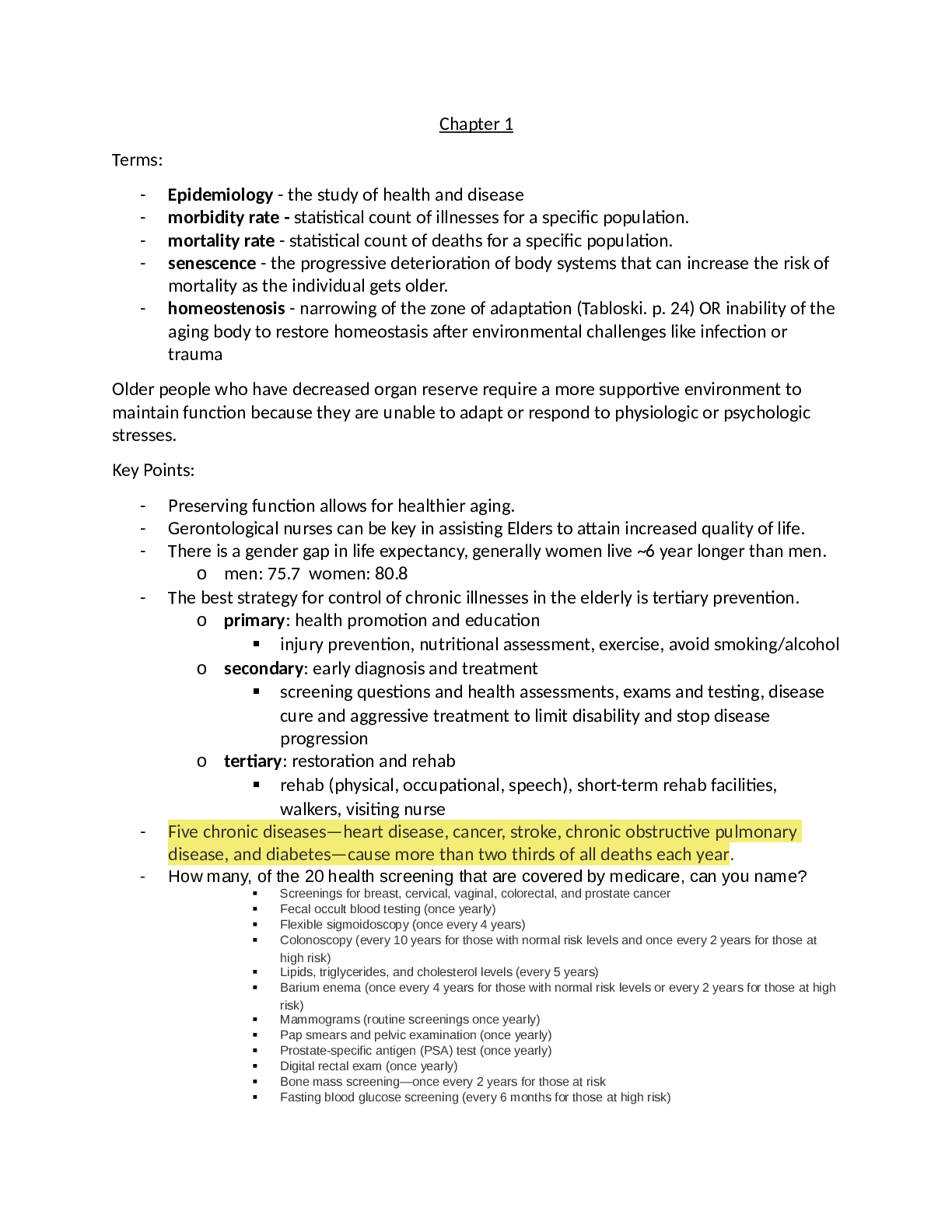
Chapter 1 Terms: - Epidemiology - the study of health and disease - morbidity rate - statistical count of illnesses for a specific population. - mortality rate - statistical count of deaths for a...
By AMAZING GRACE , Uploaded: Aug 10, 2022
$9
Medical Studies> Test Prep > The respiratory system test prep 2022 (All)

The respiratory system test prep 2022
The respiratory system test prep 2022
By Nightingale , Uploaded: Aug 05, 2022
$8
*NURSING> Test Prep > ATI Care of Children RN 2019 Proctored Exam - Level 3! QUES TION A ND A NSWERS LA TES T VERSION 2 (All)
ATI Care of Children RN 2019 Proctored Exam - Level 3! QUES TION A ND A NSWERS LA TES T VERSION 2
ATI Care of Children RN 2019 Proctored Exam - Level 3! QUES TION A ND A NSWERS LA TES T VERSION 2 022 1. A nurseisassessingaschool-agechild whohasheart failureandistakingfurosemide. Which of the f...
By markstudys , Uploaded: Aug 05, 2022
$12
*NURSING> Test Prep > Nursing_knowledge_Assessment_Practice_Exam Practice Test B (All)
Nursing_knowledge_Assessment_Practice_Exam Practice Test B
HESI Practice Test B
By Nightingale , Uploaded: Aug 01, 2022
$9
Driving Course> Test Prep > Arizona permit test 2022 complete 100% correct answers (All)

Arizona permit test 2022 complete 100% correct answers
Arizona permit test 2022 complete 100% correct answers
By CoursesExams , Uploaded: Jul 27, 2022
$3
English 106> Test Prep > Module: FMT3701 Assignment 2 (All)

Module: FMT3701 Assignment 2
Module: FMT3701 Assignment 2 Unique Number: Student number: Student name: Contents Page Question 1 Pages 3 Question 2 Page 4 Question 3… Page 5 Question 4… Page 6&7 Question1:...
By Stuvia , Uploaded: Jul 27, 2022
$8
General Questions> Test Prep > 2022 California Permit Tests (all tests1-24) Answered. (All)

2022 California Permit Tests (all tests1-24) Answered.
2022 California Permit Tests (all tests1-24) Answered. Which child would require a child passenger restraint system? - A 7-year-old who is 4 feet 8 inches tall When you tailgate other drivers (...
By Expert1 , Uploaded: Jul 26, 2022
$15
Ethical Hacking> Test Prep > UHC 2022 Ethics and Compliance Test (All)
UHC 2022 Ethics and Compliance Test
Agent John is planning to conduct a series of events. Some will be strictly educational, others will be formal presentations of specific UnitedHealthcare plans, and others will be informal marketing...
By MAYEXPERT , Uploaded: Jul 25, 2022
$11
Document information
Connected school, study & course
About the document
Uploaded On
Aug 18, 2022
Number of pages
202
Written in

Additional information
This document has been written for:
Uploaded
Aug 18, 2022
Downloads
0
Views
167