Biology > LECTURE NOTES > BIOL 102 Chapter 7-8 notes latest (2020) – Liberty University / BIOL 102 Chapter 7-8 notes latest (All)
BIOL 102 Chapter 7-8 notes latest (2020) – Liberty University / BIOL 102 Chapter 7-8 notes latest (2020)
Document Content and Description Below
BIOL 102 Chapter 7-8 notes latest (2020) – Liberty University / BIOL 102 Chapter 7-8 notes latest (2020) CHAP 7: Circulating Blood The cardiovascular system consists of a pump—the heart—and... blood vessels that carry pumped blood and substances in it throughout the body. Circulating Blood The cardiovascular system consists of a pump—the heart—and blood vessels that carry pumped blood and substances in it throughout the body. Blood Vessels Blood vessels, including arteries, arterioles, capillaries, venules, and veins, are specialized for different blood transport functions. Disorders of the Cardiovascular System Connections: The Cardiovascular System and Blood in Homeostasis Every day in the United States, in roughly 1,000 people, the heart suddenly stops pumping blood. About half the cases of this sudden cardiac arrest occur in people under 40 and often are due to inborn heart disorders. In older people, heart attack is a common culprit. Regardless, in nearly 90 percent of cases sudden cardiac arrest is fatal. Blood carries oxygen, and when the flow abruptly stops, oxygen-deprived brain cells begin to die unless the heart can be restarted. Later in this chapter you’ll learn about emergency procedures that can save the lives of cardiac arrest victims. First, however, we examine the normal functioning of the cardiovascular system—the heart and blood vessels that together perform the vital function of circulating blood throughout the body. Homeostasis Preview The cardiovascular system delivers oxygen, nutrients, hormones, and other substances to body cells. It carries away wastes and substances cells produce. Flowing blood also carries excess heat to the body surface. --- --- 7.1 The Cardiovascular System: Moving Blood through the Body The cardiovascular system is built to rapidly transport blood to every living cell in the body. The Heart and Blood Vessels Make up the Cardiovascular System “Cardiovascular” comes from the Greek kardia (heart) and the Latin vasculum (vessel). As you can see in Figure 7.1 the cardiovascular system has two main elements, the heart and blood vessels. The heart is a muscular pump that generates the pressure required to move blood throughout the body. Blood vessels are tubes of different diameters that transport blood cardiovascular system Organ system that is composed of the heart and blood vessels and that functions in the rapid transport of blood to and from tissues. heart Muscular pump that keeps blood circulating through the body. Arteries, which carry oxygenated blood to tissues, are shaded red. Veins, which carry deoxygenated blood away from tissues, are shaded blue. Notice, however, that for the pulmonary arteries and veins the roles are reversed. The heart pumps blood at high pressure into arteries, which have a large diameter. From there blood flows into smaller and narrower vessels called arterioles (ar-teer-ee-ole), which branch into even narrower capillaries [L. capillus, hair]. Fluids and solutes diffuse out of the capillaries into the extracellular fluid and eventually enter body cells. In the reverse route, fluid and solutes move from body cells into the extracellular fluid and from there into the blood in capillaries. Blood flows from capillaries into small venules, then into large-diameter veins that return blood to the heart. As you will read later on, the volume of blood flowing to a particular part of the body is adjustable. So is the rate at which it flows. This flexibility permits the cardiovascular system to deliver blood in ways that suit conditions in different parts of the body. For example, blood flows rapidly through arteries, but in capillaries it must flow slowly so that there is time for substances moving to and from cells to diffuse into and out of the extracellular fluid. arteries Any of the large-diameter blood vessels that conduct deoxygenated blood to the lungs and oxygenated blood to all body tissues. The thick, muscular artery wall allows arteries to smooth out pulsations in blood pressure caused by heart contractions. arterioles Any of the blood vessels between arteries and capillaries. They are control points where the volume of blood delivered to different body regions can be adjusted. capillaries A thin-walled blood vessel that functions in the exchange of gases and other substances between blood and interstitial fluid. venules Small blood vessel that receives blood from tissue capillaries and merges into larger-diameter veins; a limited amount of diffusion occurs across venule walls. veins Of the circulatory system, the large-diameter vessels that lead back to the heart. The Cardiovascular System is Linked to the Lymphatic System The heart’s pumping action puts pressure on blood flowing through the cardiovascular system. Partly because of this pressure, small amounts of water and some proteins dissolved in blood are forced out and become part of extracellular fluid (the fluid outside cells). A network of drainage vessels picks up excess extracellular fluid and usable substances in it and returns them to the cardiovascular system. This vessel network is part of the lymphatic system, which includes organs with major roles in body defenses. We consider it more fully in Chapter 9. Blood Circulation is Essential for Maintaining Homeostasis The heart and blood vessels are sometimes referred to as the “circulatory system.” This name is apt because blood circulates through the system, bringing body cells such essentials as oxygen and nutrients from food. Circulating blood also takes away the wastes produced by our metabolism, along with excess heat. In fact, cells depend on blood to constantly pick up and deliver an extremely diverse range of substances, including those that move into or out of the digestive system, the urinary system, and the respiratory system SUMMARY: The cardiovascular system consists of the heart and blood vessels. The system transports blood to and from all living cells in the body. Some fluid and dissolved substances in blood move from blood vessels to the extracellular fluid. The lymphatic system collects excess extracellular fluid and returns it—along with useful substances it contains—to the cardiovascular system. --- --- 7.2 The Heart: A Muscular Double Pump The heart is a durable pump that consists mainly of cardiac muscle. Your heart is located more or less in the center of your chest (Figure 7.4A). Its structure reflects its role as a long-lasting pump. The heart wall is mostly cardiac muscle tissue, the myocardium (Figure 7.4B). A tough, fibrous sac, the pericardium (peri = “around”), surrounds, protects, and lubricates it. The heart’s chambers have a smooth lining (endocardium) composed of connective tissue and a layer of epithelial cells. This kind of epithelial cell layer, known as endothelium, also lines blood vessels. myocardium The cardiac muscle tissue. Location of the heart in the chest. B The heart’s internal anatomy. Blue arrows represent blood flow into and out of the right ventricle. Red arrows represent blood flow into and out of the left ventricle. C The shapes of heart valves. The Heart Has Two Halves and Four Chambers A thick wall, the septum, divides the heart into two halves, right and left. Each half has two chambers: an atrium (ay-tree-um) (plural: atria) located above a larger ventricle (ven-tri-kul). Flaps of membrane separate the two chambers and serve as a one-way atrioventricular valve (AV valve) between them. The AV valve in the right half of the heart is called a tricuspid valve because its three flaps come together in pointed cusps (Figure 7.4C). In the heart’s left half, the AV valve consists of just two flaps; it is called the bicuspid valve or mitral valve. Tough, collagen-reinforced strands (chordae tendineae, or “heartstrings”) connect the AV valve flaps to cone-shaped muscles that extend out from the ventricle wall. When a blood-filled ventricle contracts, this arrangement prevents the flaps from opening backward into the atrium. Each half of the heart also has a valve between the ventricle and the arteries leading away from it. The pulmonary valve controls blood flow to the pulmonary artery, and the aortic valve controls blood flow to the aorta. Because both these valves are shaped like a half-moon, they are also known as “semilunar” valves. During a heartbeat, the valves open and close in ways that keep blood moving in one direction, out of the heart. atrium Upper chamber in each half of the heart; the right atrium receives deoxygenated blood (from tissues) entering the pulmonary circuit of blood flow, and the left atrium receives oxygenated blood from pulmonary veins. ventricle Of the heart, one of two chambers from which blood is pumped out. Compare atrium. atrioventricular valve One-way flow valve between the atrium and ventricle in each half of the heart. pulmonary valve Valve in the heart that opens from the right ventricle into the pulmonary artery. aortic valve Valve that opens from the left ventricle into the aorta. In a “Heartbeat,” The Heart’s Chambers Contract, Then Relax Blood is pumped each time the heart beats. It takes less than a second for a “heartbeat”—one sequence of contraction and relaxation of the heart chambers. This - - - - - - - - - - - - - CHAP 8: This chapter expands on our discussion of the cardiovascular system in Chapter 7. Red blood cells contain the oxygen-carrying protein hemoglobin (2.12), and all blood cells develop in red bone marrow (5.2). Our discussion here of blood typing shows a key function of the recognition proteins in cell plasma membranes (3.4). Section 8.7 on blood clotting provides good examples of how enzymes catalyze chemical reactions that are vital to life (2.8). Blood consists of plasma, red blood cells, white blood cells, and platelets. Red blood cells carry and , white blood cells function in defense, and platelets help clot blood. Circulating blood helps maintain proper pH and body temperature. Blood Types Surface markers on red blood cells establish each person’s blood type. Blood Clotting Mechanisms that clot blood help prevent blood loss. When Carlos needed a leaky heart valve repaired, the surgeon urged that he have some of his blood drawn and stored in case a transfusion was needed. A major reason for the precaution: Carlos had type blood. A complex gene-based system determines the characteristic we call blood type. Because relatively few people inherit type , it often is in short supply in blood banks. And if Carlos were to need blood during the surgery, only donor blood that was a near-perfect genetic match to his would be usable. Otherwise, his immune system would launch a potentially fatal attack on the replacement cells. Blood’s “type” is only one of the key characteristics of blood. As you’ll learn in this chapter, an amazingly versatile substance courses through your arteries and veins. Homeostasis Preview Blood transports oxygen, nutrients, and other materials. It also carries ions important for maintaining proper pH in extracellular fluid. Blood-borne proteins have many physiological roles. Some function in blood clotting, which prevents minor injuries from leading to dangerous blood loss. --- --- 8.1 Blood: Plasma, Blood Cells, and Platelets Blood is a sticky fluid that consists of water, blood cells, and other substances. Links to Properties of water 2.5, Proteins 2.11, Osmosis 3.10, Skeleton 5.2 It is literally true that “blood is thicker than water.” Blood consists of plasma, blood cells, and cell fragments called platelets. If you are a woman of average size, your body has about 4 to 5 liters of this unusual fluid. Men have slightly more. In all, blood amounts to about 6 to 8 percent of your body weight. Plasma is the Fluid Part of Blood If you whirl a prepared blood sample in a centrifuge, the test tube’s contents should look like what you see in Figure 8.1. About 55 percent of whole blood is plasma (plaz-muh). Plasma is mostly water. It transports blood cells and fragments called platelets, and over a hundred other substances. Most of these “substances” are different plasma proteins, which have a variety of functions. plasma Liquid portion of blood; consists of water, various proteins, ions, sugars, dissolved gases, and other substances. Blood Consists of Cells, Platelets, and Plasma. In the micrograph (A) the red disks are red blood cells. Platelets are pink. The wrinkled gold balls are white blood cells. The plasma portion (B) makes up a little over half of the volume of blood. Plasma proteins determine blood’s fluid volume—how much of it is water. Two-thirds of plasma proteins are albumin molecules made in the liver. Because there is so much of it—that is, because its concentration is so high— albumin has a major influence on the osmotic movement of water into and out of blood. Albumin also carries many substances in blood, from wastes to therapeutic drugs. Other plasma proteins include certain hormones and proteins involved in immunity and blood clotting. Lipoproteins carry lipids, and still other plasma proteins transport fat-soluble vitamins. Plasma proteins are in high demand for various therapeutic uses, and in many countries people who wish to donate plasma can do so at government-regulated collection centers. Plasma also contains ions, glucose and other simple sugars, amino acids, various communication molecules, and dissolved gases—mostly oxygen, carbon dioxide, and nitrogen. The ions (such as , , , and ) help maintain the volume and pH of extracellular fluid. The blood cells in your test tube arose from stem cells in red bone marrow. Remember from Chapter 4 that a stem cell is like a “blank slate”—it stays unspecialized and retains the ability to divide. Some of the daughter cells, however, differentiate—they become specialized to carry out particular functions. As Figure 8.2 shows, the formation of specialized blood cells begins with pluripotent (“many powers”) stem cells from which two lines of precursor cells arise. The descendants of lymphoid stem cells circulate mainly in the lymphatic system. Myeloid (“from bone marrow”) stem cells give rise to the other types of circulating blood cells. Animated! Various Types of Blood Cells Form, Each with Its Particular Function. Chemicals called growth factors stimulate the growth and specialization of the different subgroups of blood cells. Red Blood Cells Carry Oxygen and About 45 percent of whole blood—the bottom portion in the test tube in Figure 8.1—consists of erythrocytes (eh-rith-row-site), or red blood cells (RBCs). Each red blood cell is a biconcave disk, like a thick pancake with a dimple on each side. The cell’s red color comes from the iron-containing protein hemoglobin. Hemoglobin transports oxygen that body cells need for aerobic respiration. Red blood cells also carry away some carbon dioxide wastes. erythrocytes [Gk. erythros, red, and kytos, vessel] Red blood cell (RBC). red blood cells (RBCs) Erythrocyte; an oxygen-transporting cell in blood. White Blood Cells Defend and Clean up Leukocytes, or white blood cells, make up a tiny fraction of whole blood. Even so, they play crucial roles in body defense. Some remove dead or worn-out cells, or material identified as foreign to the body. Others target or destroy disease agents such as bacteria or viruses. Most white blood cells go to work after they squeeze out of blood vessels and enter tissues. How many are in the body at any one time varies, depending on your activity level and whether you are healthy or fighting an infection. In different types of white blood cells, the nucleus varies in its size and shape, and there are other differences as well. For example, some contain granules that become visible when the cells are stained for viewing under a microscope. This group (called granulocytes) includes neutrophils, eosinophils, and basophils. All have roles in body defenses that you will read more about in Chapter 9. Other leukocytes don’t have visible granules in their cytoplasm (and so are agranulocytes). Those known as monocytes develop into macrophages, “big eaters” that enter tissues and engulf and destroy invading microbes and debris. Another type, lymphocytes, operates in immune responses discussed in Chapter 9. Some types of white blood cells may live for years, but most types live for only a few days or, during a major infection, perhaps a few hours. Leukocytes, or white blood cells Leukocyte; any of the macrophages, eosinophils, neutrophils, and other cells that are the central components of the immune system. Platelets Help Clot Blood Some stem cells in bone marrow develop into “giant” cells called megakaryocytes (mega = “large”). These cells shed bits of cytoplasm enclosed in a plasma membrane. The fragments, known as platelets (playt-let), last only about a week, but millions are always circulating in our blood. Platelets release substances that begin the process of blood clotting described in Section 8.7. platelets A cell fragment in blood that releases substances necessary for blood clotting. SUMMARY: Blood consists of plasma, red blood cells, white blood cells, and cell fragments called platelets. --- --- 8.2 How Blood Transports Oxygen Transporting oxygen is a key function of blood, and the key to oxygen transport is the protein called hemoglobin. Hemoglobin is the Oxygen Carrier If you were to analyze a liter of blood drawn from an artery, you would find only a quarter teaspoon of oxygen dissolved in the plasma—just 3 milliliters. Yet, like all large, active, warm-bodied animals, we humans require a lot of oxygen to maintain the metabolic activity of our cells. The protein hemoglobin (heem-oh-glow-bin) (Hb) meets this need. In addition to the small amount of dissolved oxygen, a liter of arterial blood usually carries around sixty-five times more bound to the heme groups of hemoglobin molecules. This oxygen-bearing hemoglobin is called oxyhemoglobin. hemoglobin [Gk. haima, blood, and L. globus, ball] Iron-containing, oxygen-transporting protein that gives red blood cells their color oxyhemoglobin A hemoglobin molecule that has oxygen bound to it. What Determines How Much Oxygen Hemoglobin Can Carry? As conditions change in different tissues and organs, so does the tendency of hemoglobin to bind with and hold on to oxygen. Several factors influence this process. The most important factor is how much oxygen is present relative to the amount of carbon dioxide. Other factors are the temperature and acidity of tissues. Hemoglobin is most likely to bind oxygen in places where blood plasma contains a relatively large amount of oxygen, where the temperature is relatively cool, and where the pH is roughly neutral. This is exactly the environment in our lungs, where the blood must take on oxygen. By contrast, metabolic activity in cells uses oxygen. It also increases both the temperature and the acidity (lowers the pH) of tissues. Under those conditions, the oxyhemoglobin of red blood cells arriving in tissue capillaries tends to release oxygen, which then can enter cells. We can summarize these events this way: The protein portion of hemoglobin also carries some of the carbon dioxide wastes that cells produce, along with hydrogen ions that affect the pH of body fluids. You’ll read more about hemoglobin in Chapter 10, where we examine how the respiratory system carries out exchanges of oxygen and carbon dioxide. You can see the structure of a hemoglobin molecule in Figure 8.3. Notice that it has two parts: the protein globin and heme groups that contain iron. Globin is built of four linked polypeptide chains, and each chain is associated with a heme group. It is the iron molecule at the center of each heme group that binds oxygen. Therefore, each hemoglobin molecule can carry four oxygen atoms. This diagram represents hemoglobin, which is a globular protein that has four iron-containing heme groups. Oxygen binds to the iron in heme groups, which is one reason why humans require iron as a mineral nutrient. Oxygen in the lungs diffuses into the blood plasma and then into individual red blood cells. There it binds with the iron in hemoglobin. This oxyhemoglobin is deep red. Hemoglobin that is depleted of oxygen looks purplish, especially when it is observed through skin and the walls of blood vessels. SUMMARY: Hemoglobin in red blood cells carries oxygen. Oxygen binds to iron in heme groups in each hemoglobin molecule. The relative amounts of oxygen and carbon dioxide present in blood and the temperature and acidity of tissues affect how much oxygen hemoglobin binds—and therefore the amount of oxygen available to tissues. --- --- 8.3 Making New Red Blood Cells Red blood cells do not live long. In response to hormones, stem cells in bone marrow constantly produce new ones. Every minute, about 180 million new red blood cells enter your bloodstream. Each one gradually loses its nucleus and other organelles. Those structures are unnecessary because red blood cells don’t divide or make new proteins. Red blood cells have enough enzymes and other proteins to function for about 120 days. As they near the end of their life, die, or become damaged or abnormal, phagocytes called macrophages remove them from the blood. Much of this cleanup occurs in the spleen, which is located in the upper left abdomen. As a macrophage dismantles a hemoglobin molecule, amino acids from its proteins return to the bloodstream and the iron in its heme groups returns to red bone marrow. There the iron may be recycled in new red blood cells. The rest of the heme group is converted to the orangish pigment bilirubin. Liver cells take up this pigment, which is mixed with bile that is released into the small intestine during digestion. Steady replacements from stem cells in bone marrow keep a person’s red blood cell count fairly constant over time. A complete blood count (CBC) is a tally of the number of red blood cells, white blood cells, and platelets in a microliter of blood. On average, an adult male’s red blood cell count is around 5.4 million. In an adult female the count averages about 4.8 million red blood cells. complete blood count (CBC) The number of red blood cells, white blood cells, and platelets in a microliter of blood. Having a stable red blood cell count is important for homeostasis, because body cells need a reliable supply of oxygen. Your kidneys make erythropoietin (EPO). This hormone stimulates the production of new red blood cells when they are needed. The process relies on a negative feedback loop (Figure 8.4). In this loop, the kidneys monitor the level of oxygen in your blood. When it falls below a set point, kidney cells detect the change and soon release EPO. It stimulates stem cells in bone marrow to produce more red blood cells. As new red blood cells enter your bloodstream, the blood can carry more oxygen and the oxygen level rises in your blood and tissues. This information feeds back to the kidneys. They make less erythropoietin, and production of red blood cells in bone marrow drops. [Show More]
Last updated: 1 year ago
Preview 1 out of 62 pages
Reviews( 0 )
Recommended For You
Physiology> LECTURE NOTES > Gastrointestinal physiology- GI motility (All)
Gastrointestinal physiology- GI motility
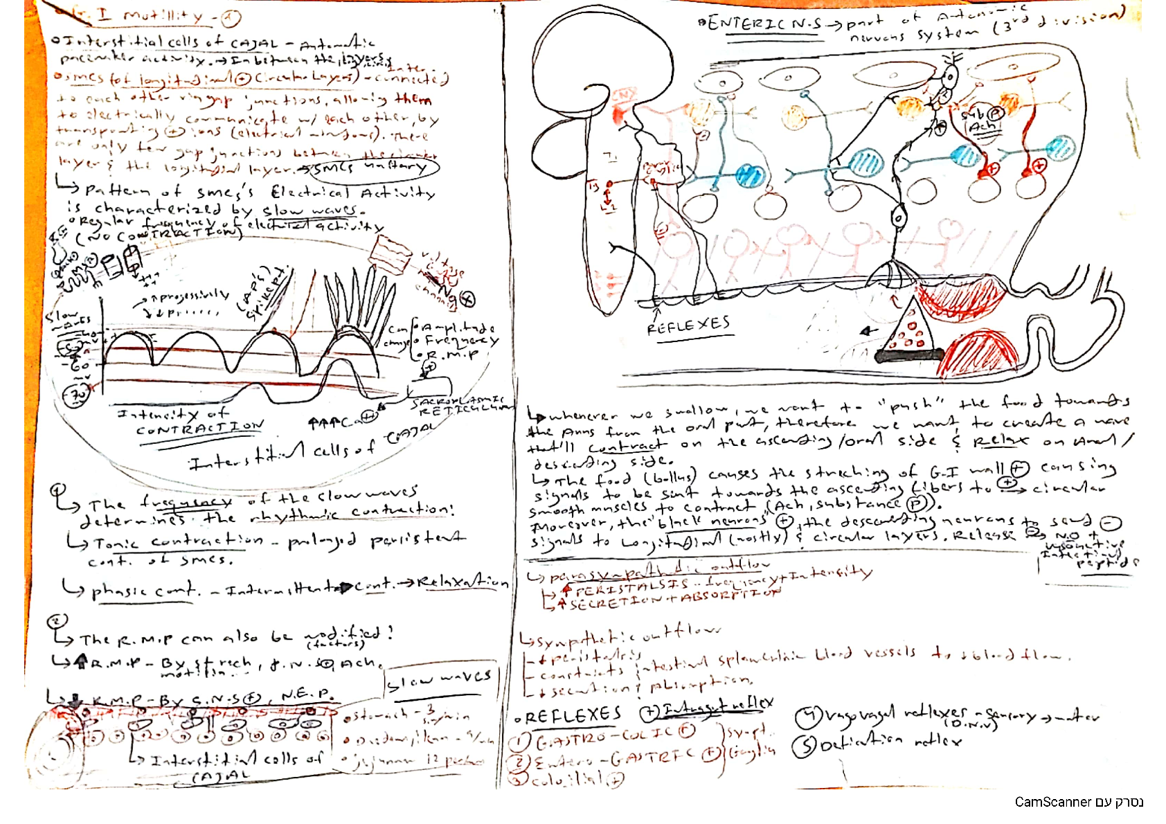
This document is regarding gastrointestinal physiology- stomach, small intestinal, large intestinal and enteric nervous system. GI motility is defined by the movements of the digestive system, and...
By Dan18268 , Uploaded: Jan 17, 2024
$7
BioChemistry> LECTURE NOTES > C785 BIOCHEMISTRY NOTES (WESTERN GOVERNORS UNIVERSITY) (All)
C785 BIOCHEMISTRY NOTES (WESTERN GOVERNORS UNIVERSITY)
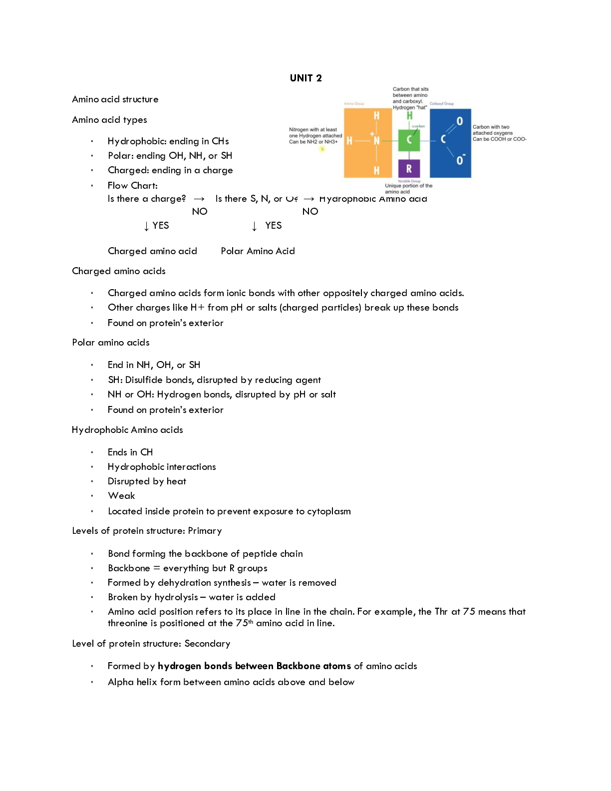
Amino acid structure Amino acid types • Hydrophobic: ending in CHs • Polar: ending OH, NH, or SH • Charged: ending in a charge • Flow Chart: Is there a charge? → Is there S, N, or O? → Hydrophob...
By Professor Marjorie Barker , Uploaded: Sep 25, 2022
$5.5
Management> LECTURE NOTES > Mnb1601 Summarise - Lecture Notes Unisa (All)
Mnb1601 Summarise - Lecture Notes Unisa
MNB1601 NOTES OPERATIONS MANAGEMENT The operations function is that function of the business aimed at executing the transformation process. The importance of operations management: It can reduce the...
By ACADEMICTUTORIAL , Uploaded: Nov 29, 2021
$1.5
*NURSING> LECTURE NOTES > Mark Klimek Lecture Notes NCLEX REVIEW (All)

Mark Klimek Lecture Notes NCLEX REVIEW
Mark Klimek Lecture Notes NCLEX REVIEW
By ACADEMICTUTORIAL , Uploaded: Nov 30, 2021
$3
Psychology> LECTURE NOTES > PSYCH 104 Lecture Notes Chapters-1-7 & 11 (All)
PSYCH 104 Lecture Notes Chapters-1-7 & 11
PSYCH 104 Lecture Notes Chapters-1-7 & 11
By ACADEMICTUTORIAL , Uploaded: Nov 30, 2021
$2.5
*NURSING> LECTURE NOTES > NR 283 Pathophysiology notes ch 1,2, 21 (All)

NR 283 Pathophysiology notes ch 1,2, 21
NR 283 Pathophysiology notes ch 1,2, 21
By ACADEMICTUTORIAL , Uploaded: Mar 16, 2022
$3.5
*NURSING> LECTURE NOTES > NR 509 Adv Physical Assessment- Midterm Notes (All)
NR 509 Adv Physical Assessment- Midterm Notes
NR 509 Adv Physical Assessment- Midterm Notes Soap note example for the patient. This is a great start to your learning and practice.
By ACADEMICTUTORIAL , Uploaded: Mar 20, 2022
$4
*NURSING> LECTURE NOTES > Mark klimek lecture 1-12 notes (All)
Mark klimek lecture 1-12 notes
Mark Klimek 1-12 notes for NCLEX test review.
By ACADEMICTUTORIAL , Uploaded: Mar 16, 2023
$4.5
*NURSING> LECTURE NOTES > Mark Klimek Test taking strategies (All)
.png)
Mark Klimek Test taking strategies
Mark Klimek Test taking strategies Lab Values: DEADLY DANGEROUS: Elevated K+ ( >6) - Hold K+, Assess heart, Prepare Kayexalate/D5W, Call Dr. Elevated pH ( >6) - Assess Vitals, Call doctor ...
By Kirsch , Uploaded: Mar 14, 2023
$9
Biology> LECTURE NOTES > Lecture Materials > University of Louisville BIO 240 Ch 11 Cell Signaling Notes (All)

Lecture Materials > University of Louisville BIO 240 Ch 11 Cell Signaling Notes
University of Louisville BIO 240 Ch 11 Cell Signaling Notes Lecture Notes Local and Long Distance Signaling o Communication between cells using gap junctions (animal cells) or plasmodesmata (p...
By QuizMaster , Uploaded: Aug 02, 2022
$4
Document information
Connected school, study & course
About the document
Uploaded On
May 28, 2020
Number of pages
62
Written in

Additional information
This document has been written for:
Uploaded
May 28, 2020
Downloads
0
Views
163