*NURSING > STUDY GUIDE > Chamberlain College of Nursing: NR 283 Patho Exam 2,100% CORRECT (All)
Chamberlain College of Nursing: NR 283 Patho Exam 2,100% CORRECT
Document Content and Description Below
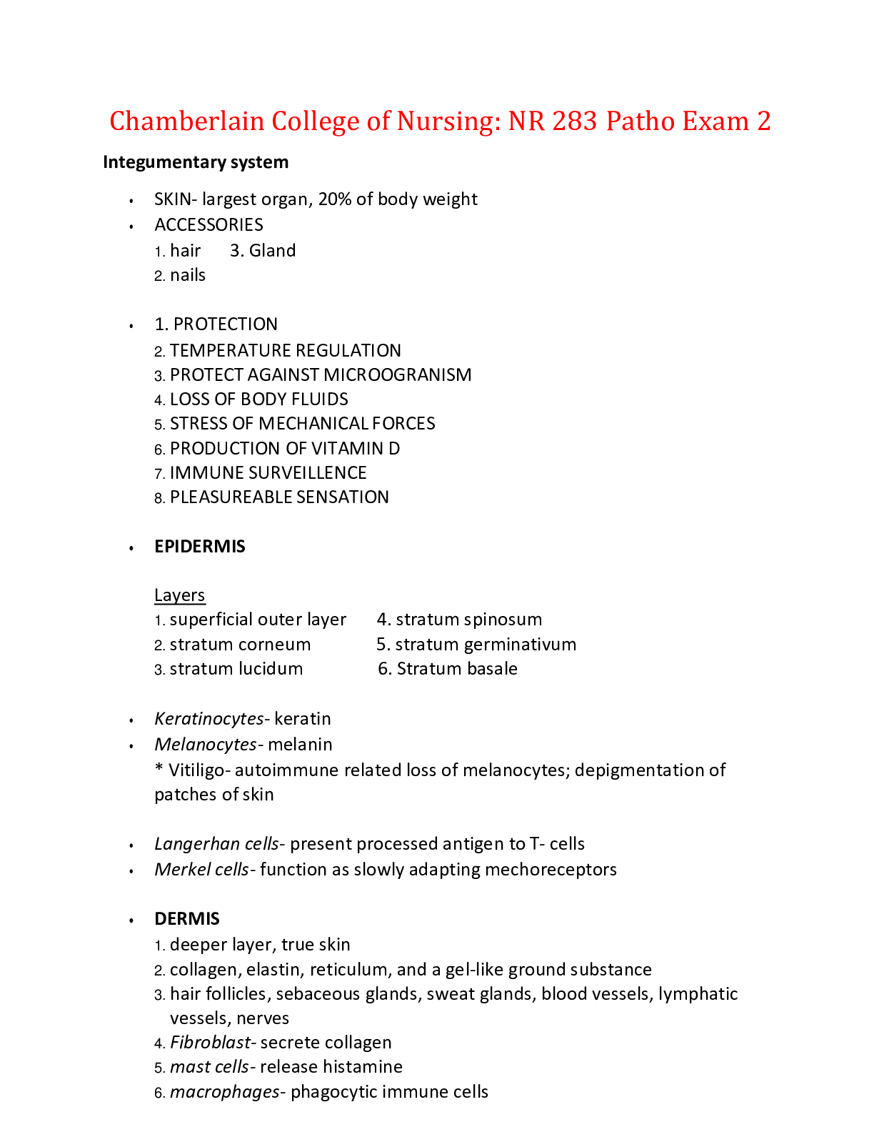
Chamberlain College of Nursing: NR 283 Patho Exam 2 Integumentary system • SKIN- largest organ, 20% of body weight • ACCESSORIES 1. hair 3. Gland 2. nails • 1. PROTECTION 2. TEMPERATURE... REGULATION 3. PROTECT AGAINST MICROOGRANISM 4. LOSS OF BODY FLUIDS 5. STRESS OF MECHANICAL FORCES 6. PRODUCTION OF VITAMIN D 7. IMMUNE SURVEILLENCE 8. PLEASUREABLE SENSATION • EPIDERMIS Layers 1. superficial outer layer 4. stratum spinosum 2. stratum corneum 5. stratum germinativum 3. stratum lucidum 6. Stratum basale • Keratinocytes- keratin • Melanocytes- melanin * Vitiligo- autoimmune related loss of melanocytes; depigmentation of patches of skin • Langerhan cells- present processed antigen to T- cells • Merkel cells- function as slowly adapting mechoreceptors • DERMIS 1. deeper layer, true skin 2. collagen, elastin, reticulum, and a gel-like ground substance 3. hair follicles, sebaceous glands, sweat glands, blood vessels, lymphatic vessels, nerves 4. Fibroblast- secrete collagen 5. mast cells- release histamine 6. macrophages- phagocytic immune cells * Histocytes- loose connective tissue; phagocytize pigments and debris of inflammation • SUBCUTANEOUS LAYER 1. fat cell or adipocytes and connective tissue 2. dermal collagen is continuous w/ the subcutaneous collagen 3. macrophages, fibroblast, fat cells, nerves, fine muscles, blood vessels, lymphatics, and hair root follicles • DERMAL APPENDAGES 1. hair, nails 2. Sebaceous glands- secrete sebum 3. Eccrine sweat glands- thermoregulate and cool the body through evaporation 4. Apocrine sweat glands – limited function • Blood supply and innervation 1. papillary capillaries provide a rich supply of blood 2. sympathetic nervous system regulates vasoconstriction and vasodilation through a- adrenergic receptors • PRIMARY LESIONS 1. Macule- flat, circumscribed area that a change in the color of skin (freckles, measles) 2. wheal- transient lesion w/ well defined and often changing borders caused by edema of the dermis ( insect bites, allergic reactions) 3. Papule- elevated, firm, and circumscribed area, measures less than 1 cm (warts) 4. Pustule- elevated, superficial lesion, filled with fluid ( acne, impetigo) • SECONDARY LESIONS 1. Excoriation- loss of epidermis, linear, hollowed, crusted (scabies, scratches) 2. Fissure- linear crack or break from the epidermis to the dermis, maybe moist and dry (athlete’s foot) 3. Erosion- loss of a part of the epidermis, depressed area, moist, glisten, rupture of a vesicle or bulla (chicken pox, diaper dermatitis) 4. Ulcer- loss of epidermis and dermis, concave, varies in size (pressure sore) 5. pressure ulcer- result of an unrelieved pressure on skin, causing underlying tissue damage * shearing forces, friction, moisture * occlude capillary blood flow w/ resulting ischemia and necrosis * STAGE 1 – nonblanchable erythema of intact skin * STAGE 2- partial thickness, skin loss, involves epidermis or dermis * STAGE 3- full thickness, skin loss, involving damage or subcutaneous tissue * STAGE 4- Full thickness, skin loss w/ damage to muscle, bone, or supporting structures • Decubitis ulcer- results when an individual lies or sits in one position for an extended period of time • Dermatitis or Eczema 1. most common 2. characterized by pruritus, lesions w/ distinct borders, epidermal changes 3. Chronic eczema- thickened, lethargy, hyperpigmented skin from recurrent irritation and scratching • Psoriasis- chronic, relapsing, proliferative skin disorder 1. T- cell autoimmune-mediated skin disease 2. scaly, thick, silvery, elevated lesions 3. epidermal turnover- 26 to 30 day, 3 to 4 days 4. cells don’t have time to mature and keratinize 5. Ultraviolet (B), Keratolytic agents, corticosteroids, maintain skin moisture • Cellulitis- infection of dermis and subcutaneous tissue 1. staphylococcus, group B, streptococci 2. systemic antibiotics, Burrow soaks, corticosteroids • Impetigo- superficial lesion of skin * common in children * coagulase – positive staphylococcus or a a-hemolytic streptococci • Herpes simplex virus (HSV) 1. HSV1 * contact w/ infected saliva * cold sores, fever blisters * antiviral topical agents 2. HSV2 * Genitial infections * skin to skin mucous membrane contact during viral shedding * vertical transmission from mother to neonate * associated w/ neonate morbity and mortality * antiviral topical agents • Herpes zoster (shingles) and Varicella (chickenpox) *varicella- zoster virus ( VZV) * intitial infection w/ varicella, followed yrs later by herpes zoster 1. pain and paresthesia localized to dermatom 2. vesicular eruptions along a facial, cervical, or thoracic lumbar dermatome * compresses, calamine lotion, baking soda * anti-viral drugs w/in 72 hours * Topical lidocaine patch, anticonvulsant, tricyclic antidepressants • Basal cell carcinoma and squamous cell carcinoma (most common) • Malignant melanoma (most serious) most common cause of skin cancer 1. chronic UV radiation 2. sunscreen, protective clothing • Kaposi sarcoma vascular malignancy * drug-induced immunosuppression (after kidney transplant) * exhibited on lower legs of man (classic form) * epidemic and nonepidemic form related AIDS *Kaposi- associated herpes virus 8 (HHV-8) found in all forms *purpleish – brown macules, developing into plaques and nodules *local lesions- surgical removal * multiple disseminated- combination of immunomodulatory, cytotoxic, and antiviral drugs *highly active antiretroviral therapy (HAART) for AIDS • Aging skin *reflects environmental and genetic changes *thinner, dryer, wrinkles, and changes in pigmentation *# of capillary loops shorten and decrease * melanocytes and Langerhan cells decrease *sebaceous, eccrine, and apocrine gland atrophy *wound healing delayed *temp. regulation delayed * pressure, touch receptors, free nerve endings decrease • Pityriasis rosea- *Circular single lesion *well demorcrated *salmon pink * secondary lesions extend over trunk and extremities • Vegetans pemphigus- large blisters in tissue folds of the axilla and groin area • Erythmea multiforme- “BULLS EYE” or target lesions • Acne Rosacea *inflammation of the skin *bulbous nose *conjunctivitis • Furuncles- inflammation of the hair follicles • Tinea capitis- fungal infection of the scalp • Scleroderma- massive deposits of collagen w/ fibrosis • Pulmonary disease *dyspen and cough * cyanosis * altered breathing * chest pain * Hyperventilation * clubbing * hemoptysis * abnormal sputum • Dyspnea *subjective sensation of uncomfortable breathing * severe dyspnea 1. flaring nostrils 2. use of accessory muscles of respiration 3.retraction of intercostal spaces * dyspnea on exertion 1. shortness of breath w/ activity *orthopynea- dyspynea w/ lying down *paroxysmal nocturnal dyspnea- awaking at night and gasping for air (need to sit or stand up) • Cough- protective reflex that helps clear the airways by explosive expiration *acute cough- resolves in 2 to 3 weeks *chronic cough- lnger than 3 wks *abnormal sputum – changes in the amount, consistency, color, odor, provide information about disease • Hemoptysis- coughing up blood or bloody secretions • Eupnea- normal breathing patterns • Abnormal breathing patterns are adjustments made by the body to minimize the work of respiratory muscles • Kussmal respiratory (hyperpnea) *slightly increased ventilatory rate * large tidal volume * no expiratory pause • Labored breathing- increased work of breathing • Restricted breathing- disorders that stiffens the lungs or chest wall and decrease compliance • Cheyne- stokes respirations- alternating periods of deep and shallow breathing *apnea lasting for 15 to 60 seconds, followed by ventilations that increase in volume until a peak is reached, after which ventilation decreases again to apnea • Hypoventilation * alveolar ventilation leads to acidosis *hypercapnia * airway obstruction, chest wall restriction, altered neurologic control of breathing • Hyperventilation *alveolar ventilation leads to alkalosis *hypocapnia *anxiety, hypoxemia, head injury • Cylindrical bronchial dilation = symmetrically dilated airways • Cyanosis- bluish discoloration of skin and mucous membranes *develops when there is 5 grams of desaturated hemoglobin * Peripheral cyanosis- (nail beds) poor circulation *central cyanosis – decrease arterial oxygenation (low partial pressure of oxygen PaO2) (buccal mucous membranes, lips) • Chest trauma may result in increased pressure in chest cavity w/ resulting collapse of right lung • Parietal pleura infected may result friction rub • Pleural Pain *most common pain in pulmonary disease *sharp or stabbing pain *infection and inflammation of parietal pleura accompanied by pleura stretch during inspiration and pleura friction rub • Chest wall pain may due to airway, muscle, or rib pain • Hypercapnia- increased carbon dioxide (Co2) in arterial blood *occurs from decreased drive to breathe or an inadequate ability to respond to ventililatory stimulation. * caused by 1. disease of medulla 2. large airway obstruction 3. thoracic cage abnormalities 4. depression of respiratory center • Hypocapnia *assessed for sever anxiety *results in alkalosis *confirmed by PaCO<sub>2/sub>36mmHg • Hypoxemia- *shunting * alvelolar dead space – area where alveoli are ventilated but not perfused • Pneumonia- infection of lower respiratory tract *responsible for more disease and death than any other infection *streptococcus pneumonia- community acquired pneumonia) * healthcare associated pneumonia *ventilator – associated pneumonia * ROUTES OF INFECTION- aspiration, inhalation, endotracheal tubes, and suctioning • Pneumoccal pneumonia- acute lung injury (ALI), resulting in inflammatory cytokines and cells, causes alveolar edema *edema aids in the spread of infection into adjacent portions of lung *involved lobe undergoes consolidation *PHASES 1. consolidation 2. red hepatization 3. grey hepatization 4. Resolution • Viral Pneumonia *seasonal, mild, self-limiting * can set the stage for secondary bacterial infection *Influenza *chills, malaise, pleuritic chest pain, cough, fever, dyspnea, proceeded upper respiratory infection * vaccination, antibiotics, isolate, deep breathing, hydration, adequate O2, anti-viral/anti-fungal in severe cases • Nosocomial pneumonia- pseudomas aeruginosa ( pathogen) • Aspiration pneumonia risk factors *amount, size, pH, of aspirate *bacterial content present in aspirate • Small cell carcinoma= antidiuretic hormone producing lung tumor • Acute respiratory distress causes *TB, inhalation of excessive amounts of coal dust, Reumatoid arthritis(pulmonary fibrosis) • Chronic cough resulting from viral infection: *return for treatment if cough persists after 8 wks *acess for GERD *acess for allergic rhinitis • Risks for chronic upper airway obstruction *congenital malformations *subglottic stenosis • Tuberculosis *Myobacterium TB *acid-fast bacillus *leading cause of death from a curable infectious disease * air borne *granulomatous lesion *causeous necrosis (cheese-like material) *may remain dormant or cause disease *isolation of bacilli *latent TB = asymptomatic *fatigue, weight loss, anorexia, low grade fever in after noon, night sweats, purulent cough * PPD, chest x- ray, sputum culture *Isoniazid, Rifampin, Pyrazinamide, Ethambutol (for 18 mths, review at 6) • Obstructive pulmonary disease *airway obstruction is worse w/ respiration *emptying lungs is slowed, more force/ time is required to expire *wheezing, dyspnea *increased work of breathing *perfusion mismatching *decreased in FEV (Forced expiratory volume) in 1 sec *can lead to asthma, chronic bronchitis, emphysema, COPD • Asthma- chronic inflammatory disorder of the bronchial mucosa *causes bronchial hyperrepsoniveness, constriction of airways, obstruction * one half of all cases develop in childhood *hygiene hypothesis *familial disorder *bronchospasm attacks, bronchial inflammation, mucosal edema, increased mucosal production *Early asthmatic response ***IgE responses 1. vasodialation 2. increased capillary permeability 3. mucosal edema 4. bronchospasm 5. tenacious mucous secretions 6. antige exposure to bronchial mucosa activates dendritic cells to present antigento CD4+cells 7. IL-4 stimulates B-cells activation and of production of antigen-specific IgE 8. IL-5 stimulates eosinophils, which contribute to increase bronchial hyperresponsiveness 9. IL-8 activates neutrophils 10. IL-13 impairs mucocillary clearance, enhances fibroblast secretions, increase broncho constriction 11. IL 17 increases neutrophilic inflammation 12. IL- 22 stimulates airway epithelial cells, causing further innate and adaptive immune responses *Late asthmatic response 1. 4 to 8 hrs after early response 2. chemotactic recruitment of lymphocytes, eosinophils, and neutrophils 1. prolonged smooth muscle contraction 2. airway scarring 3. increase bronchial hyperresponsiveness 4. impaired mucocillary functions 5. loads to airway remodeling if left untreated 3. increase work of breathing 4. Hypoxemia 5. Hyperinflation * Tachycardia, tachypnea * pulsus paradoxus * O2, bronchodilators, corticosteroids, beta-agonist inhalers • COPD- chronic bronchitis and emphysema *airflow limitation that is not fully reversible * progressive *3rd cause in U.S., 6th worldwide * Tobacco smoke, dusts, chemicals, pollution, *genetics (alpha 1 antitrypsin gene) • Chronic bronchitis- hypersecretion of mucus and chronic productive cough that last for 3 mths of a yr and at least 2 consecutive yrs *hypertrophied bronchial smooth muscle *hypoxemia and hypercapnia *Increase mucous, size, # of mucous glands, bronchial edema, thick mucous * airways collapse thus trapping gas in lungs * decrease exercise tolerance *wheezing, shortness of breath * copious cough (smokers cough) *polycythemia * decrease FEV1 * quit smoking, bronchodilators, expectorants, chest therapy, antibiotics, steroids, O2 • Emphysema- abnormal permanent enlargement of gas exchange air ways accompanied by destruction of alvelolar walls w/ fibrosis *loss of elastic recoil *primary emphysema (genetic) *secondary ( smoke, environment) * destruction of alveoli * oxidative stress *apoptosis of lung’s structural cell *alveolar destruction produces large air spaces w/ in lung parenchyma (bullae) and air spaces adjacent to pleurae (bleb) *dyspnea, tachypnea, BARRELL CHEST, leans forward to increase lung capcity * O2, inhaled bronchodilators, oral or inhaled corticosteroids, antibiotics, anticholinergenics, beta agonist • Pulmonary vascular disease- any condition that affects the blood vessels along the route between the heart and lungs. *causes may be due to pulmonary arterial hypertension, pulmonary venous hypertension, pulmonary embolism, chronic thromboembolic disease • RIGHT HEART FUNCTION 1. pumps blood throughout the lungs (pulmonary circulation) 2. delivers bloods to the lung for O2 3. low pressure system • LEFT HEART FUNCTION 1. pumps blood (oxygenated) through the circulation 2. delivers metabolic waste products to lungs, kidneys, and liver 3. high pressure system • Arteries- carry blood away from the heart (thick walls, oxygenated) • Veins- carry blood to the heart (deoxygenated) • Capillaries- exchange fluids between the blood and interstitial space • Pericardium *double walled membranous sac *parietal- surface layer *visceral- inner layer (epicardium) *prevents displacement, acts as a physical barrier, pain & mechanoreceptors, brings force to normalize BP • Pericardial cavity *space between parietal and visceral layers *contains pericardial fluids (20mL) • Thoracic duct receives lymph from most of the body • Angiotension II - vasoconstriction • CHAMBERS OF THE HEART *right atrium *left atrium *interatrial septum *right ventricle *left ventricle *interventricular septum *thickness of each chamber depends on pressure or resistance it must over come to eject blood • Atrioventricular valves (AV’S)- one way blood flow from the atria to ventricles • Semilunar valves- one way flow from ventricles to either pulmonary artery or aorta • Distole- relaxation (ventricles fill) • Systole- contraction (blood leaves ventricles) • CARDIAC CYCLE *PHASE1- atrial systole or ventricular diastole *PHASE2- isovolumetric ventricular systole *PHASE3- ventricular ejection ( semilunar valves open) *PHASE4- isometric ventricular relaxation (aortic valves close) *PHASE5- passive ventricular filling (mitral and tricuspid valves open) • S1- “LUB” • S2-“DUB” • S3-ventricular gallop (early diastole) • S4- atrial gallop (late diastole) • Caridiac output- HR X STROKE VOLUME = CO • BLOODFLOW 1. right atrium from superior vena cavae 8. left ventricle 2. tricuspid valve 9. aortic valve 3. right ventricle 10. systemic 4. pulmonary trunk 11. inferior vena cavae 5. pulmonary arteries 6. Pulmonary viens 7. left atrium • Coronary circulation – supplies oxygen and other nutrients to the myocardium *coronary arteries protect form ischemia and formed by arteriogenesis or angiogenesis * coronary capillaries- where gas exchange of O2 and other nutrients takes place *coronary viens- coronary sinus, great cardia vein, posterior vein of left ventricle • CONDUCTING SYSTEM *network of specialized tissue that stimulates contraction *modified cardiac myocytes * 1. SA node (60-100 bpm) 2. AV node 3. Bundle of his (AV bundle) 4. Bundle branches 5. purkinje fibers • QRS complex *P wave- depolarization of atria *QRS complex – depolarization of vetricles *T wave- repolarization of ventricles * repolarization of the atria is covered by the complex • EKG *S wave- negative deflection following Rwave *Rwave- initial positive deflection *Q wave- 1st downward wave of QRS complex (Q is often absent) * T wave- ventricular repolarization *p wave –atrial depolarization • Autoregulation *blood flow regulate their own blood flow *coronary circulation= pressure between 60 &180 *ensures constant coronary blood flow *originates in vascular smooth muscles of arterioles • MAP= diastolic+ 1/3 (systolic –diastolic) • Vasa vasarum- source of nutrients for blood vessels • Autonomic nervous system *Influences rate of impulse (firing), depolarization, and repolarization of myocardium *influences strength of atrial and ventricular contraction * produces changes in the heart and circulatory system faster than metabolic or humoral agents • CARDIAC INNERVATION *sympathetic nerves- increase electrical conductivity and the strength of myocardial contraction *parasympathetic nerves- slows conduction of action potentials through the heart and decrease the strength of the contraction • Myocardial contraction- represents the intrinsic ability of the heart/myocardium to contract. Changes in the ability to produce force during contraction result from incremental degrees of binding between myosin (thick) and actin (thin) filaments. • Excitation contraction coupling- process where action potential triggers the cycle of events, leading to a cross bridge activity and contraction *requires calcium * calcium troponin complex facilitates contraction • Myocardial relaxation- *calcium, troponin, tropomysin needed to facilitate * troponin release of calcium begins relaxation *vital to optimal cardiac function • Cardiac output- volume of blood flowing through wither systemic or pulmonary circuit *HR X STROKE VOLUME= CO * ejaction fraction- amount of blood ejected per minute ( 55% or higher in normal adult) 1. indicator of ventricular function 2. stroke volume/ end diastolic volume * preload- pressure generated at the end of diastole 1. AKA- LEFT VENTRICULAR END DIASTOLIC PRESSURE (LVED) 2. determined by 2 factors 1. amount of venous return to ventricle 2. blood left in ventricle after systole or end systolic volume 3. when preload exceeds physiologic range, further muscle stretching causes a decline in CO2. * Afterload 1. resistance to ejection during systole 2. aortic systolic pressure is a good index of afterload for the left ventricle 3. decrease afterload, heart contracts more rapidly 4. increase afterload, slows contractions and increase work load • Frank’s starling law *volume of blood at the end of diastole *myocardial stretch determines force (more stretch= increase force of contraction) *major way that the right and left ventricles maintain equal minute outputs, despite stroke output variation • Myocardial contractibility *stroke volume *changes in stretching of ventricular myocardium caused by changes in ventricular volume (preload) *alterations in nervous system input to the ventricles *adequate of myocardial O2 supply: 1. inotropic agent- increase the force of contraction 1. norepinephrine – from sympathetic nerves 2. epinephrine – from adrenal cortex 3. thyroid hormone and dopamine 2. negative inotropic agents- decrease the force of contraction 1. acetycholine- releases from vagus nerve 3. hypoxia- decreases contractility • Hear rate- 70 BPM *cardiovascular control center *neural reflexes 1. sinus arrhythmia 2. baroreceptors reflex- when BP fall, heart rate increases, arterioles constrict 3. brainbridge reflex- changes in heart rate from IV infusions * atrial receptors *hormones and biochemical • Cardio inhibitory center- parasympathetic excitatory neurons • Systemic circulation *arteries *viens *arterioles *peripheral vascular system – systemic circulation that *capillaries supplies skin and extremities *venules • Angiogenesis- growth of new vessels that branch from existing vessels (branches from small vessels) • Arteriogenesis- branching from larger vessels • Vasculgenesis- growth of vessels from progenitor or stemlike cells that originate in bone marrow and other body tissue • Cigarette smoke can increase thrombotic state • Endothelium Roles *transport *coagulation *antithrombogenesis and fibrolysis *immune system function * tissue growth and wound healing * vasomotion – contraction and relaxation of vessels • Velocity and viscosity affect blood flow • Hypertension – (high BP)- increase cardiac output or total peripheral resistances or both *120-130 systolic/ 80-90 diastolic *older age *hypertension can cause damage to the arteries *can cause heart disease, stroke, kidney disease, bone loss *risk factors- obesity, race, gender, high sodium, sedentary lifestyle, stress, age, family history, congestive heart failure (secondary occurenc) • CORONARY ARTERY DISEASE (atherosclerosis) *fatty streak, raised fibrous plaque, smooth cell proliferation *insufficient blood supply to the heart, the heart muscle does not receive the oxygenated blood it requires *antiplatelets- nitrates, beta adrenergic blockers, calcium channel blockers *chest x ray, lipid level, ECG, stress test, LDH, coronary angioplasty, tomography, cardiac troponin *die (serum lipid levels)t, smoking, lack of exercise, diabetes, age, gender, family history * exercise, B vitamins, lopid, Zocor, Lipitor * can cause atherosclerotic stenosis, coronary artery spasm, coronary thrombosis ( all can cause silent or agina ischemia) *“Raynauds” disease • Angina- (pain) *substernal, GI, Referred *feeling anxious, shortness of breath, weakness, cold sweat * cause dysrhythmia, decrease myocardial contractibility • Prinzmetal’s angina- long duration and may wake a person from sleep • Myocardial infarction- (heart attack) *percutaneous transluminal coronary angioplasty *stent replacement *atheroectomy *longer angioplasty *coronary artery bypass graft • Acute myocardial infarction *arrhythmia (common cause) *substernal pain *diaphoresis * cool, clammy skin *fever • Peripheral artery disease (PVD) *narrow and hardening of arteries that supply blood to the legs and feet *decrease blood flow results in nerve and tissue damage to extremeties *common in men over 50 *gradual onset and asymptomatic *atherosclerosis is a common cause of PVD *smoking, diabetes, hyperlipidemia, A-fib, CAD, CVD, renal disease *leg pain, cyanosis, pallor, weak or absent peripheral pulse *numbess, cool extremities, minimal hair growth * ambulation, properly fitted shoes, keep feet clean and dry, avoid fatty foods, ankle rotation, support hose, radiation or vein stripping • Arteriosclerosis- chronic disease of arterial system *thickening or hardening of vessel walls * smooth muscle cells and collagen fibers migrate to tunica intima • Aneurysm- local dilation or outpouching of a vessel wall or cardiac chamber (aorta is highly susceptible) • Embolism- bolus of matter that is circulation in the blood stream *dislodged thrombus * bacteria *air bubble * cancer cells *amniotic fluid * foreign substance *Aggregate of fat • Varicose veins- vein is which blood has pooled ( prolonged standing, pregnancy, trauma) • Chronic venous insufficiency- inadequate venous return over a long period of time due to varicose veins or valvular incompetence ( venous stasis ulcers) • Deep vein thrombosis- destruction of venous flow leading to increase venous pressure ( venous stasis can be factor) • CONGESTIVE HEART FAILURE- cardiac dysfunction that result in inadequate perfusion of tissues w/ blood borne nutrient *systolic heart failure- pulmonary congestion, left EJ will be greater than 40, filling defect *right heart failure- commonly caused by a diffuse hypoxic pulmonary disease *high output failure- heart can’t keep up w/ demand • Mitral regurgitation- *can be due to abnormalities of valves, annulus, chordae, tendinae, papillary muscle of LV *common causes can be degenerative (myxomatous) disease, ischemia heart disease, infectious endocarditis • Pericardial effusion- (blood effusion) *endocardium inflamed by staphylococcus aureus *resulting in the presence of frank blood (coagulation defect) • Infectious endocarditis *SLE, Marfan’s syndrome, Danlos syndrome *A-fib *forceful, displaced, systolic thrill *soft S1, S3 *Pansystolic *Barlow’s syndrome- floppy mitral valve • Aortic regurgitation *asymptomatic for yrs *develop LV failure (dyspnea, orthopnea, fatigue) *collapsing pulse, laterally displaced apex beat (thrusting), wide pulse pressure *CORRIGAN’S SIGN- carotid pulsations *DE MUSSET’S SIGN- head nodding w/ each beat *QUINCKE’S SIGN- capillary pulsations * TRAUBE’S- “pistol shot” sound over a femoral A. *AUSTIN FINT MUMUR- mid diastolic murmur over cardiac murmur • Aortic stenosis- orifice of the aortic semilunar valves narrow causing diminishing blood flow from left V into aorta • Mitral stenosis- impaired blood flow from left atrium to left ventricle • Dyslipidemia- low lipoproteins (VLDL) triglycerides and proteins • Cellular injury of myocardium – myocardial cells remain viable if blood returns in 20 min. • Hemoglobin carries oxgen • Hematopoiesis- production of blood cells and platelets • Agranulocytes- lymphocytes and monocytes • Granulocytes- neutrophils, basophils, eosinophils • Erythropoietin – a hormone released by the kidneys that stimulate RBC production • Reticulocyte- immature erythrocyte • Thrombopoiesis- endothelial cells primary produced in the liver • Red bone marrow- primary site for production and maturation of blood cells and platelets in adults • Leukocytosis- increase WBC’s • Neutrophilia- increase neutrophil • Neutropenia- decrease neutrophil • Lymphocytosis- increase lymphocytes • Leukopenia- low white blood cell count • Ansiocytosis- RBC’s vary in size • Polychromasia- insufficient hemoglobin w/in the cell • Basophilic stippling- caused by intense regenerative anemia or lead poisioning • Rouleaux- grouping of RBC’s in stacks, when increase globulin and fibrinogen • Anemia *fewer circulating RBC’S * hemmorage *decreased PCV *decreased HgB *decreased production • Regenerative anemia- bone marrow able to respond, increase production, releases reticulocytes • Non- regenerative anemia- bone marrow doesn’t respond, no reticulocytes • Hypoxia- blood loss/anemia, decrease in RBC OR Hb • Polycythemia- excess EPO production by kidneys • Micocytic hypochromic anemia- *iron metabolism *globin synthesis *parphyrin synthesis *heme production • B12- required for nerve myelination, extended deficiency can cause neurological damage 1. absorption of B12 1. complex w/ intrinsic factor 2. transported to ileum 3. B12 combines w/ transcobalamin II to be transported to PB and tissues 4. stored in liver * distributed by TC1 (plasma), TC2 ( cells), TC3 ( liver) * causes of B12 deficiency 1. pernicious anemia 6. Ideal resection and 2. lack of intrinsic factor and Coron’s diease 3. gastrectomy 7. protienuria 4.intestinal stagnant loop syndrome 8. tapeworm 5.chronic tropical sprue • Folate *deficiency causes impaired cell division, altered protein synthesis *folate absorption takes place in the ileum *poluglutamate hydrolyzed by pteroyl polyglutamate *hydrolase * falate deficiency 1. celiac disease 4. Hemolytic diseases 2. sprue 5. Malignant disease 3. pregnancy and lactation 6. Inflammatory disease • Increase folic acid increases BP • Lack of folic acid triples dementia • Old platelets are destroyed by splenic phagocytosis or by Kupffer cells in liver • Thrombathenia- decrease platelet fuction • Clot formation *Factor XIIa (hagemen) activates prothrombin to thrombin *thrombin converts fibrinogen to fibrin • Clot dissolution *Factor VIIa activates plasminogen to plasmin which dissolves fibrin • Vitamin K dependent factors *II, VII, IX, X • Prostacyclin inhibits platelet aggregation • Tissue factor initiates coagulation • Coagulation steps *formation of prothrombin activator *PTA converts prothrombin to thrombin *thrombin converts fibrinogen to fibrin Factor XIIa stabilizes fibrin • Fibrinolytic system acitvators – Factors XIIa (kallikrien), streptokinase, tPA urokinase like A • PT test to monitor oral coagulation treatment • Iron absorption occurs in the Duodeum • Disorders associated w/ thymoma *Myasthenia gravis *pernicious anemia (SLE, ra, aplasia) autoimmune disorders • Newmann Pick disease- sphingomyelinase deficiency • Gaucher’s disease- accumulation of glcusycoramide in lysosomes of reticuoendothelial cells due to glucocerebrosidase deficiency • Kostmann’s syndrome- neutropenic disease • Thalassemia major- anemias w/ bone deformities • Stromtolites present in alcoholism and liver disease • Factors in increase Iron absorption *acids * iron deficiency *vitamin C * primary hemochromatosis *inorganic iron *sugars/ amino acids • Factors in decrease Iron absorption *alkalis * excess iron *Antacids * decrease utilization *organic iron *gastroectomy *Ferric iron *achlohydria * phytates *intestinal mucosal abnormalities *tea • Duodeum and upper jejunum – site of iron absorption • Iron deficiency- caused by altered heme synthesis in erythiod cells *hemoglobin of 7g/dl to 8g/dl accompanied by pallor *common in children due to their extremely high need for iron for normal growth • Infectious mononucleosis is commonly caused by EBV ( Epstein Barr virus) • Lymphoma- palpable and tender nodes • Hodgkins lymphoma- peak incidence occurs in the early 20’s through 30’s and then later in life • Burkitt lymphoma- fast growing tumor of the jaw and facial bones • Spontaneous bleeding w/out trauma is possible when platelet count is less than 10,000 • Heparin is most common drug induced thrombosytopenia • Thrombocytopenic Pupura (TTP) *more common in females * caused by platelet aggregates *occurring more frequently • Analgesics – used for chest pain • Triad of Virchow *injury to endothelium *abnormalities to blood flow *hypercoagulability of the blood • Spinal cord damage will result in VASOGENIC SHOCK [Show More]
Last updated: 1 year ago
Preview 1 out of 33 pages
Instant download
Buy this document to get the full access instantly
Instant Download Access after purchase
Add to cartInstant download
Reviews( 0 )
Document information
Connected school, study & course
About the document
Uploaded On
Oct 25, 2021
Number of pages
33
Written in

Additional information
This document has been written for:
Uploaded
Oct 25, 2021
Downloads
0
Views
32
.png)