NURS 807 Chapter 7: The Head and Neck 100% (Correct And Verified Solution)
Document Content and Description Below
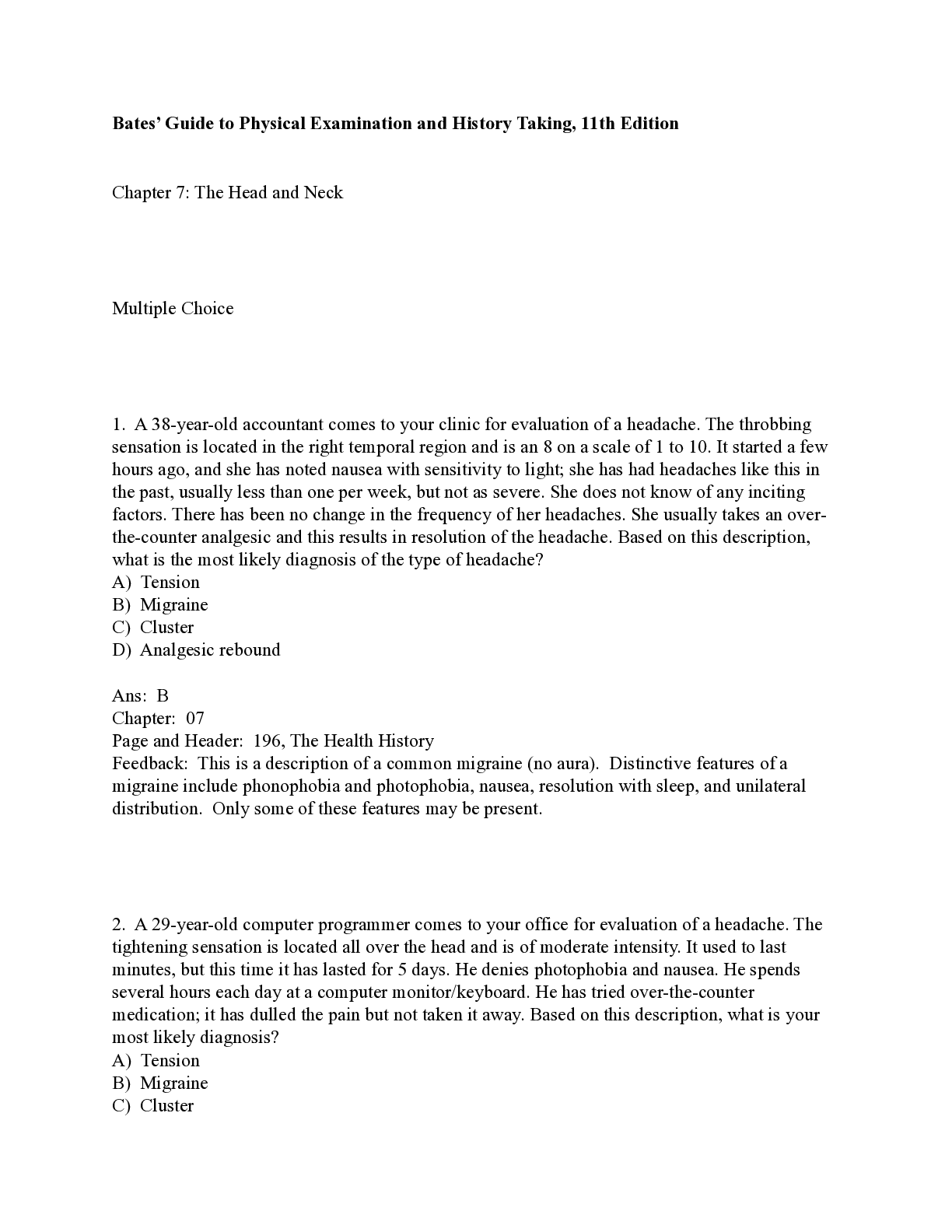
Bates’ Guide to Physical Examination and History Taking, 11th Edition Chapter 7: The Head and Neck Multiple Choice 1. A 38-year-old accountant comes to your clinic for evalu... ation of a headache. The throbbing sensation is located in the right temporal region and is an 8 on a scale of 1 to 10. It started a few hours ago, and she has noted nausea with sensitivity to light; she has had headaches like this in the past, usually less than one per week, but not as severe. She does not know of any inciting factors. There has been no change in the frequency of her headaches. She usually takes an over-the-counter analgesic and this results in resolution of the headache. Based on this description, what is the most likely diagnosis of the type of headache? A) Tension B) Migraine C) Cluster D) Analgesic rebound Ans: B Chapter: 07 Page and Header: 196, The Health History Feedback: This is a description of a common migraine (no aura). Distinctive features of a migraine include phonophobia and photophobia, nausea, resolution with sleep, and unilateral distribution. Only some of these features may be present. 2. A 29-year-old computer programmer comes to your office for evaluation of a headache. The tightening sensation is located all over the head and is of moderate intensity. It used to last minutes, but this time it has lasted for 5 days. He denies photophobia and nausea. He spends several hours each day at a computer monitor/keyboard. He has tried over-the-counter medication; it has dulled the pain but not taken it away. Based on this description, what is your most likely diagnosis? A) Tension B) Migraine C) Cluster D) Analgesic rebound Ans: A Chapter: 07 Page and Header: 196, The Health History Feedback: This is a description of a typical tension headache. 3. Which of the following is a symptom involving the eye? A) Scotomas B) Tinnitus C) Dysphagia D) Rhinorrhea Ans: A Chapter: 07 Page and Header: 196, The Health History Feedback: Scotomas are specks in the vision or areas where the patient cannot see; therefore, this is a common/concerning symptom of the eye. 4. A 49-year-old administrative assistant comes to your office for evaluation of dizziness. You elicit the information that the dizziness is a spinning sensation of sudden onset, worse with head position changes. The episodes last a few seconds and then go away, and they are accompanied by intense nausea. She has vomited one time. She denies tinnitus. You perform a physical examination of the head and neck and note that the patient's hearing is intact to Weber and Rinne and that there is nystagmus. Her gait is normal. Based on this description, what is the most likely diagnosis? A) Benign positional vertigo B) Vestibular neuronitis C) Ménière's disease D) Acoustic neuroma Ans: A Chapter: 07 Page and Header: 252, Table 7–3 Feedback: This is a classic description of benign positional vertigo. The vertigo is episodic, lasting a few seconds to minutes, instead of continuous as in vestibular neuronitis. Also, there is no tinnitus or sensorineural hearing loss as occurs in Ménière's disease and acoustic neuroma. You may choose to learn about Hallpike maneuvers, which are also helpful in the evaluation of vertigo. 5. A 55-year-old bank teller comes to your office for persistent episodes of dizziness. The first episode started suddenly and lasted 3 to 4 hours. He experienced a lot of nausea with vomiting; the episode resolved spontaneously. He has had five episodes in the past 1½ weeks. He does note some tinnitus that comes and goes. Upon physical examination, you note that he has a normal gait. The Weber localizes to the right side and the air conduction is equal to the bone conduction in the right ear. Nystagmus is present. Based on this description, what is the most likely diagnosis? A) Benign positional vertigo B) Vestibular neuronitis C) Ménière's disease D) Acoustic neuroma Ans: C Chapter: 07 Page and Header: 252, Table 7–3 Feedback: Ménière's disease is characterized by sudden onset of vertiginous episodes that last several hours to a day or more, then spontaneously resolve; the episodes then recur. On physical examination, sensorineural hearing loss is present. The patient does complain of tinnitus. 6. A 73-year-old nurse comes to your office for evaluation of new onset of tremors. She is not on any medications and does not take herbs or supplements. She has no chronic medical conditions. She does not smoke or drink alcohol. She walks into the examination room with slow movements and shuffling steps. She has decreased facial mobility and a blunt expression, without any changes in hair distribution on her face. Based on this description, what is the most likely reason for the patient's symptoms? A) Cushing's syndrome B) Nephrotic syndrome C) Myxedema D) Parkinson's disease Ans: D Chapter: 07 Page and Header: 253, Table 7–4 Feedback: This is a typical description for a patient with Parkinson's disease. Facial mobility is decreased, which results in a blunt expression—a “masked” appearance. The patient also has decreased blinking and a characteristic stare with an upward gaze. In combination with the findings of slow movements and a shuffling gait, the diagnosis of Parkinson's is almost clinched. 7. A 29-year-old physical therapist presents for evaluation of an eyelid problem. On observation, the right eyeball appears to be protruding forward. Based on this description, what is the most likely diagnosis? A) Ptosis B) Exophthalmos C) Ectropion D) Epicanthus Ans: B Chapter: 07 Page and Header: 255, Table 7–6 Feedback: Exophthalmos is the condition when the eyeball protrudes forward. If it is bilateral, it suggests the presence of Graves' disease. If it is unilateral, it could still be caused by Graves' disease. Alternatively, it could be caused by a tumor or inflammation in the orbit. 8. A 12-year-old presents to the clinic with his father for evaluation of a painful lump in the left eye. It started this morning. He denies any trauma or injury. There is no visual disturbance. Upon physical examination, there is a red raised area at the margin of the eyelid that is tender to palpation; no tearing occurs with palpation of the lesion. Based on this description, what is the most likely diagnosis? A) Dacryocystitis B) Chalazion C) Hordeolum D) Xanthelasma Ans: C Chapter: 07 Page and Header: 256, Table 7–7 Feedback: A hordeolum, or sty, is a painful, tender, erythematous infection in a gland at the margin of the eyelid. 9. A 15-year-old high school sophomore presents to the emergency room with his mother for evaluation of an area of blood in the left eye. He denies trauma or injury but has been coughing forcefully with a recent cold. He denies visual disturbances, eye pain, or discharge from the eye. On physical examination, the pupils are equal, round, and reactive to light, with a visual acuity of 20/20 in each eye and 20/20 bilaterally. There is a homogeneous, sharply demarcated area at the lateral aspect of the base of the left eye. The cornea is clear. Based on this description, what is the most likely diagnosis? A) Conjunctivitis B) Acute iritis C) Corneal abrasion D) Subconjunctival hemorrhage Ans: D Chapter: 07 Page and Header: 257, Table 7–8 Feedback: A subconjunctival hemorrhage is a leakage of blood outside of the vessels, which produces a homogenous, sharply demarcated bright red area; it fades over several days, turning yellow, then disappears. There is no associated eye pain, ocular discharge, or changes in visual acuity; the cornea is clear. Many times it is associated with severe cough, choking, or vomiting, which increase venous pressure. It is rarely caused by a serious condition, so reassurance is usually the only treatment necessary. 10. A 67-year-old lawyer comes to your clinic for an annual examination. He denies any history of eye trauma. He denies any visual changes. You inspect his eyes and find a triangular thickening of the bulbar conjunctiva across the outer surface of the cornea. He has a normal pupillary reaction to light and accommodation. Based on this description, what is the most likely diagnosis? A) Corneal arcus B) Cataracts C) Corneal scar D) Pterygium Ans: D Chapter: 07 Page and Header: 258, Table 7-9 Feedback: A pterygium is a triangular thickening of the bulbar conjunctiva that grows slowly across the outer surface of the cornea, usually from the nasal side. Reddening may occur, and it may interfere with vision as it encroaches on the pupil. Otherwise, treatment is unnecessary. 11. Which of the following is a “red flag” regarding patients presenting with headache? A) Unilateral headache B) Pain over the sinuses C) Age over 50 D) Phonophobia and photophobia Ans: C Chapter: 07 Page and Header: 196, The Health History Feedback: A unilateral headache is often seen with migraines and may commonly be accompanied by phonophobia and photophobia. Pain over the sinuses from sinus congestion may also be unilateral and produce pain. Migraine and sinus headaches are common and generally benign. A new severe headache in someone over 50 can be associated with more serious etiologies for headache. Other red flags include: acute onset, “the worst headache of my life”; very high blood pressure; rash or signs of infection; known presence of cancer, HIV, or pregnancy; vomiting; recent head trauma; and persistent neurologic problems. 12. A sudden, painless unilateral vision loss may be caused by which of the following? A) Retinal detachment B) Corneal ulcer C) Acute glaucoma D) Uveitis Ans: A Chapter: 07 Page and Header: 196, The Health History Feedback: Corneal ulcer, acute glaucoma, and uveitis are almost always accompanied by pain. Retinal detachment is generally painless, as is chronic glaucoma. 13. Sudden, painful unilateral loss of vision may be caused by which of the following conditions? A) Vitreous hemorrhage B) Central retinal artery occlusion C) Macular degeneration D) Optic neuritis Ans: D Chapter: 07 Page and Header: 196, The Health History Feedback: In multiple sclerosis, sudden painful loss of vision may accompany optic neuritis. The other conditions are usually painless. 14. Diplopia, which is present with one eye covered, can be caused by which of the following problems? A) Weakness of CN III B) Weakness of CN IV C) A lesion of the brainstem D) An irregularity in the cornea or lens Ans: D Chapter: 07 Page and Header: 196, The Health History Feedback: Double vision in one eye alone points to a problem in “processing” the light rays of an incoming image. The other causes of diplopia result in a misalignment of the two eyes. 15. A patient complains of epistaxis. Which other cause should be considered? A) Intracranial hemorrhage B) Hematemesis C) Intestinal hemorrhage D) Hematoma of the nasal septum Ans: B Chapter: 07 Page and Header: 196, The Health History Feedback: Although the source of epistaxis may seem obvious, other bleeding locations should be on the differential. Hematemesis can mimic this and cause delay in life-saving therapies if not considered. Intracranial hemorrhage and septal hematoma are instances of contained bleeding. Intestinal hemorrhage may cause hematemesis if there is obstruction distal to the bleeding, but this is unlikely. 16. Glaucoma is the leading cause of blindness in African-Americans and the second leading cause of blindness overall. What features would be noted on funduscopic examination? A) Increased cup-to-disc ratio B) AV nicking C) Cotton wool spots D) Microaneurysms Ans: A Chapter: 07 Page and Header: 201, Health Promotion and Counseling Feedback: It is important to screen for glaucoma on funduscopic examination. The cup and disc are among the easiest features to find. AV nicking and cotton wool spots are seen in hypertension. Microaneurysms are seen in diabetes. 17. Very sensitive methods for detecting hearing loss include which of the following? A) The whisper test B) The finger rub test C) The tuning fork test D) Audiometric testing Ans: D Chapter: 07 Page and Header: 201, Health Promotion and Counseling Feedback: While it is important to screen for hearing complaints with methods available to you, it should be realized that some physical examination techniques are limited. Nonetheless, you should be comfortable performing these tests, as audiometric testing is not always available. 18. Which area of the fundus is the central focal point for incoming images? A) The fovea B) The macula C) The optic disk D) The physiologic cup Ans: A Chapter: 07 Page and Header: 205, The Eyes Feedback: The fovea is the area of the retina which is responsible for central vision. It is surrounded by the macula, which is responsible for more peripheral vision. The optic disc and physiologic cup are where the optic nerve enters the eye. 19. A light is pointed at a patient's pupil, which contracts. It is also noted that the other pupil contracts as well, though it is not exposed to bright light. Which of the following terms describes this latter phenomenon? A) Direct reaction B) Consensual reaction C) Near reaction D) Accommodation Ans: B Chapter: 07 Page and Header: 205, The Eyes Feedback: The constriction of the contralateral pupil is called the consensual reaction. The response of the ipsilateral eye is the direct response. The dilation of the pupil when focusing on a close object is the near reaction. Accommodation is the changing of the shape of the lens to sharply focus on an object. 20. A patient is assigned a visual acuity of 20/100 in her left eye. Which of the following is true? A) She obtains a 20% correct score at 100 feet. B) She can accurately name 20% of the letters at 20 feet. C) She can see at 20 feet what a normal person could see at 100 feet. D) She can see at 100 feet what a normal person could see at 20 feet. Ans: C Chapter: 07 Page and Header: 205, The Eyes Feedback: The denominator of an acuity score represents the line on the chart the patient can read. In the example above, the patient could read the larger letters corresponding with what a normal person could see at 100 feet. 21. On visual confrontation testing, a stroke patient is unable to see your fingers on his entire right side with either eye covered. Which of the following terms would describe this finding? A) Bitemporal hemianopsia B) Right temporal hemianopsia C) Right homonymous hemianopsia D) Binasal hemianopsia Ans: C Chapter: 07 Page and Header: 211, Techniques of Examination Feedback: Because the right visual field in both eyes is affected, this is a right homonymous hemianopsia. A bitemporal hemianopsia refers to loss of both lateral visual fields. A right temporal hemianopsia is unilateral and binasal hemianopsia is the loss of the nasal visual fields bilaterally. 22. You note that a patient has anisocoria on examination. Pathologic causes of this include which of the following? A) Horner's syndrome B) Benign anisocoria C) Differing light intensities for each eye D) Eye prosthesis Ans: A Chapter: 07 Page and Header: 211, Techniques of Examination Feedback: Anisocoria can be associated with serious pathology. Remember to exclude benign causes before embarking on an intensive workup. Testing the near reaction in this case may help you to find an Argyll Robertson or tonic (Adie's) pupil. 23. A patient is examined with the ophthalmoscope and found to have red reflexes bilaterally. Which of the following have you essentially excluded from your differential? A) Retinoblastoma B) Cataract C) Artificial eye D) Hypertensive retinopathy Ans: D Chapter: 07 Page and Header: 211, Techniques of Examination Feedback: Hypertensive retinopathy requires a careful examination of the optic fundus. It cannot be diagnosed or excluded merely from the red reflex. Typically, the red reflex would be normal in this case. The other conditions are all associated with an abnormal red reflex. 24. A patient presents with ear pain. She is an avid swimmer. The history includes pain and drainage from the left ear. On examination, she has pain when the ear is manipulated, including manipulation of the tragus. The canal is narrowed and erythematous, with some white debris in the canal. The rest of the examination is normal. What diagnosis would you assign this patient? A) Otitis media B) External otitis C) Perforation of the tympanum D) Cholesteatoma Ans: B Chapter: 07 Page and Header: 225, Techniques of Examination Feedback: These are classic history and examination findings for a patient suffering from external otitis. Otitis media would not usually have pain with movement of the external ear, nor drainage unless the eardrum was perforated. In this case the examination of the eardrum is recorded as normal. Cholesteatoma is a growth behind the eardrum and would not account for these symptoms. Otitis media would classically be accompanied by a bulging, erythematous eardrum. 25. A patient with hearing loss by whisper test is further examined with a tuning fork, using the Weber and Rinne maneuvers. The abnormal results are as follows: bone conduction is greater than air on the left, and the patient hears the sound of the tuning fork better on the left. Which of the following is most likely? A) Otosclerosis of the left ear B) Exposure to chronic loud noise of the right ear C) Otitis media of the right ear D) Perforation of the right eardrum Ans: A Chapter: 07 Page and Header: 271, Table 7–21 Feedback: The above pattern is consistent with a conductive loss on the left side. Causes would include: foreign body, otitis media, perforation, and otosclerosis of the involved side. 26. A young man is concerned about a hard mass he has just noticed in the midline of his palate. On examination, it is indeed hard and in the midline. There are no mucosal abnormalities associated with this lesion. He is experiencing no other symptoms. What will you tell him is the most likely diagnosis? A) Leukoplakia B) Torus palatinus C) Thrush (candidiasis) D) Kaposi's sarcoma Ans: B Chapter: 07 Page and Header: 274, Table 7–23 Feedback: Torus palatinus is relatively common and benign but can go unnoticed by the patient for many years. The appearance of a bony mass can be concerning. Leukoplakia is a white lesion on the mucosal surfaces corresponding to chronic mechanical or chemical irritation. It can be premalignant. Thrush is usually painful and is seen in immunosuppressed patients or those taking inhaled steroids for COPD or asthma. Kaposi's sarcoma is usually seen in HIV-positive individuals and is classically a deep purple. 27. A young woman undergoes cranial nerve testing. On touching the soft palate, her uvula deviates to the left. Which of the following is likely? A) CN IX lesion on the left B) CN IX lesion on the right C) CN X lesion on the left D) CN X lesion on the right Ans: D Chapter: 07 Page and Header: 231, Mouth and Pharynx Feedback: The failure of the right side of the palate to rise denotes a problem with the right 10th cranial nerve. The uvula deviates toward the properly functioning side. 28. A college student presents with a sore throat, fever, and fatigue for several days. You notice exudates on her enlarged tonsils. You do a careful lymphatic examination and notice some scattered small, mobile lymph nodes just behind her sternocleidomastoid muscles bilaterally. What group of nodes is this? A) Submandibular B) Tonsillar C) Occipital D) Posterior cervical Ans: D Chapter: 07 Page and Header: 236, The Neck Feedback: The group of nodes posterior to the sternocleidomastoid muscle is the posterior cervical chain. These are common in mononucleosis. 29. You feel a small mass that you think is a lymph node. It is mobile in both the up-and-down and side-to-side directions. Which of the following is most likely? A) Cancer B) Lymph node C) Deep scar D) Muscle Ans: B Chapter: 07 Page and Header: 236, The Neck Feedback: A useful maneuver for discerning lymph nodes from other masses in the neck is to check for their mobility in all directions. Many other masses are mobile in only two directions. Cancerous masses may also be “fixed,” or immobile. 30. You are conducting a pupillary examination on a 34-year-old man. You note that both pupils dilate slightly. Both are noted to constrict briskly when the light is placed on the right eye. What is the most likely problem? A) Optic nerve damage on the right B) Optic nerve damage on the left C) Efferent nerve damage on the right D) Efferent nerve damage on the left Ans: B Chapter: 07 Page and Header: 211, Techniques of Examination Feedback: Because both pupils can constrict, efferent nerve damage is unlikely. When the light is placed on the left eye, neither a direct nor a consensual response is seen. This indicates that the left eye is not perceiving incoming light. [Show More]
Last updated: 1 year ago
Preview 1 out of 14 pages
Buy this document to get the full access instantly
Instant Download Access after purchase
Add to cartInstant download
We Accept:

Reviews( 0 )
$17.00
Document information
Connected school, study & course
About the document
Uploaded On
Dec 30, 2020
Number of pages
14
Written in
Additional information
This document has been written for:
Uploaded
Dec 30, 2020
Downloads
0
Views
26



.png)

 (1).png)

