Health Care > STUDY GUIDE > NUR 265 Exam 2 Study Guide: Latest updated : Guaranteed A+ Guide Solution (All)
NUR 265 Exam 2 Study Guide: Latest updated : Guaranteed A+ Guide Solution
Document Content and Description Below
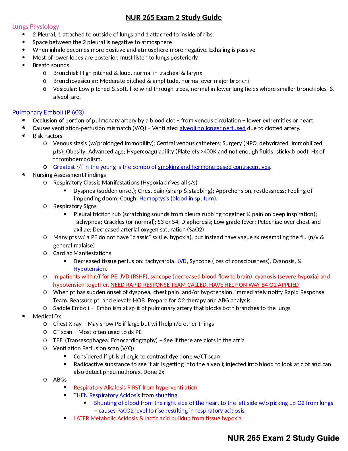
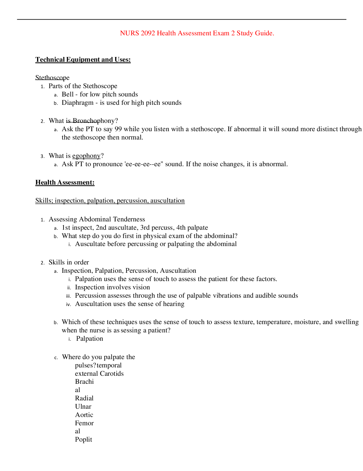
Lungs Physiology 2 Pleural, 1 attached to outside of lungs and 1 attached to inside of ribs. Space between the 2 pleural is negative to atmosphere When inhale becomes more positive and ... atmosphere more negative. Exhaling is passive Most of lower lobes are posterior, must listen to lungs posteriorly Breath sounds o Bronchial: High pitched & loud, normal in tracheal & larynx o Bronchovesicular: Moderate pitched & amplitude, normal over major bronchi o Vesicular: Low pitched & soft, like wind through trees, normal in lower lung fields where smaller bronchioles & alveoli are. Pulmonary Emboli (P 603) Occlusion of portion of pulmonary artery by a blood clot – from venous circulation – lower extremities or heart. Causes ventilation-perfusion mismatch (V/Q) – Ventilated alveoli no longer perfused due to clotted artery. Risk Factors o Venous stasis (w/prolonged immobility); Central venous catheters; Surgery (NPO, dehydrated, immobilized pts); Obesity; Advanced age; Hypercoagulability (Platelets >400K and not enough fluids; sticky blood); Hx of thromboembolism. o Greatest r/f in the young is the combo of smoking and hormone based contraceptives. Nursing Assessment Findings o Respiratory Classic Manifestations (Hypoxia drives all s/s) Dyspnea (sudden onset); Chest pain (sharp & stabbing); Apprehension, restlessness; Feeling of impending doom; Cough; Hemoptysis (blood in sputum). o Respiratory Signs Pleural friction rub (scratching sounds from pleura rubbing together & pain on deep inspiration); Tachypnea; Crackles (or normal); S3 or S4; Diaphoresis; Low grade fever; Petechiae over chest and axillae; Decreased arterial oxygen saturation (SaO2) o Many pts w/ a PE do not have “classic” sx (i.e. hypoxia), but instead have vague sx resembling the flu (n/v & general malaise) o Cardiac Manifestations Decreased tissue perfusion: tachycardia, JVD, Syncope (loss of consciousness), Cyanosis, & Hypotension. o In patients with r/f for PE, JVD (RSHF), syncope (decreased blood flow to brain), cyanosis (severe hypoxia) and hypotension together, NEED RAPID RESPONSE TEAM CALLED. HAVE HELP ON WAY B4 O2 APPLIED o When pt has sudden onset of dyspnea, chest pain, and/or hypotension, immediately notify Rapid Response Team. Reassure pt. and elevate HOB. Prepare for O2 therapy and ABG analysis o Saddle Emboli – Embolism at split of pulmonary artery that blocks both branches to the lungs Medical Dx o Chest X-ray – May show PE if large but will help r/o other things o CT scan – Most often used to dx PE o TEE (Transesophageal Echocardiography) – See if there are clots in the atria o Ventilation Perfusion scan (V/Q) Considered if pt is allergic to contrast dye done w/CT scan Radioactive substance to see if air is getting into the alveoli; injected into blood to look at clot and can [Show More]
Last updated: 1 year ago
Preview 1 out of 23 pages
Reviews( 0 )
Document information
Connected school, study & course
About the document
Uploaded On
Feb 02, 2023
Number of pages
23
Written in

Additional information
This document has been written for:
Uploaded
Feb 02, 2023
Downloads
0
Views
52


 Study Guide.png)
 Exam Study Guide.png)


.png)