NURSING. > STUDY GUIDE > NCSBN –Lesson 8E: Genitourinary System Study Guide,100%CORRECT (All)
NCSBN –Lesson 8E: Genitourinary System Study Guide,100%CORRECT
Document Content and Description Below
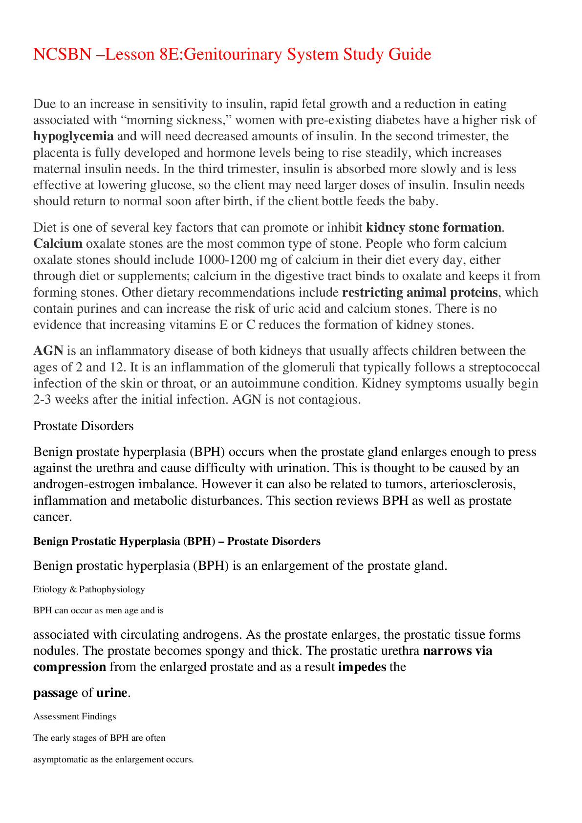
NCSBN –Lesson 8E:Genitourinary System Study Guide Due to an increase in sensitivity to insulin, rapid fetal growth and a reduction in eating associated with “morning sickness,” women with pre... -existing diabetes have a higher risk of hypoglycemia and will need decreased amounts of insulin. In the second trimester, the placenta is fully developed and hormone levels being to rise steadily, which increases maternal insulin needs. In the third trimester, insulin is absorbed more slowly and is less effective at lowering glucose, so the client may need larger doses of insulin. Insulin needs should return to normal soon after birth, if the client bottle feeds the baby. Diet is one of several key factors that can promote or inhibit kidney stone formation. Calcium oxalate stones are the most common type of stone. People who form calcium oxalate stones should include 1000-1200 mg of calcium in their diet every day, either through diet or supplements; calcium in the digestive tract binds to oxalate and keeps it from forming stones. Other dietary recommendations include restricting animal proteins, which contain purines and can increase the risk of uric acid and calcium stones. There is no evidence that increasing vitamins E or C reduces the formation of kidney stones. AGN is an inflammatory disease of both kidneys that usually affects children between the ages of 2 and 12. It is an inflammation of the glomeruli that typically follows a streptococcal infection of the skin or throat, or an autoimmune condition. Kidney symptoms usually begin 2-3 weeks after the initial infection. AGN is not contagious. Prostate Disorders Benign prostate hyperplasia (BPH) occurs when the prostate gland enlarges enough to press against the urethra and cause difficulty with urination. This is thought to be caused by an androgen-estrogen imbalance. However it can also be related to tumors, arteriosclerosis, inflammation and metabolic disturbances. This section reviews BPH as well as prostate cancer. Benign Prostatic Hyperplasia (BPH) – Prostate Disorders Benign prostatic hyperplasia (BPH) is an enlargement of the prostate gland. • Etiology & Pathophysiology o BPH can occur as men age and is associated with circulating androgens. As the prostate enlarges, the prostatic tissue forms nodules. The prostate becomes spongy and thick. The prostatic urethra narrows via compression from the enlarged prostate and as a result impedes the passage of urine. • Assessment Findings o The early stages of BPH are often ▪ asymptomatic as the enlargement occurs. o As it progresses, clients will complain of changes in micturition (day or night) ▪ often reporting difficulty starting or stopping stream, ▪ a smaller than usual stream ▪ less frequency urinating and dribbling ▪ They may also complain of nocturia. • Diagnostic Studies o Digital rectal examination o Labs: ▪ Urinalysis ▪ Serum creatinine and blood urea nitrogen (BUN) studies ▪ Serum prostate-specific antigen (PSA) more for prostate cancer ▪ Transrectal ultrasound (TRUS) • Health Promotion & Health Screening o The following programs and health screenings are currently recommended and/or mandated as part of a healthy person's regular health assessment in the U.S. ▪ Everyone • Dental exam: o Regular visits will help to identify any tooth or gum problems before they progress o Should begin within six months of a child's first tooth and no later than the first birthday o Regular check-ups and cleanings should be performed every six months • Hearing test: o Ear problems can be signs of health, development or communication issues: ▪ Electrophysiologic test: used to measure newborn's hearing ability based on electrical information from the auditory nervous system ▪ Pure tone audiometry: used for children aged 4 years and older o Specific candidates for screening includes: perinatal infection (rubella, herpes, cytomegalovirus), chronic ear infections, Down syndrome, low birth weight infants, family history of hearing impairment o Mandated by school districts or a state's education or health department o Recommended every 10 years; every three years after age 50 • Vision test: o Regular examinations can prevent many leading causes of blindness and can help correct poor vision o The American Optometric Association suggests that: ▪ Children under the age of 3-years-old should be screened during regular pediatric appointments ▪ School-age children have their vision check every two years ▪ Adults up to age 40 should be checked every 2-3 years ▪ Adults after age 40 should have their vision checked every other year or more frequently if they have diabetes or hypertension o Basic vision testing typically includes: ▪ Visual acuity: tested using the Snellen eye chart, using either letter of the alphabet or the letter "E" for younger children ▪ Glaucoma screening o Mandated by school districts or a state's education or health department • Blood pressure test: o According to the American Heart Association, men and women aged 18 and older should be screened for high blood pressure at least once every two years (unless there is a family history of cardiovascular disease) o Screening for children and adolescents is also recommended but an optimal interval has yet to be determined o Recommended screening method ▪ Auscultatory method with a properly calibrated sphygmomanometer and correctly fitting cuff ▪ Person should be seated quietly in a chair for at least five minutes with feet on the floor and arms supported at heart level ▪ At least two measurements are taken, two minutes apart ▪ Be aware of "white coat hypertension" o Prehypertensive individuals (systolic pressure 120-139 mm Hg and diastolic pressure 80-89 mm Hg) should be counseled on lifestyle modifications such as weight reduction, exercise, diet and smoking cessation o Systolic pressure greater than 140 mm Hg and/or diastolic greater than 90 mm Hg should be referred to a health care provider for possible antihypertensive drug therapy • Cholesterol test: baseline at age 20; every five years if normal • Well-child care: o Well-child care (birth to age 6 years) includes routine care, comprehensive health promotion and disease prevention exams; vision and hearing screenings; height, weight, and head circumference; routine immunizations; and developmental and behavioral appraisal in accordance with the American Academy of Pediatrics (AAP) and the Centers for Disease Control and Prevention (CDC) guidelines o Scoliosis screening: ▪ Early detection and intervention is important because untreated scoliosis can lead to disfigurement, impaired mobility, and cardiopulmonary complications ▪ Recommendations vary but generally performed at onset of adolescence ▪ Screenings (typically in 6th grade) are mandated by school districts or a state's education or health department • Physical exam: o Every 1-5 years depending on risk factors and health concerns o Rectal exam: annually over age 40 o Stool check for blood (Stool Occult Blood): annually • Skin cancer screening and self-exam o The American Cancer Society encourages periodic self- examinations by visually inspecting any new, misshapen or discolored moles or lesions o Regular screenings are included in a routine physical exam • Colonoscopy: o Screening used to check for cancer or precancerous growths in the colon or rectum o The average person should have a colonoscopy once every 10 years after turning 50 (unless there is a family history of colon cancer) • Immunizations (non-childhood): o Tetanus immunization booster: every 10 years o Influenza vaccine: annually o Pneumococcal vaccine: at age 65 (or all persons aged 19-64 years with chronic or immunosuppressive medical conditions, e.g., asthma) ▪ Men • Testicular self-exam: o Testicular cancer is the most common type of cancer in men between the ages of 15-24 and is highly curable when caught early o Men should visually inspect and palpate the skin on the scrotum and testicles in front of a mirror, following a warm bath or shower • Digital rectal exam: o The most direct way for a health care provider to screen for prostate and colorectal cancer o Men age 50 and older (or earlier for those at high risk for cancer) may benefit from an annual digital rectal exam as part of the routine physical exam • Prostate-specific antigen (PSA) test: o This blood test measures the amount of PSA in a man's blood: ▪ As men age, PSA levels naturally rise ▪ Elevated PSA levels means there is an enlarged prostate, which may be an indicator of prostate cancer o Typically combined with the digital rectal exam o Formerly an annual screening for all men over 50 was recommended; routine screening is no longer recommended unless a risk exists ▪ Women • Pap smear: o Detects the earliest signs of cervical cancer by checking for any changes in the cells of the cervix o The American College of Obstetricians and Gynecologists (ACOG) recommends that women should have their first Pap test three years after first having sex, but no later than age 21 ▪ The test should be performed yearly until the age of 30 ▪ Women ages 30-65 should have the test every 2-3 years after three consecutive normal Pap smears ▪ Women 70 years and older can stop having Pap smears after three consecutive normal Pap smears without any abnormal Pap smears in the last 10 years • Clinical breast exam: o Helps health care providers discover breast cancer in its early stages o Women in their 20s and 30s should have a clinical breast exam as part of the regular, routine physical, at least every three years o Women ages 40 and older should have yearly clinical breast exams • Mammogram: o Used to detect and diagnose breast cancer o The American Cancer Society recommends that "women age 40 and older should have a screening mammogram every year and should continue to do so for as long as they are in good health" • Self breast exam: o Monthly breast exams should be performed to detect any changes in their breasts and underarm areas o Should be performed throughout one's life, beginning in the 20s o Should be done at the same time each month (preferably seven days after onset of the menstrual cycle, when the breasts are less tender) o It should be emphasized that self-exams are not a substitute for mammography or regular exams conducted by a health care professional • Bone density test: o Used for screening for osteoporosis, the test uses bones that are more likely to break due to osteoporosis, e.g. hip and lower spine o Most popular bone density test is dual energy X-ray absorptiometry (DEXA) o A baseline bone density test should be done at age 50 or at a time coinciding with menopause • Management o If asymptomatic, follow with annual diagnostics o If symptomatic, treatment may include the following: ▪ Antihypertensives – relax smooth muscles in the prostate and bladder neck but will not decrease the prostate size ▪ Prazosin – decreases urinary urgency, hesitancy, dribbling, retention and nocturia ▪ Doxazosin – decreases urinary urgency, hesitancy, dribbling, retention and nocturia ▪ Terazosin – decreases urinary urgency, hesitancy, dribbling, retention and nocturia o Hormonal manipulation – finasteride, which decreases prostate size, urinary urgency, hesitancy, dribbling, retention and nocturia o Balloon dilation – offers temporary relief of urinary urgency, hesitancy, dribbling, retention and nocturia o Surgery, if indicated (transurethral resection of prostate (TURP), open prostatectomy, laser surgery or insertion of prostatic stent) • Complimentary & Integrative Health o There is some evidence that urtica dioica (stinging nettle) and pygeum africanum may improve some symptoms of BPH. • Complications o Complications can include acute urinary retention, involuntary bladder spasms, hydronephrosis (swelling of kidney d/t build-up of urine), UTIs and gross hematuria. • Nursing Interventions o Assess and evaluate clients for: ▪ Presence of urinary urgency, hesitancy, dribbling, retention, nocturia, infection and hematuria ▪ Presence of bladder distention ▪ Presence of post-void residual (evaluate with straight catheter or bladder scan, if ordered) ▪ Orthostatic blood pressure, if antihypertensives are being used for BPH o Facilitate urinary elimination o Provide privacy for clients during elimination o Encourage the client to talk to their health care provider prior to taking any prescription, over-the-counter (OTC) or herbal medications o Monitor intake and output (I/O) and weight on a daily basis o Post-op care for surgical treatment: ▪ Maintain urinary catheter patency ▪ Monitor urine output for volume and color every 1-2 hours ▪ Maintain continuous bladder irrigation system with normal saline as prescribed ▪ Irrigate continuous bladder irrigation system as needed for obstruction or as ordered by the health care provider ▪ Medicate the client for pain and bladder spasms as needed ▪ Instruct the client to do Kegel exercises after catheter removal to aid in urinary control Prostate Cancer – Prostate Disorders Prostate cancer is the most common cause of cancer death in men. Black men are twice as likely than white men to develop prostate cancer. Most of these cancers originate in the posterior prostate gland, while the rest can grow near the urethra. An adenocarcinoma is reported to be the most common type of prostate tumor. • Etiology & Pathophysiology o Age (65 or older), diet high in saturate fats, and ethnicity (black men) are risk factors that increase the incidence of prostate cancer. o Prostate cancer is typically a slow growing cancer, however it can spread beyond the prostate gland. o Without metastasis, the prognosis is good, however after metastasis the survival rate is at about 35%. o Bone metastasis, statistically speaking, contributes to the low survival rate. • Assessment Findings o Since early stages of prostate cancer produce little to no symptoms, consistent screening for prostate cancer is critical. o In advanced stages clients will present with: ▪ a weak urine streams ▪ hematuria ▪ urinary hesitancy ▪ incomplete bladder emptying ▪ dysuria • These symptoms are usually a result of tumor progression. • Diagnostic Studies o A digital rectal exam helps determine the size and location of the prostate as well as nodular presents. o Laboratory tests will show elevated levels of prostate-specific antigen (PSA). o A biopsy of the prostate may be used to distinguish between benign or malignant tumors. o Additionally, MRI and CT scans will be ordered to determine the tumors extent. • Management/Nursing Interventions o Radical prostatectomy (can cause urinary incontinence and erectile dysfunction) nerve-sparing procedure, and cryotherapy (can cause damage to the urethra) are among the surgical techniques considered for prostate cancer. o Radiation therapy such as external beam radiation and brachytherapy are commonly used to treat prostate cancer. o Chemotherapy is used when clients are diagnosed with hormone-refractory prostate cancer. o Finally, drug therapy such as androgen deprivation therapy (can cause osteoporosis and fractures), androgen synthesis inhibitors and androgen receptor blockers are used in treatment. o Again, encouraging prostate cancer screening is one of the most important roles health care providers can play. Early detection is key to survival. o Nursing interventions will include care for clients who undergo radiation and chemotherapy (if applicable). o It is important to assess the client's psychological response to their diagnosis (this is true of all cancers) and provide resources and support to them and their family. o If the client is discharged with an indwelling catheter, then the nurse will always provide teaching on how to clean the urethral meatus and instruct them to keep the collection bag lower than the bladder. o The client will maintain a high fluid intake and report any signs of a bladder infection. If the client is discharged without a catheter, urinary incontinence may be a problem. o Kegel exercises can improve incontinence and providing resources for incontinence products will also help these clients. o Palliative care to control pain is an important part of treatment. Pain control requires an ongoing assessment and evaluation of current pain management techniques and often palliative care teams are helpful (Palliative care can assist all of your clients suffering from poor pain management). Female Reproductive Disorders The female reproductive organs are the ovaries, fallopian tubes, uterus, cervix, vagina and vulva. Since the combined primary function of these organs is reproduction, disorders affecting them can result in infertility. This section reviews common female reproductive disorders, as well as associated cancers. Cystocele – Female Reproductive Disorders A cystocele occurs when the bladder herniates into the vaginal canal. • Etiology & Pathophysiology o Cystocele is associated with obstetrical trauma or may be due to a congenital defect. Findings may appear after a hysterectomy. o With age, it may appear as genitalia atrophy. • Assessment Findings o In the early stages, the client will be asymptomatic. o As it progresses, the client will complain of pelvic pressure, changes in micturition such as frequency, urgency, stress incontinence and the inability to empty the bladder. o Reoccurring urinary tract infections (UTI) will also occur. • Diagnostic Studies o A pelvic examination and a urinalysis with urine culture will be performed. • Management o In postmenopausal clients – possibly estrogen replacement therapy o Insertion of vaginal pessary to support the pelvic organs o Surgical intervention (if indicated): ▪ To restore bladder function ▪ To repair the anterior vaginal wall • Complications o Complications can include infection and urinary incontinence. • Nursing Interventions o Assess clients for: ▪ A history of obstetrical trauma, abdominal surgery, menopause or estrogen therapy ▪ Micturition, with attention to recent changes in micturition pattern ▪ Presence of pain ▪ Presence of a bulge from the vagina while standing upright or bearing down ▪ Provide pain management as needed ▪ Teach post-op clients pelvic rest for six weeks ▪ Teach actions to enhance control of incontinence (Kegel exercises) • Begin by emptying your bladder. • Tighten the pelvic floor muscles and hold for a count of 10. • Relax the muscles completely for a count of 10. • Do 10 repetitions, 3 to 5 times a day (morning, afternoon, and night). ▪ Discuss actions for clients to prevent urinary retention Pelvic Inflammatory Disease (PID) – Female Reproductive Disorders Pelvic inflammatory disease (PID) is an infection of the cervix ascending to the fallopian tubes and broad ligaments. ▪ Etiology & Pathophysiology o Increased incidence in clients with sexually transmitted infections (STI) o Causative agents: Neisseria gonorrhoeae, Chlamydia trachomatis (C. trachomatis) or Mycoplasma hominis o A history of multiple sexual partners o The use of IUDs (intrauterine devices) o History of therapeutic abortion o Douching ▪ Assessment Findings o Pelvic pain o Fever o Abnormal cervical discharge o Cervical motion tenderness o Irregular cervical bleeding o Nausea, vomiting and acute abdominal pain o Dysuria, frequent urination o Chlamydia, gonorrhea or other sexually transmitted infection ▪ Diagnostic Studies o Endocervical culture o CBC with differential o Laparoscopy to view fallopian tubes o Culdocentesis (peritoneal fluid is obtained from the cul de sac of a female patient) ▪ Management o Pharmacologic Intervention: ▪ Anti-infectives: tetracyclines, penicillins, quinolones or cephalosporins (used singly or in combination, depending on the causative organism) ▪ Analgesics o Surgical intervention to drain the abscess may be required. ▪ Complications o Complications of PID include ectopic pregnancy, infertility, rupture or abscess, sepsis and chronic pelvic pain. ▪ Nursing Interventions o Assess the client for: ▪ A history of menses issues, contraceptive use and sexual habits • Level of pain • Vital signs for hypotension, hypovolemia and fever • How an STD will impact the client socially and emotionally • Pain management • Need to restore fluid balance o Client teaching will include: ▪ Take all antibiotics prescribed, not just until symptoms subside ▪ Have yearly pelvic exams with STI screening ▪ Avoid transmission of STIs via latex condoms ▪ All the client's sexual partners should be treated for STIs also to prevent re-infection Endometriosis – Female Reproductive Disorders Endometriosis occurs when the endometrium tissue grows in cysts at various sites throughout the pelvis and/or abdominal wall. ▪ Etiology & Pathophysiology o Endometriosis occurs at any age, although it is most commonly seen in women between 25-45 years old. There is a higher incidence in white women than in African American women. It is a response to ovarian hormonal stimulation. Progestin decreases endometriosis and estrogen increases endometriosis. ▪ Assessment Findings o May be asymptomatic o May present with pelvic pain (typically before or during menstruation) o Dyspareunia (pain during sexual intercourse) o Painful defecation o Abnormal uterine bleeding o Persistent infertility o Hematuria, dysuria and flank pain if the bladder is involved ▪ Diagnostic Studies o Pelvic examination o Digital rectal examination o Laparoscopy o Abdomen: ultrasound, CT scan and barium studies ▪ Management o Pharmacologic Intervention: ▪ Danazol – atrophy of ectopic endometrial tissue ▪ Leuprolide acetate – reduction of pain and lesions in endometriosis ▪ Progestins – decreases endometriosis ▪ Oral contraceptives o Surgical Interventions: ▪ Laparoscopic surgery ▪ CO2 laser laparoscopy ▪ Laparotomy ▪ Presacral neurectomy ▪ Hysterectomy ▪ Complications o Complications include infertility. ▪ Nursing Interventions o Assess the client for: ▪ Presence of uterine bleeding, pelvic pain, painful intercourse and painful defecation ▪ Level and characteristics of pain ▪ Psychosocial impact of infertility (especially in the child-bearing age group) ▪ Pain reduction ▪ Referrals, including those to help your client maintain and/or increase self- esteem Breast Cancer – Female Reproductive Disorders Breast cancer is the most common cancer in women. Breast cancer typically affects clients after the age of 50, which accounts for 70% of breast cancer cases. However the incidence of breast cancer before the age of 50 has increased because of early detection. ▪ Etiology & Pathophysiology o The exact cause of breast cancer remains unknown. Medical researchers have determined that genes such as BRCA1 and BRACA2 have been linked to breast cancer, confirming there is a hereditary component. o Other risk factors include: ▪ Family history ▪ Radiation exposure ▪ Premenopausal ▪ Obesity ▪ Age ▪ Use of hormonal contraceptives ▪ Early onset of menses or late menopause ▪ Nulligravida or first pregnancy after 30 ▪ High fat diet ▪ Colon, endometrial or ovarian cancer ▪ Postmenopausal women on progestin and estrogen therapy ▪ Alcohol use ▪ Benign breast disease ▪ Assessment Findings o Typically, a client will present with a thickening of breast tissue or a painless lump or mass. o A mass can also be detected on a mammogram before it is palpable. o Skin around the nipple area may be scaly and there may be clear, milky or bloody discharge from the nipple. ▪ The nipple may also be retracted. ▪ Edema in the arm may indicate advanced nodal involvement. ▪ Diagnostic Studies o Self-breast exams should be performed monthly, followed by a yearly clinical exam for screening of breast cancer. o Mammography is a screen method used to detect tumors that are to small to palpate. o A fine needle aspiration and biopsy provide cells to confirm a diagnosis and hormone receptor assay can pinpoint if the tumor is estrogen or progesterone dependent. o Ultrasound, chest X-ray, scans of the bone, brain, liver, etc. will be used to detect metastasis. ▪ Management/Nursing Interventions o Common treatments for breast cancer include a combination of surgery, radiation, chemotherapy and hormonal therapy, depending of the stage and type of cancer. o There are a variety of surgical procedures used to treat breast cancer. ▪ A lumpectomy is used to remove small, well defined lesions and nearby lymph nodes in early stages of breast cancer. ▪ A partial mastectomy removes a tumor, section of normal tissue, and axillary lymph nodes. ▪ A total mastectomy involves the removal of the breast tissue. ▪ A modified radical mastectomy is an intervention that removes the entire breast, axillary lymph nodes and the lining of the chest muscles. ▪ Typically, radiation and/or chemotherapy follows after a surgical procedure. o Phantom breast pain is common and important for the nurse to recognize after mastectomy. o Remember that regardless of the surgery planned, the client should be taught the plan for pain control and after-surgery recovery expectations such as reporting complications, dressing and drain care, coughing and deep breathing. o Upper extremity lymphedema can occur at any point after treatment for breast cancer. o Teach treatment for lymphedema including no blood pressure, venipunctures or injections on the affected arm. ▪ Acute lymphedema often requires complete decongestive therapy. ▪ Finally, psychosocial support is critical for the client and family via promoting communication, providing resources and information. Genitourinary Disorders The renal system's primary function is to collect the body's waste and expel the waste as urine. It also serves as a filtration system for electrolytes. The kidneys filter about 45 gallons of fluid each day. This section reviews common disorders associated with the renal system. Urinary Tract Infections (UTI) – Genitourinary Disorders Urinary tract infections (UTI) are infections, caused by various agents, in different parts of the urinary system. • Etiology & Pathophysiology o In women, acute UTI is often caused by Escherichia coli (E coli). o In men, the cause is usually obstructive abnormalities. o Catheter-associated urinary tract infection (CAUTI) is caused by the formation of biofilms by urinary pathogens common on the surfaces of catheters and collecting systems. • Assessment Findings o Dysuria, frequency, urgency and nocturia o Suprapubic pain o Findings of hematuria more often with kidney involvement • Diagnostic Studies o Urinalysis (use clean, voided specimen) o Urine culture (use clean-catch midstream process for urine specimen collection or aspirate specimen from retention catheter) • Management o The preferred pharmacologic intervention for a UTI is antimicrobial therapy as indicated. • Complications • Pyelonephritis (inflammation of the kidney) and sepsis are potential complications. • Nursing Interventions • Assess the client for: • A history of the frequency of urinary tract infections (UTIs) • Voiding habits, personal hygiene and contraceptive methods • A history of vaginal discharges, itching, irritation and dysuria • Manage pain: • Systemic analgesics • Urinary analgesics/antispasmodics • Client teaching will include: • With female clients, discuss the need to void after intercourse • Cleanse the perineum from front to back • Wear cotton underwear • Increase water intake • Avoid carbonated and caffeinated fluids, e.g., coffee, tea, alcohol and colas • Prevent catheter-associated urinary tract infections (CAUTIs) by: • Insert catheters only for appropriate reasons • Leave catheters in place only as long as needed • Insert catheters using aseptic technique and sterile equipment • Maintain a closed drainage system • Maintain unobstructed urine flow Renal Calculi (Urolithiasis) – Genitourinary Disorders Renal calculi (kidney stones or renal lithiasis) are small, hard deposits made of mineral and acid salts that form inside the kidneys. • Etiology & Pathophysiology o Hypercalcemia, hypercalciuria and hyperuricemia o Chronic dehydration and high purine diet (organ meats, yeast, etc.) o Cystinuria (genetic disorder) o Chronic infections (Proteus vulgaris) or chronic obstruction with urinary stasis o Environmental factors, i.e., living in a warm, humid climate o More prevalent in men, with peak age of onset between 20-30 years o Can be found anywhere in the urinary system o Spontaneous passage occurs in vast majority of clients o Calculi can lodge and cause obstruction; common sites for obstruction are ▪ bladder neck, renal pelvis and ureters o Often recurs in clients with a history of two or more stones • Assessment Findings o Severe pain in low back/flank pain– the site is dependent on location of obstruction o Increased hydrostatic pressure o Renal colic and ureteral colic o Findings can mimic cystitis o With obstruction: when stones (calculi) block urine flow, the client will show findings of UTI with fever and chills o Gastrointestinal findings: ▪ Nausea and vomiting ▪ Diarrhea ▪ Abdominal discomfort • Diagnostic Studies o An intravenous pyelogram (IVP) will be conducted to determine the site and degree of the obstruction. o Other diagnostic studies include a retrograde or antegrade pyelography, analysis of the stone, urinalysis with culture and sensitivity and a 24-hour urine test. • Management o Pharmacologic Intervention: ▪ Diuretics – to control hypercalciuria and thus prevent calcium stones (hydrochlorothiazide [HCTZ]) ▪ Allopurinol – to help prevent calcium and uric acid stones ▪ Opioid narcotics – pain relief ▪ Antibiotics – if infection present o Surgical Interventions: ▪ Extracorporeal shock wave lithotripsy (ESWL) ▪ Percutaneous nephrolithotomy (PCNL) ▪ Ureteroscopic stone removal ▪ Percutaneous stone dissolution (chemolysis) ▪ Ureteroscopy ▪ Temporary or permanent ureteral stent placement ▪ Pyelolithotomy, nephrolithotomy or ureterolithotomy ▪ Cystolithotomy ▪ Nephrectomy (surgical removal of a kidney) • Complications o An obstruction can occur from residual stone material (fragments). o Infections from bacteria or infected stone fragments are also a potential complication of renal calculi. o Additionally, chronic renal function impairment can occur if obstruction persists. • Nursing Interventions o Assess the client for: ▪ A history of UTIs, dietary habits and a family history of kidney stones ▪ Pain, location and intensity – is typically severe • Priority is asking for pain (intensity) vasovagal response hypotension and syncope ▪ Findings of UTI ▪ Findings of urinary obstruction ▪ Pain management – narcotics often indicated ▪ Maintenance of urine flow – strain urine ▪ Infection control ▪ Client teaching will include: • Increase fluid intake so that client produces at least two quarts of urine every 24 hours • Increase intake of foods high in calcium (note: in the past, clients were told to avoid dairy products) • Avoid taking calcium in pill form, including antacids with calcium base • Collaborate with nutrition therapy based on type of stone Acute Kidney Injury (AKI) – Genitourinary Disorders Acute kidney injury (AKI), formerly known as acute renal failure, is the abrupt loss of kidney function resulting in the retention of urea and other nitrogenous waste products, and in the dysregulation of extracellular volume and electrolytes. • Etiology & Pathophysiology o Prerenal – decreased renal blood flow (due to systemic assault, such as hemorrhage, trauma or burns) o Intrarenal – injury to renal tissue due to toxins, intrarenal ischemia, vascular disorders and immunologic processes o Postrenal – inflamed or obstructed kidney stops or slows urine flow anywhere in the urinary tract • Assessment Findings o Prerenal – hypotension, hypoperfusion, reduced urine output, shriveled skin and dry mucous membranes o Intrarenal – history of glomerulonephritis, edema, rash and changes in kidney function (both output and chemistry) o Postrenal – history of urinary obstruction, difficulty voiding and changes in micturition • Diagnostic Studies o Labs: ▪ Urinalysis and urine for culture and sensitivity ▪ Serum creatinine and blood urea nitrogen (BUN) ▪ Urea and electrolytes ▪ Inflammatory markers ▪ Calcium and phosphate ▪ CBC and arterial blood gases o Additionally, a chest X-ray, ECG and renal ultrasound may be performed. • Management o To identify and treat the cause of AKI: ▪ Discontinue all nephrotoxic drugs, such as aminoglycosides, angiotensin converting enzyme (ACE) inhibitors, NSAIDs and radiocontrast media ▪ Eliminate exposure to nephrotoxins ▪ Postrenal causes, including anything that obstructs outflow from the kidneys (prostatic hypertrophy, tumors, strictures and neurogenic bladder) o To treat life-threatening issues: ▪ Administer intravenous (IV) fluids if acute kidney failure is caused by a lack of fluids ▪ Medications to control serum potassium: calcium, glucose and sodium polystyrene sulfonate o Medications to restore serum calcium levels o Hemodialysis • Complications o Complications of AKI include: ▪ systemic infections ▪ arrhythmias secondary to hyperkalemia ▪ electrolyte imbalances ▪ gastrointestinal (GI) bleeding due to stress ulcers ▪ multiple organ system failure. • Nursing Interventions o Assess/monitor the client for: ▪ A history of cardiac disease, malignancy, sepsis or recent infection ▪ Exposure to nephrotoxic drugs ▪ 24-hour urine output ▪ Serum labs to achieve fluid and electrolyte balance ▪ Prevent infection ▪ Serum electrolytes ▪ Minimize stress to prevent GI bleeding ▪ Neurologic function ▪ Daily weight o Maintain adequate nutrition: ▪ Regulate protein intake ▪ Offer high-carbohydrate options ▪ Restrict (as needed) foods high in potassium, phosphorus and sodium ▪ If required, provide care for the dialysis patient Chronic Kidney Disease (CKD) – Genitourinary Disorders Chronic kidney disease (CKD) is a progressive, irreversible deterioration in renal function, in which the body cannot balance metabolism and fluid/electrolytes. CKD results in uremia. • Etiology & Pathophysiology • Several disease processes have been found to cause CKD. Hypertension that is severe and prolonged, diabetes mellitus, glomerulopathy, interstitial nephritis, polycystic disease and obstructive uropathy. CKD may also be caused by a congenital disorder. • Assessment Findings • Respiratory – pulmonary edema, pleural effusion and pleural rub • Cardiovascular – HTN, hyperkalemia (with ECG changes), pericardial effusion and tamponade • Neuromuscular – sleep disorders, headache, lethargy, peripheral neuropathies, seizures and coma • Metabolic/endocrine – hyperlipidemia, decreased libido, impotence, amenorrhea and glucose intolerance glucose intolerance • Acid/base – water retention, metabolic acidosis, hyperkalemia, hypocalcemia, hypermagnesemia and hyperphosphatemia • GI – anorexia, nausea, vomiting and gastric ulcerations • Blood – anemia from decreased or no erythropoietin production, increased bleeding and platelet defects • Skeletal – renal osteodystrophy and osteomalacia from decreased calcium levels • Skin – pruritus, uremic frost, hyperpigmentation, ecchymosis, xerosis and half-and-half nails • Psychosocial – changes in cognition, behavior and personality • Diagnostic Studies • Arterial blood gases – to determine acid-base balance • Elevated serum creatinine, potassium, phosphorus and BUN • Complete blood count – to detect anemia • Decreased serum levels of bicarbonate, calcium and proteins (albumin) • Management • Pharmacologic Intervention: • To treat hypertension: angiotensin-converting enzyme inhibitors, angiotensin II receptor blockers • To lower cholesterol: statins • To treat anemia: epoetin or epoetin alfa • To reduce swelling: diuretics • To protect the bones: calcium and vitamin D • Adequate Nutrition: • Adopt a low-protein diet • Avoid products with added salt • Restrict dietary potassium • Restrict dietary phosphorus by reducing intake of chicken, milk, legumes and carbonated drinks • Dialysis. • Nursing Interventions • Assess and monitor the client for: • Fistula for thrill and bruit for a client receiving hemodialysis • Oral mucous membranes for halitosis and ulcers • Skin • Nutritional status, including anorexia, weight loss and nausea and vomiting • Breath sounds, respiratory rate and pattern • Neurologic status, particularly any confusion or difficulty concentrating • Findings of impaired inflammatory and infectious response • Peripheral edema and findings of circulatory overload • Implement seizure precautions • Labs: • Serum calcium and phosphorus levels, indicated by paresthesia, tetany, muscle cramps, hypotension and prolonged QT interval • Hematocrit (HCT) and hemoglobin (Hgb) for signs of anemia, including shortness of breath, pallor, fatigue and tachycardia • Serum electrolytes and arterial blood gases • Additionally, the nurse will evaluate the client's response to illness and compliance with treatment. They will assist the client with identifying support systems and resources. Glomerulonephritis – Genitourinary Disorders Glomerulonephritis is a bilateral inflammation of the glomeruli. • Etiology & Pathophysiology • Glomerulonephritis is commonly seen following a streptococcal infection. • It can occur at any age but is often seen in boys ages 3-7. • Children and adults recover fully, however, older adult clients can progress to renal failure. • Acute, rapidly progressive glomerulonephritis commonly occurs after the age of 50 and can be idiopathic. • It has also been associated with proliferative glomerular disease. • Chronic glomerulonephritis is a slow, progressive disease that causes inflammation, sclerosis, scarring and renal failure. • It is difficult to diagnosis and is often found when the disease is irreversible. • In nearly all types of glomerulonephritis, the epithelial layer of the glomerular membrane is disturbed. Glomerular injury occurs as a result of inflammation in response to immune complex deposits. • Assessment Findings • Clients may present with: • decreased urination or oliguria • coffee-colored urine • SOB • Orthopnea • periorbital edema • hypertension • crackles • nausea, malaise and weight loss. • Diagnostic Studies • The BUN and creatinine levels will be elevated and the serum protein levels will decrease. • Antistreptolysis-o titers are elevated in many clients. • Serum phosphorous levels are increased while serum calcium levels are decreased. • A urinalysis may show white blood cells, a common indicator of infection or inflammation, and increased protein, which can indicate nephron damage. • A kidney-ureter-bladder radiography can show kidney enlargement • and a renal biopsy will confirm the diagnosis. • Management/Nursing Interventions • Management focuses on symptomatic relief. • Rest is important allowing the inflammation and hypertension to subside. • Edema is treated with a low-salt diet and fluid restrictions as well as diuretics. • A low protein diet is also recommended if there is nitrogenous wastes present. A course of antibiotics will need to be completed if there is evidence of a streptococcal infection. Sexually Transmitted Diseases Sexually transmitted infections (STIs), also known as sexually transmitted diseases (STDs), are transmitted from one person to another through sexual contact. This section reviews common STDs and how they impact the general population. Sexually Transmitted Diseases (STD) Overview • Sexually Transmitted Diseases (STDs) are a group of diseases resulting from a sexual encounter with an infected individual. They are also referred to as sexually transmitted infections (STIs). • Some of the more common STDs include: • Bacterial: chlamydia, gonorrhea, syphilis • Viral: genital herpes (HSV-2), genital warts, hepatitis B (HBV) and HIV/AIDS • Parasitic: pubic lice (crabs) • Nursing Interventions o Assess the client for the presence of lesions and a history of lesions or other sexually transmitted diseases. o Minimize any fear and anxiety through education and help the client cope with any altered body image. o Teach the client methods of reducing the transmission of STDs and discuss the disclosure of the client's disease status to all sexual partners (sexual partners need treatment also). o Instruct the client to take all prescribed medication as ordered. o STDs are reportable to the CDC so check your organizations policy for your reporting mandate. Bacterial – Chlamydia – STD Chlamydia is the most common sexually transmitted disease. It is caused by infection with Chlamydia trachomatis. • Etiology & Pathophysiology o The infection is caused by the bacteria Chlamydia trachomatis. • Assessment Findings o Chlamydia is often called the "silent epidemic" because most people do not know they are infected. o Women are usually asymptomatic, although they may experience ▪ lower abdominal pain ▪ burning pain with urination ▪ vaginal discharge. o Most men are also asymptomatic, but some may experience ▪ discharge pain or burning with urination ▪ Inflammation or an infection in a duct in the testicles. • Diagnostic Studies o Molecular testing of urethral discharge in males or cervical secretions in females (also known as nucleic acid amplification tests or NAAT) for Chlamydia trachomatis. o Testing for gonorrhea is typically done at the same time. • Management o Pharmacologic Intervention: ▪ Azithromycin and doxycycline can be used ▪ In newborns, prophylactic erythromycin eye ointment will be used o The prognosis is good. Up to 95% of clients are cured after one course of antibiotics. o However, in women it can lead to pelvic inflammatory disease, chronic pelvic pain, ectopic pregnancy or infertility. o Men may develop sexually acquired reactive arthritis or painful swelling of the testicles. • Nursing Interventions o All sexually active women should be screened yearly for chlamydia. o Teach options for safer-sex practices to sexually active men and women. Bacterial – Gonorrhea – STD Gonorrhea is one of the most common and oldest known STDs. It is caused by Neisseria gonorrhea. • Assessment Findings • Women are usually asymptomatic, but may present with • itching and burning of the vagina • usually with a thick yellow-green discharge • bleeding between menstrual periods • the need to urinate often. • Men often present with • pain or burning during urination • thick, yellow penile discharge • inflammation or infection of a duct in the testicles • inflammation or infection of the prostate gland • sore throat • rectal pain and discharge. • Newborns present with • an irritation of the mucous membranes in the eyes. • Diagnostic Studies • Molecular testing for Neisseria gonorrhoeae culture. • Testing for chlamydia is usually done at the same time. • Management • Pharmacologic Intervention: • Cephalosporins (ceftriaxone) • Newborns: prophylactic erythromycin eye drops or ointment • Prognosis is excellent, with up to 99% cured after one course of antibiotics. • Complications • Meningitis and perihepatitis (peritoneal coating of the liver), arthritis • Women: may cause pelvic inflammatory disease, ectopic pregnancy and infertility • Men: may develop sexually acquired reactive arthritis, painful swelling of the testicles, epididymitis • Newborns: can cause blindness if untreated • Nursing Interventions • Emphasize the need for regular Pap smears and pelvic examinations. • Listen to and support feelings and concerns about the diagnosis and teach options for safer-sex practices. Bacterial – Syphilis – STD Syphilis is a chronic STD caused by the spirochete Treponema pallidum. It is sometimes called "the great impostor" because it has a variety of findings that can mimic many other infections. • Etiology & Pathophysiology • The infection is caused by Treponema pallidum. • Assessment Findings • Syphilis has four distinct stages: o Primary phase • usually starts with a sore (a lesion called a chancre) at the site of infection o Secondary phase • 4-10 weeks after the appearance of chancres, the client may experience flu-like symptoms (fever, joint pain, muscle aches, headache and sore throat) • rash (involving palms and soles) • patchy hair loss o Latent (dormant) phase • occurs one year or more after the first chancre with occasional relapses back to previous S/S. o Tertiary syphilis • 4-20 years after primary phase, may have lesions, cardiovascular findings and neurological findings • Diagnostic Studies • Darkfield microscopy can be used in early stages • Blood tests include o rapid plasma reagin (RPR) o venereal disease research laboratory (VRDL) o fluorescent treponemal antibody absorption (FTA-ABS) o microhemagglutination assay for T pallidum (MHA-TP). • Management • Penicillin is the drug of choice; tetracyclines (doxycycline) or erythromycin if penicillin allergy. • Prognosis is good in the first two stages. The majority of individuals are cured with antibiotics. • Individuals with tertiary syphilis have a poor prognosis. • Nursing Interventions • Emphasize the importance of abstaining from sexual activity until the client and all partners are cured • Reinforce need for follow-up testing • Listen to and support feelings and concerns about the diagnosis • Teach options for safer-sex practices to sexually active men and women Viral – Genital Herpes – STD Genital herpes (HSV-2) is an STD usually caused by the herpes simplex viruses type 2 (HSV- 2). • Etiology & Pathophysiology • The infection is caused by HSV-2. • Assessment Findings • The client will present with o clustered painful vesicles o ulcers on or around the genitals or rectum o They may also have mild lymphadenopathy. o The virus can be reactivated as a result of stress, infection, pregnancy or sunburn. • Diagnostic Studies • A visual inspection during an outbreak and a herpes simplex virus culture will be performed. • Management • Antiviral medications (acyclovir, famciclovir or valacyclovir) can shorten and prevent outbreaks and daily suppressive therapy for symptomatic herpes (acyclovir, famciclovir or valacyclovir) can reduce transmission to partners. • Genital herpes is a chronic, life-long viral infection. • The client will have recurrent genital sores and often have psychological stress. • Pregnant women with active genital lesions may require a C-section because the virus can be fatal to fetuses or newborns. • Nursing Interventions • Reinforce information about the disease, including medications to treat an outbreak and symptomatic relief • Avoid tight-fitting clothing • Avoid petroleum products cause virus to spread • Keep blisters or sores clean and dry • Local application of ice packs to reduce pain and swelling • Listen to and support feelings and concerns about the diagnosis • Teach options for safer-sex practices and how to prevent transmission of the disease during an outbreak Viral – Genital Warts – STD Genital warts (also known as condyloma acuminata or venereal warts) is a highly contagious and common sexually transmitted disease caused by the human papillomavirus (HPV). • Etiology & Pathophysiology • The infection is caused by HPV, which is the leading cause of cervical cancer in women. • Assessment Findings • Clients will present with o flesh-colored or gray growths found on or around the genitals or rectum. o Most clients will report painless bumps, itching and discharge. • Diagnostic Studies • Lesions may be visible only using an enhancing technique called acetowhite test. • Colposcopy and biopsy are necessary with a positive test for HPV. • Management • Prevention through HPV vaccination (Gardasil) • Treatment using cryotherapy, laser treatment and electrodesiccation (caution – the resulting smoke plume may be infectious) • Pharmacologic Intervention: o Topical treatments: podophyllum resin, podofilox, trichloroacetic acid, 5-fluorouracil and imiquimod o Interferon alpha-n3 • Nursing Interventions • Provide information about the disease, including the fact that while cryotherapy and creams may treat the visible warts, the virus remains in the body forever. • Listen to and support the client's feelings and concerns about the diagnosis. • Teach options for safer-sex practices and how to prevent transmission of disease during an outbreak. Viral – HIV/AIDS – STD • Etiology & Pathophysiology • HIV infection can range from a brief acute retroviral syndrome to a multi-year chronic and clinically latent illness that eventually progresses to a symptomatic, life-threatening immunodeficiency disease known as AIDS. • The disease process progressively depletes CD4 T-lymphocytes. o When CD4 T-lymphocyte (T-cell) count falls below 200 cells/microliter, clients are at risk for developing life-threatening AIDS-defining opportunistic infections or unusual malignancies, e.g., Pneumocystis pneumonia, Toxoplasma gondii encephalitis, disseminated Mycobacterium avium complex disease, tuberculosis and bacterial pneumonia. • Transmission occurs when bodily fluids (blood, semen, vaginal secretions and breast milk) come in contact with a mucous membrane or damaged tissue, or are directly injected into the bloodstream. • Assessment Findings • Disease progression varies greatly and some people may be without symptoms for more than 10 years. • It may take up to six months after exposure for someone to test positive on an HIV antibody test. • Common findings in HIV: o Lack of energy o Weight loss o Frequent fevers and sweats o Persistent skin rashes or flaky skin o Short-term memory loss o Mouth, genital or anal sores o (from herpes infections) • Common finding in AIDS: o Cough and SOB o Seizures and lack of coordination o Difficult or painful swallowing o Mental symptoms such as confusion and forgetfulness o Nausea, abdominal cramps, vomiting, severe and persistent diarrhea o Severe headaches with neck stiffness o Pneumocystis carinii pneumonia (PCP) o Kaposi's sarcoma: • A malignant tumor of the endothelium lining the heart, blood vessels, lymphatic system and serous cavities • Most benign form limited to the skin (particularly the lower extremities) • Characterized by diffuse cutaneous lesions • Diagnostic Studies • History and physical exam • Testing for other STDs, including N. gonorrhoeae, C. trachomatis, HBV • Complete blood and platelet counts, blood chemistry profile and lipid profile • CD4 T-cell analysis and determination of HIV plasma viral load o when CD4 T-cell count is less than 200 Dx CRITERIA • Bronchoscopy for PCP • Antibody screening testing using conventional or rapid enzyme immunoassay (EIA) or ELISA or polymerase chain reaction (PCR) • Reactive screening tests must be confirmed by a supplemental antibody test (Western blot [WB] and indirect immunofluorescence assay [IFA]) or virologic test (the HIV-1 RNA assay) • Polymerase chain reaction (PCR) – confirmatory test • Tissue biopsy for Kaposi's • Management • There is no known cure at the present time. However clients can live full lives with pharmacologic management. • Pharmacologic Intervention: o Antiviral agents: azidothymidine – usually in combination therapy o Postexposure prophylaxis (PEP) or postexposure chemoprophylaxis for health care workers (following a sharp injury and exposure to body fluids with high viral load) o Pneumocystis: trimethoprim/sulfamethoxazole, pentamidine o Kaposi's: vinblastine, vincristine and interferon Alpha-2A o Highly active antiretroviral therapy (HAART): a combination of at least three antiretroviral drugs that attack different parts of HIV or stop the virus from entering blood cells • Nursing Interventions • HIV is a reportable disease to the CDC • Initiate standard precautions: o Wear gloves o Use eye/face protection if performing an aerosol-generating procedure or if contact with respiratory secretions is anticipated o Initiate airborne plus contact precautions if cough, fever or pulmonary infiltrate is present in any lung location in an HIV-infected client or client at high risk for HIV infection • Use personal protective equipment when splashing of body fluids may occur • Use postural drainage and percussion only when secretions are present and coughing does not adequately clear lungs • Administer oxygen as ordered and provide a restful environment • Monitor for signs of dehydration and maintain a diet high in calories and protein, low in residue • Encourage fluids and provide supplemental feedings as ordered • Administer medications as ordered, provide skin care as indicated and weigh the client daily • Provide care of the client on mechanical ventilation (see Lesson 7: Safety and Infection Control on Safety and Procedures – Ventilators) • Assess cognitive impairment (see Lesson 3: Health Promotion and Maintenance for assessment of the neurological system) • Provide emotional support for the client and caregivers • Care of the HIV-associated cancer client undergoing chemotherapy (see Lesson 6: Pharmacological and Parenteral Therapies for information about chemotherapy) • Care of the client on total parenteral nutrition (TPN) (see Lesson 6: Pharmacological and Parenteral Therapies for information on TPN) • Maintain confidentiality of clients per established regulations • Client and family/caregiver teaching should include: o Avoid persons with known infections or large crowds o Practice safer-sex o Energy-conserving techniques o Wear MedicAlert® identification o Report infections immediately to a health care provider o Not to donate blood, serum or semen o Not to share toothbrushes, razors or other items that may draw blood o The importance of compliance with medication regimen [Show More]
Last updated: 1 year ago
Preview 1 out of 30 pages
Instant download
Buy this document to get the full access instantly
Instant Download Access after purchase
Add to cartInstant download
Reviews( 0 )
Document information
Connected school, study & course
About the document
Uploaded On
Jun 16, 2023
Number of pages
30
Written in
Additional information
This document has been written for:
Uploaded
Jun 16, 2023
Downloads
0
Views
44


.png)
