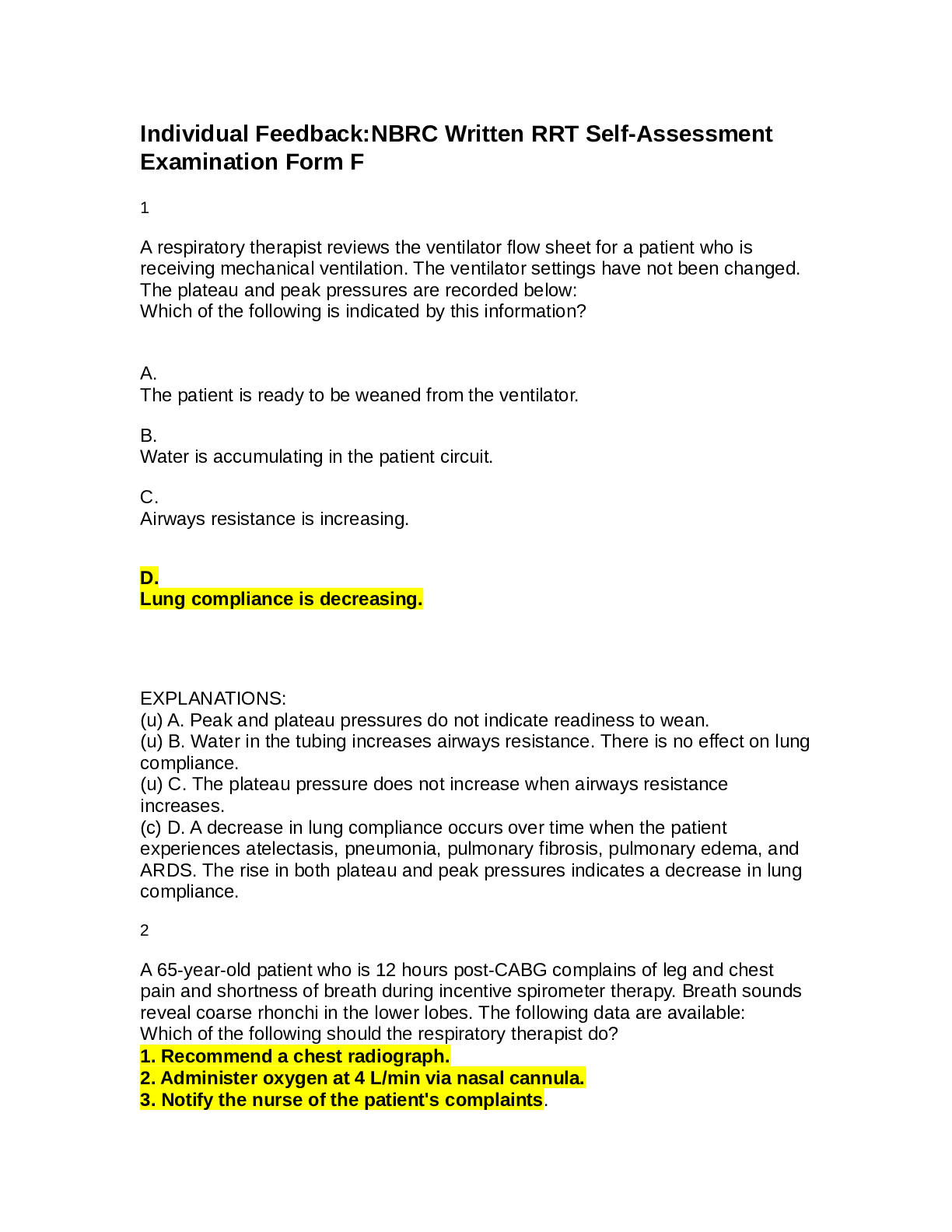
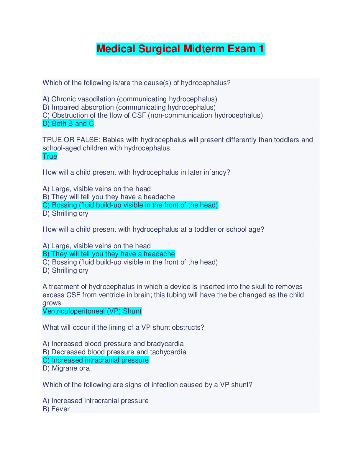
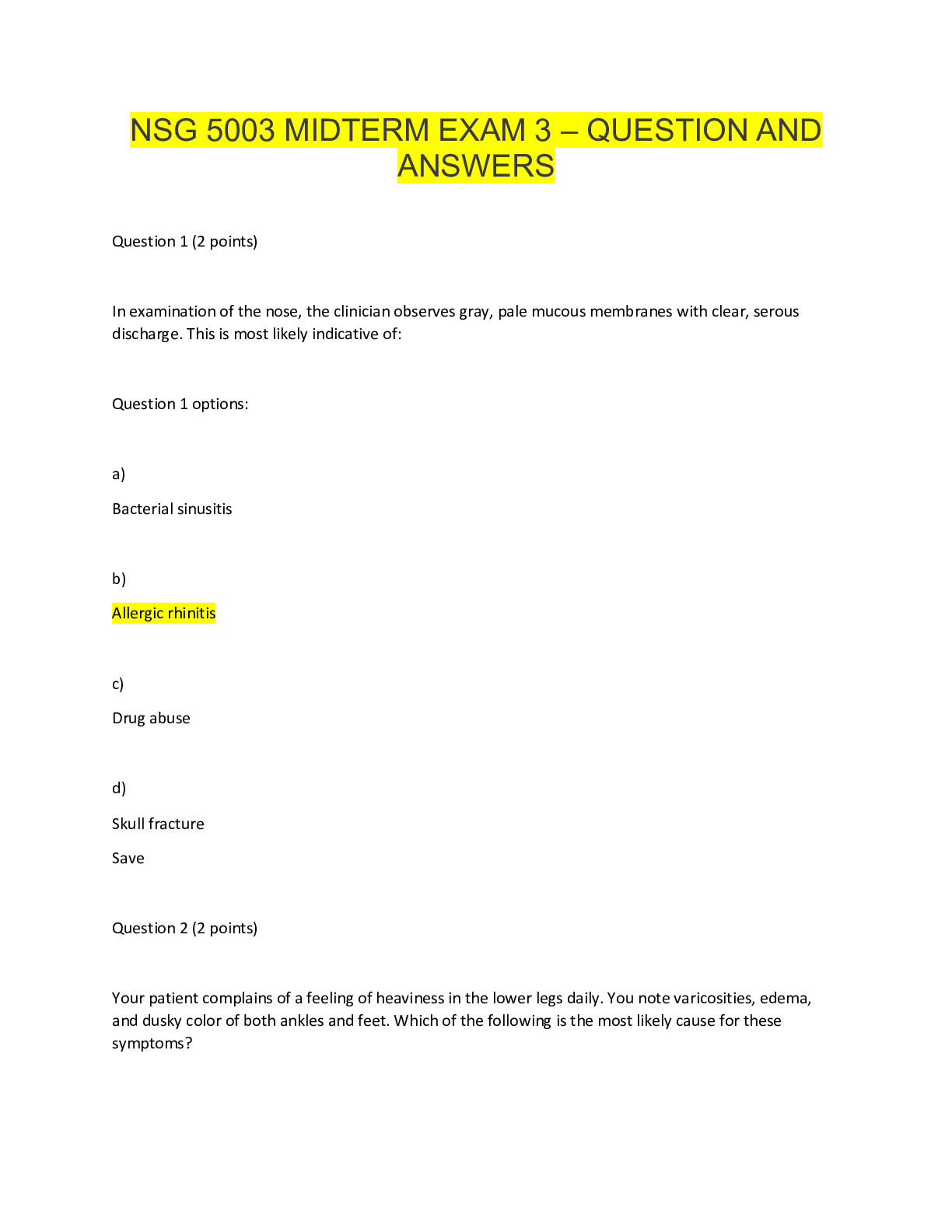
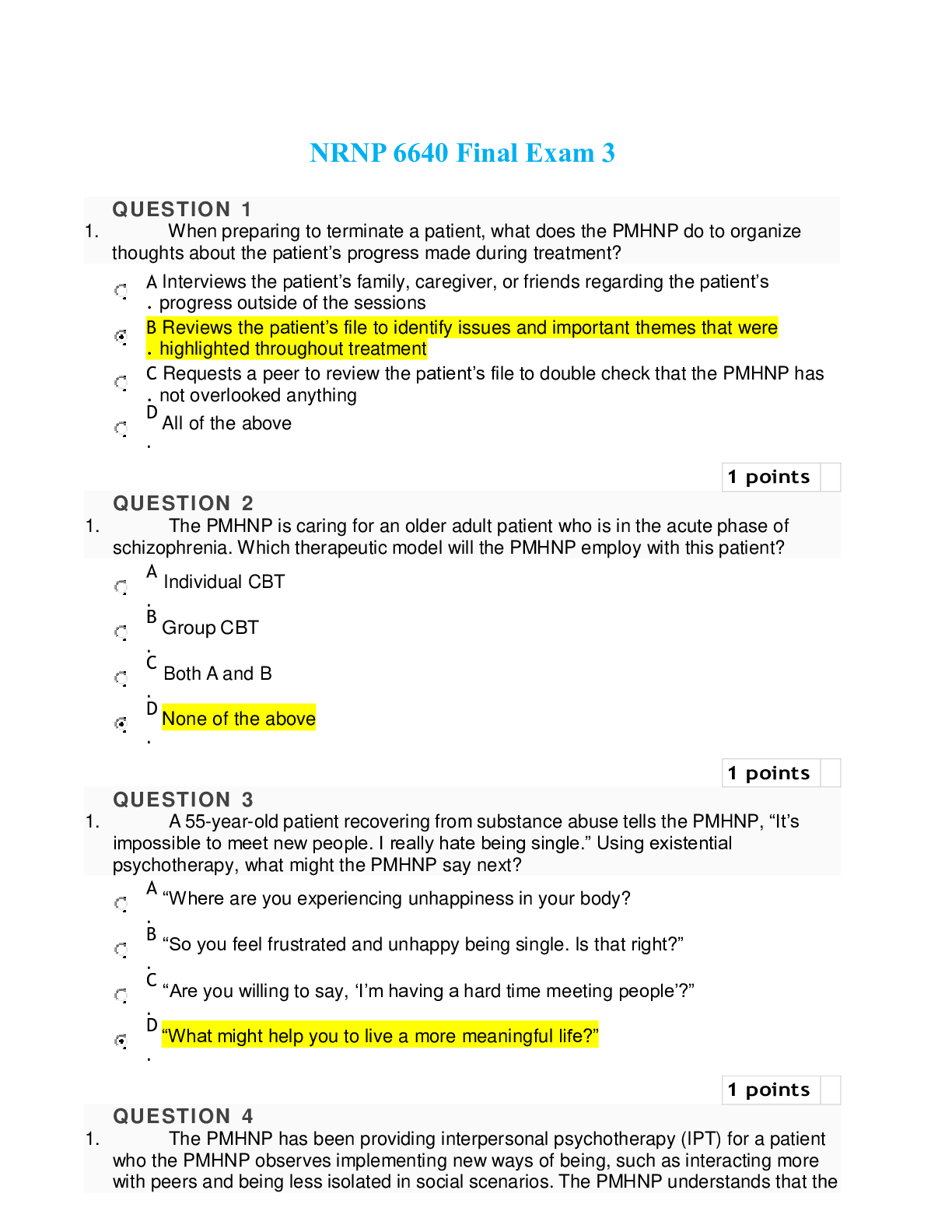
NSG 222 Family Nursing Exam 3 - Questions and Answers
Document Content and Description Below
NSG 222 Family Nursing Exam 3 - Questions and Answers Mild Shock Mild: 20% blood loss, Symptoms: diaphoresis, increased cap refill, cool extremities, maternal anxiety Moderate Shock Moderate: 20-40%... blood loss Symptoms: Tachycardia, postural hypotension, oliguria Severe Shock Severe: over 40% blood loss Symptoms: Hypotension, agitation/confusion, hemodynamic instability Pathophysiology of Ectopic Pregnancy Normally, the fertilized ovum implants in the uterus. In ectopic pregnancy, the journey along the fallopian tube is arrested or altered in some way. With an ectopic pregnancy, the ovum implants outside the uterus. The most common site for implantation is the fallopian tubes (96%), but some ova may implant in the ovary, the intestine, the cervix, or the abdominal cavity Risk factors of ectopic pregnancy Chlamydia infection resulting in tubal damage Other associated risk factors for ectopic pregnancy include previous tubal surgery, infertility, PID, previous pregnancy loss (induced or spontaneous), use of an intrauterine contraceptive system, previous ectopic pregnancy, uterine fibroids, sterilization, smoking (which alters tubal motility), history of multiple sexual partners, use of progestin-only oral contraceptives, douching, and exposure to diethylstilbestrol Gestational Trophoblastic Disease Definition The WHO classification of gestational trophoblastic disease (GTD) includes disorders of placental development (hydatidiform mole) and neoplasms of the trophoblast (choriocarcinoma) Gestational Trophoblastic Disease Therapeutic Management Treatment consists of immediate evacuation of the uterine contents as soon as the diagnosis is made and long-term follow-up of the client to detect any remaining trophoblastic tissue that might become malignant. D&C is used to empty the uterus. The tissue obtained is sent to the laboratory for analysis to evaluate for choriocarcinoma. Serial levels of hCG are used to detect residual trophoblastic tissue for 1 year. If any tissue remains, hCG levels will not regress. As a result of the increased risk for cancer, the client is advised to receive extensive follow-up therapy for the next 12 months. The follow-up protocol may include: Baseline hCG level, chest radiograph, and pelvic ultrasound Quantitative hCG levels every week until undetectable for three consecutive weeks; then serial hCG levels monthly for 1 year Chest radiograph every 6 months to detect pulmonary metastasis Regular pelvic examinations to assess uterine and ovarian regression Systemic assessments for symptoms indicative of lung, brain, liver, or vaginal metastasis Strong recommendation to avoid pregnancy for 1 year because the pregnancy can interfere with the monitoring of hCG levels Use of a reliable contraceptive for at least 1 year Gestational Trophoblastic Disease Nursing Assessment The nurse plays a crucial role in identifying and bringing this condition to the attention of the health care provider based on sound knowledge of the typical clinical manifestations and astute prenatal assessments. Clinical manifestations of GTD are similar to those of spontaneous abortion at about 12 weeks of pregnancy. Assess the woman for potential clinical manifestations at each prenatal visit. Be alert for the following: Report of early signs of pregnancy, such as amenorrhea, breast tenderness, fatigue Brownish vaginal bleeding/spotting Anemia Inability to detect a fetal heart rate after 10 to 12 weeks' gestation Fetal parts not evident with palpation Bilateral ovarian enlargement caused by cysts and elevated levels of hCG Persistent, often severe nausea and vomiting (due to high hCG levels) Fluid retention and swelling Uterine size larger than expected for pregnancy dates Extremely high hCG levels present; no single value considered diagnostic Early development of preeclampsia (usually not present until after 24 weeks) Absence of fetal heart rate or fetal activity Expulsion of grape-like vesicles (possible in some women) The diagnosis is made by high hCG levels and the characteristic appearance of the vesicular molar pattern in the uterus via transvaginal ultrasound. Cervical Insufficiency patio The exact mechanism contributing to cervical insufficiency is not known. The cervix may have less elastin, less collagen, and greater amounts of smooth muscle than the normal cervix and thus results in loss of sphincter tone. Structural cervical weakness is the likely cause of many recurrent second-trimester losses, but not the only etiology. Cervical length has also been associated with cervical insufficiency and subsequently preterm birth. Recent studies have examined the association between a short cervical length and the risk of preterm birth. Cervical Insufficiency Therapeutic Management Cervical insufficiency may be treated with bed rest; pelvic rest; avoidance of heavy lifting; progesterone supplementation in women at risk for preterm birth; placement of a cervical pessary (a round, silicone device at the mouth of the cervix); or surgically via a cervical cerclage procedure in the second trimester. Cerclage was created more than 50 years ago based on the hypothesis that for some women, weakness or malfunction of the cervix has a causative role in the pathway to preterm birth. It can either be performed transvaginally or transabdominally. Cervical cerclage involves using a heavy purse-string suture to secure and reinforce the internal os of the cervix Placenta Previa Monitoring Maternal and Fetal Status Avoid doing vaginal examinations in the woman with placenta previa because they may disrupt the placenta and cause hemorrhage. Assess the degree of vaginal bleeding; inspect the perineal area for blood that may be pooled underneath the woman. Estimate and document the amount of bleeding. Perform a peripad count on an ongoing basis, making sure to report any changes in amount or frequency to the health care provider. If the woman is experiencing active bleeding, prepare for blood typing and cross-matching in the event a blood transfusion is needed. Monitor maternal vital signs and uterine contractility frequently for changes. Have the client rate her level of pain using an appropriate pain rating scale. Assess fetal heart rates via Doppler or electronic monitoring to detect fetal distress. Monitor the woman's cardiopulmonary status, reporting any difficulties in respirations, changes in skin color, or complaints of difficulty in breathing. Have oxygen equipment readily available should fetal or maternal distress develop. Encourage the client to lie on her side to enhance placental perfusion. If the woman has an intravenous (IV) line inserted, inspect the IV site frequently. Alternately, anticipate the insertion of an intermittent IV access device such as a saline lock, which can be used if quick access is needed for fluid restoration and infusion of blood products. Obtain laboratory tests as ordered, including complete blood count (CBC), coagulation studies, and Rh status if appropriate. Administer pharmacologic agents as necessary. Give Rh immunoglobulin if the client is Rh-negative at 28 weeks' gestation. Monitor tocolytic (anticontraction) medication if prevention of preterm labor is needed. Patho of Spontaneous Abortion Miscarriage The causes of spontaneous abortion are varied and often unknown. The most common cause for first-trimester abortions is fetal genetic abnormalities, usually unrelated to the mother. Cervical Insufficiency Nursing Assessment Spontaneous Abortion Supportive Care When a pregnant woman calls and reports vaginal bleeding, she must be seen as soon as possible by a health care professional to ascertain the etiology. Varying degrees of vaginal bleeding, low back pain, abdominal cramping, and passage of products of conception tissue may be reported. Ask the woman about the color of the vaginal bleeding (bright red is significant) and the amount—for example, question her about the frequency with which she is changing her peripads (saturation of one peripad hourly is significant) and the passage of any clots or tissue. Instruct her to save any tissue or clots passed and bring them with her to the [Show More]
Last updated: 3 months ago
Preview 1 out of 19 pages
Instant download
Buy this document to get the full access instantly
Instant Download Access after purchase
Add to cartInstant download
Reviews( 0 )
Document information
Connected school, study & course
About the document
Uploaded On
Mar 13, 2024
Number of pages
19
Written in

Additional information
This document has been written for:
Uploaded
Mar 13, 2024
Downloads
0
Views
17