Medicine > EXAM > HESI Radiography Practice Test 1 - Questions, Answers and Rationales (Complete Solutions) (All)
HESI Radiography Practice Test 1 - Questions, Answers and Rationales (Complete Solutions)
Document Content and Description Below
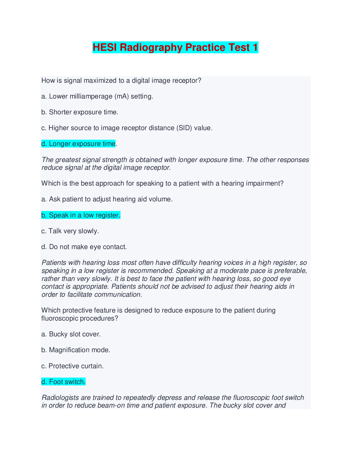
HESI Radiography Practice Test 1 - Questions, Answers and Rationales (Complete Solutions) How is signal maximized to a digital image receptor? a. Lower milliamperage (mA) setting. b. Shorter exposur... e time. c. Higher source to image receptor distance (SID) value. d. Longer exposure time. The greatest signal strength is obtained with longer exposure time. The other responses reduce signal at the digital image receptor. Which is the best approach for speaking to a patient with a hearing impairment? a. Ask patient to adjust hearing aid volume. b. Speak in a low register. c. Talk very slowly. d. Do not make eye contact. Patients with hearing loss most often have difficulty hearing voices in a high register, so speaking in a low register is recommended. Speaking at a moderate pace is preferable, rather than very slowly. It is best to face the patient with hearing loss, so good eye contact is appropriate. Patients should not be advised to adjust their hearing aids in order to facilitate communication. Which protective feature is designed to reduce exposure to the patient during fluoroscopic procedures? a. Bucky slot cover. b. Magnification mode. c. Protective curtain. d. Foot switch. Radiologists are trained to repeatedly depress and release the fluoroscopic foot switch in order to reduce beam-on time and patient exposure. The bucky slot cover and protective curtain are designed to reduce exposure to personnel, rather than to the patient. While the magnification mode is helpful in certain situations, it does increase exposure to the patient. How can digital radiography contribute to patient dose reduction today, in spite of generally being slower in system speed than 400 speed film screen systems? a. Lower kilovolts peak (kVp) should be used with digital. b. Lower milliampere-seconds (mAs) should be used with digital. c. Higher milliampere-seconds (mAs) should be used with digital. d. Higher kilovolts peak (kVp) should be used with digital Higher kVp should be used with digital radiography, because lost contrast can be replaced during image processing. The net effect is a patient dose reduction compared to film-screen systems for the same body part. Lower kVp should not be used. Lower mAs cannot be assured due to the speed of the imaging system, but it may result from the choice of higher kVp. Higher mAs may be required due to the speed of the imaging system, but it certainly does not contribute to patient dose reduction. Which position should a patient be placed in immediately following a seizure? a. Dorsal recumbent. b. Sitting. c. Lateral recumbent. d. Ventral recumbent. Lateral recumbent is the preferred position for patients to be placed after having a seizure to avoid aspiration of secretions. Dorsal recumbent inhibits breathing and does not prevent aspiration. Ventral recumbent hides the patient's face from being observed for further complications. Sitting is contraindicated due to the patient weakness following the seizure. Who judges radiographic images for quality? (Select the four that apply.) a. Ordering physicians. b. Lawyers. c. Radiologists. d. Radiographers. e. Quality control technologists Radiographs are judged for quality by essentially all quality control technologists, ordering physicians, radiographers, and radiologists who see them. Lawyers depend upon hired expert radiologists to judge radiographs for them. How does the Density log Exposure (D log E) curve appear for a digital imaging receptor (IR)? a. A straight line at a 45 degree angle. b. An S curve. c. A shallow slope. d. A steep slope. D log E curves for digital imaging demonstrate the wide dynamic range of the image receptor as a straight line at a 45 degree angle. As an image is windowed, the data is moved up and down the 45 degree line. D log E curves are bell curves with the left half of the bell demonstrated. A steep slope and shallow slope pertain to film/screen imaging. Following administration of contrast media, the radiologist asks the radiographer to obtain a conus projection. Which patient position and centering point should be used for the image? a. Lateral centered to L3-L4. b. Anteroposterior (AP) centered to L3-L-4. c. Lateral centered to T12-L-1. d. Anteroposterior (AP) centered to T-12-L The conus projection is used to image the conus medullaris or terminal end of the spinal cord. It terminates at L1-L2. An AP position centered to T12-L1 is obtained to best image this structure. An AP centered to L3-L4 is below the conus medullaris. A lateral position centered to T12-L1 does not demonstrate the conus medullaris as well as in the AP position. Using a lateral centered to L3-L4 is below the level of the conus medullaris. Which term describes stray radiation that is emitted through the x-ray tube housing? a. Exit. b. Off-focus. c. Scatter. d. Leakage. Leakage radiation is radiation emitted through the housing of the x-ray tube that results in additional exposure to the patient and radiographer. Exit radiation is the portion of the primary beam that passes through the patient to form the image on the image receptor. Scatter is a change in direction of x-rays as the beam passes through an object in its path. Off-focus radiation describes x-rays produced within the tube, but outside of the focal spot. What is the longest field size dimension allowable for 10 inches by 12 inches collimation at a 40 inch source to image distance (SID)? a. 12.8 inches. b. 13.2 inches. c. 10.6 inches. d. 11.4 inches. Field size may be off +/- 2% of the SID, which in this case is 0.8 inches. 12.8 inches is in the acceptable range. 10.6 inches and 11.4 inches have collimation-field size discrepancies that are within the acceptable range. 13.2 inches exceeds the acceptable range. A radiographer prepares to transfer a patient from a wheelchair to the x-ray table. The patient recently suffered a stroke, exhibits left-sided weakness, and is able to bear some weight. How should the radiographer move the patient to the table? a. Position the left side of the wheelchair next to the table. b. Have one radiographer lift the torso while another lifts the feet. c. Use a hydraulic lift. d. Position the right side of the wheelchair next to the table Because the patient’s strongest side is the right side, the radiographer should position the right side of the wheelchair next to the table. Hydraulic lifts are used for patients who cannot bear weight and are too heavy to lift. Positioning the left side of the wheelchair to the table places the patient’s strong side away from the table. Two radiographers to move the patient is called a two-person lift and is used when the patient cannot bear weight but can be lifted manually. How does the central ray (CR) for an anteroposterior (AP) scapula differ from an AP shoulder x-ray? a. CR is the same. b. One inch lower. c. One inch higher. d. Same level, medial. The CR for an AP scapula is one inch lower than the CR for an AP shoulder, or two inches inferior to the coracoid process. The CR is not the same for these two projections. One inch higher is at the level of the coracoid process, too high. At the same level, but medial, will be one inch too high. Where is the central ray (CR) directed for an anteroposterior (AP) projection of the lower leg? a. At the level of the medial malleolus. b. Two inches distal to the medial condyle of the tibia. c. Mid-lower leg. d. 0.5 inch inferior to patellar apex. An AP projection of the lower leg requires the CR to enter mid-lower leg. Half an inch inferior to the patellar apex, the level of the medial malleolus, and two inches distal to the medial condyle of the tibia are all either too high or too low. What should the radiographer do if a radiologist's instructions are unclear? a. Question the lead technologist. b. Perform the exam in the usual way. c. Read the protocol book. d. Ask for clarification The radiographer should ask for clarification. This will allow for any miscommunication to be corrected and improve the patient outcome. The other options are incorrect. A metallic artifact from an object that can be clearly seen as the patient is positioned is clearly the responsibility of the radiographer. For those who would argue that the radiographer is not responsible for caps on teeth, notice that the head is tipped too far forward. The occipital bone is not even visualized on this image. This cap need not have obscured the tip of the odontoid process. No one else is responsible in this case. Who is responsible for the metallic artifact seen in the open mouth odontoid image? a. The ordering physician. b. The receptionist. c. The radiographer. d. The patient Which step will reduce digital image noise, assuming no other changes? a. Longer exposure time. b. Image magnification. c. Decreased kilovolts peak (kVp). d. Small pixel size. A longer exposure time results in a greater amount of photons reaching the image receptor, which reduces image noise. Decreased kVp reduces the quantity of x-rays that reach the image receptor, which increases image noise. A matrix with a large number of small pixels results in greater image noise due to the availability of fewer photons per pixel. Magnification of the digital image does not affect image noise, but it may make what noise is there more visible. Which system can store images? a. Picture archival and communication system (PACS). b. Hospital information system (HIS). c. Digital imaging and communication in medicine (DICOM). d. Radiology information system (RIS). The PACS can store images. The HIS contains much information, including billing information and patient treatment plans, but does not contain images. Likewise, the RIS does not contain images, though it has radiology-specific information, such as a radiology schedule history. The DICOM standard allows images to be exchanged between different devices. Which action can improve recorded detail? a. Use the largest focal spot. b. Increase the source-image receptor-distance (SID). c. Select a larger kilovolts peak (kVp). d. Increase the object-image receptor-distance (OID). Increasing the SID will cause an increase in recorded detail on the image. Increasing the OID, or using a larger focal spot will all cause a decrease in recorded detail. KVp does not affect recorded detail. What is the maximum speed of filament electrons as they travel toward the anode? a. One-eighth the speed of light. b. Half the speed of light. c. One-fourth the speed of light. d. The speed of light. Filament electrons may reach speeds of about half the speed of light. The other answers are too fast or too slow. This lumbar image shows loss of resolution due to blurring. Since the single greatest cause of loss of image sharpness is patient motion, immobilizing the patient will likely have the greatest improvement on image quality. Adjusting mAs will not affect image sharpness. Selecting a faster image receptor speed and decreasing SID will degrade image sharpness further. Consider this lateral lumbar spine image. Which strategy will have the greatest improvement on image quality? a. Immobilize the patient. b. Decrease the source to image receptor distance (SID). c. Select a faster speed image receptor. d. Increase milliampere-seconds (mAs). Which x-ray beam characteristic is reduced by adding aluminum filtration? a. Quantity of x-rays. b. Half-value layer. c. Energy of the x-ray beam. d. X-ray beam quality. Added aluminum filtration results in a reduction in the quantity of x-rays in the primary beam. The half-value layer (HVL) is a numerical value used to identify the quality of the beam; added filtration results in an increase in HVL, not a reduction. The quality of the x-ray beam refers to its penetrability, which increases as a result of added filtration, not a reduction. Because most of the x-rays removed by the filter are of low energy, there is an increase in the average energy of the primary x-ray beam, not a reduction. On an accurately positioned posteroanterior (PA) chest radiograph, the manubrium of the sternum is seen at the level of which thoracic vertabrae when the patient is sthenic? a. Second. b. Fourth. c. Fifth. d. Third. The manubrium of the sternum is projected at the level of the fourth thoracic vertebra on a properly positioned PA chest radiograph. When the upper midcoronal plane of the patient is tilted backwards (away from the film), the manubrium is positioned between the first and third thoracic vertebra. If the upper midcoronal plane of the patient is tilted toward the image receptor, the manubrium will be seen at the level of the fifth thoracic vertebra or lower. The bucky slot cover should offer at least what lead thickness? a. 0.25 mm. b. 0.35 mm. c. 1 mm. d. 0.5 mm. The bucky slot cover should be at least 0.25 mm lead equivalent. Therefore, the rest of the answers are incorrect. Which is an advantage of using a optically stimulated luminescent (OSL) dosimeter as opposed to a film badge? [Show More]
Last updated: 3 weeks ago
Preview 1 out of 71 pages
Instant download
Buy this document to get the full access instantly
Instant Download Access after purchase
Add to cartInstant download
Reviews( 0 )
Document information
Connected school, study & course
About the document
Uploaded On
May 26, 2024
Number of pages
71
Written in

Additional information
This document has been written for:
Uploaded
May 26, 2024
Downloads
0
Views
13


.png)