*NURSING > STUDY GUIDE > DESCRIBING THE AIRWAY DIFFERENCES IN A PAEDIATRIC with Well Explained Answers Graded- A (All)
DESCRIBING THE AIRWAY DIFFERENCES IN A PAEDIATRIC with Well Explained Answers Graded- A
Document Content and Description Below
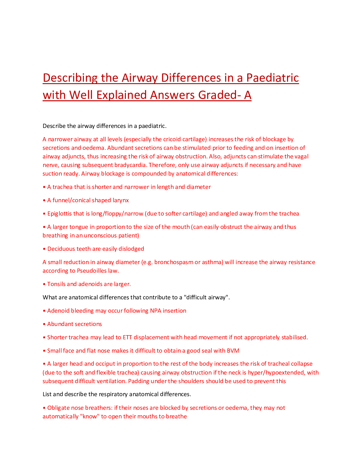
Describe the airway differences in a paediatric. What are anatomical differences that contribute to a "difficult airway". List and describe the respiratory anatomical differences. • Obligate nose... breathers: if their noses are blocked by secretions or oedema, they may not automatically "know" to open their mouths to breathe • Muscles are poorly developed. They have a lower % of slow twitch (type 1) skeletal muscle fibres in their intercostal muscles, therefore the chest and respiratory muscles tire easily making paediatrics more prone to respiratory fatigue and failure • Bones are soft, pliable and primarily cartilaginous, thus the ribs offer less protection to organs • Lungs are more fragile thus pulmonary contusions are more common in paediatric patients who have been subjected to major trauma. Pneumothorax is common following barotrauma e.g. aggressive PPV. Atelectasis is more common in paediatrics with respiratory distress. • Mediastinum: not so rigid, thus will shift more with a tension pneumothorax • Chest wall is thin thus breath sounds easily transmitted. This may result in perception of breath sounds from elsewhere in the chest, which may cause you to miss a pneumothorax or misplaced intubation. • TV is fixed (thus can only increase RR in times of need) between 6 to 8ml/kg. • FRC is lower, thus they are more susceptible to desaturation [Show More]
Last updated: 1 year ago
Preview 1 out of 8 pages
Reviews( 0 )
Document information
Connected school, study & course
About the document
Uploaded On
Aug 23, 2021
Number of pages
8
Written in

Additional information
This document has been written for:
Uploaded
Aug 23, 2021
Downloads
0
Views
55


.png)