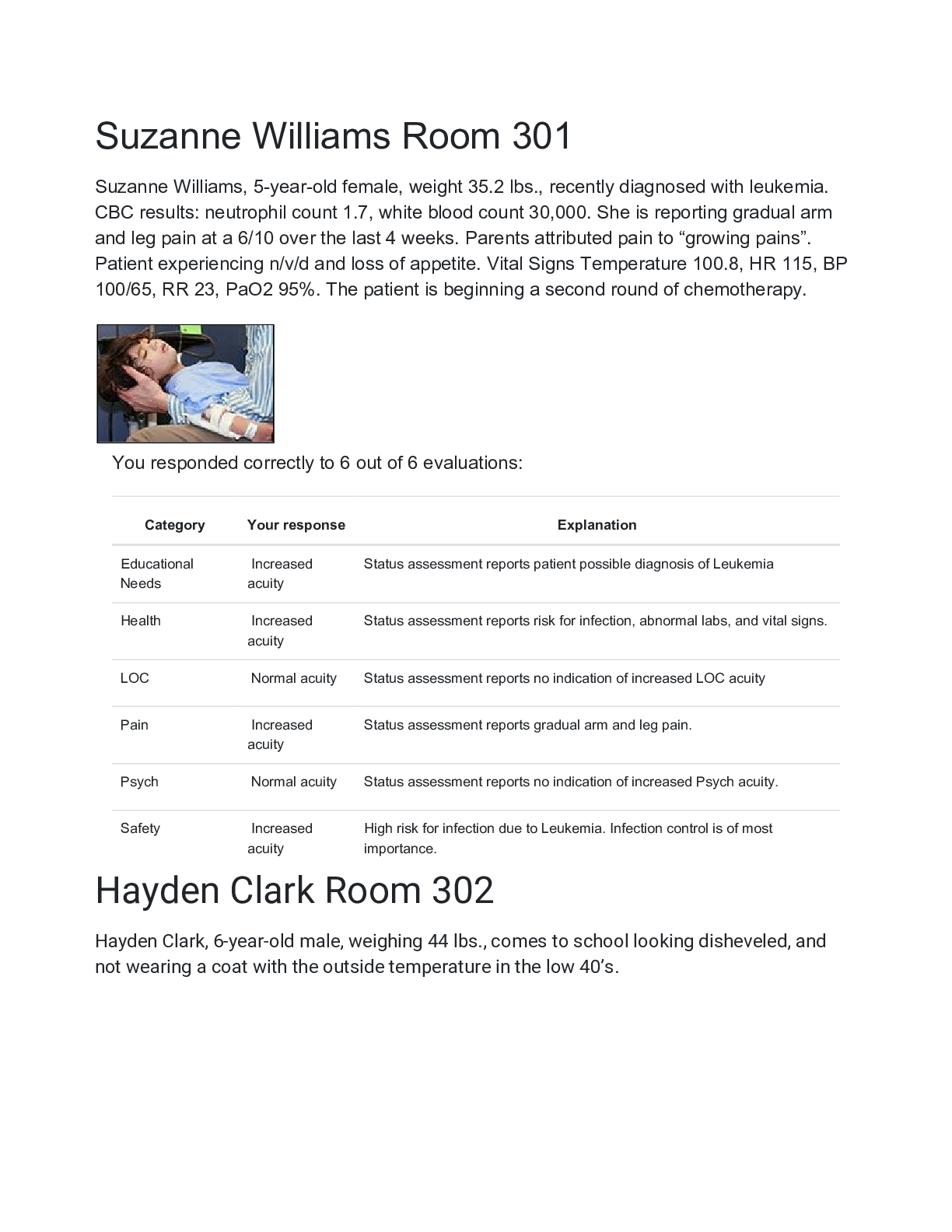
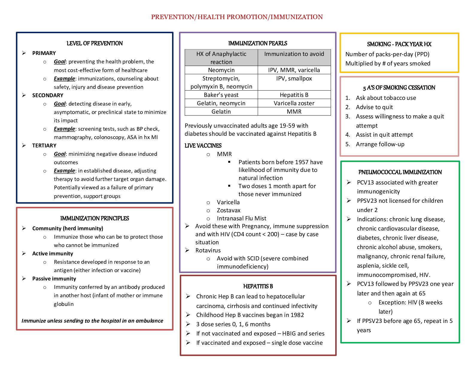
Biology > STUDY GUIDE > BIO 171 Microbiology Module 3. Portage Learning (All)
BIO 171 Microbiology Module 3. Portage Learning
Document Content and Description Below
BIO 171 Microbiology Module 3. Portage Learning.Module 3.1: Microscopy - The micrometer (µm) is one-millionth of a meter and is commonly designated at 10^-6 m - The nanometer (nm) equals 10^-9 m or... one-billionth of a meter. - For perspective, the unaided eye can resolve (see clearly) objects typically > 100 µm. However, cellular components, organelles, etc. can be as small as 0.2 µm in diameter. - Two critical factors influence our ability to see an object: the resolution and the contrast. • The resolution refers to the distance between two objects at which the objects still can be seen as separate. • The contrast is the difference in light absorbance between two areas (objects). The lower the contrast between an object and its background, the harder it will be to see that object. - Brightfield Microscopy • Brightfield microscopes are the simplest form of light, or optical, microscopes. Light, most often emitted from a standard halogen bulb, enters the microscope from the base (bottom) and is reflected via mirrors towards the sample. • Before the light reaches the sample, it first passes through a condenser converging the light beams into a focused area on the sample. • The iris diaphragm controls the amount of light that passes through the sample and into the objective lens. The objective lens is the lens closest to the sample and yields the greatest amount of magnification. (Note: The degree of magnification is directly proportional to the amount of light 1 needed. Thus, to image samples clearly at higher magnifications, more light is required). • Once light passes through the sample and the objective lens, it is directed through the ocular lens, or eyepiece, to your eye. The most common power of an ocular lens is 10x. For a microscope using two lenses (objective and ocular) the total magnification of a specimen is multiplicative. Thus, a 40x objective and a 10x ocular result in a total magnification of 400x. • In order to visualize cells, samples are most often stained with specific combinations of dyes that are taken up by the cell. Staining is often required due to the limitation of resolution on unstained cells because the flat and transparent regions of a cell may appear invisible under bright field conditions. [Show More]
Last updated: 1 year ago
Preview 1 out of 9 pages
Instant download
Buy this document to get the full access instantly
Instant Download Access after purchase
Add to cartInstant download
Reviews( 0 )
Document information
Connected school, study & course
About the document
Uploaded On
Dec 01, 2021
Number of pages
9
Written in

Additional information
This document has been written for:
Uploaded
Dec 01, 2021
Downloads
0
Views
62