Health Care > EXAM > BIO 152 A&P II Final Exam- Portage Learning-2021 (All)
BIO 152 A&P II Final Exam- Portage Learning-2021
Document Content and Description Below
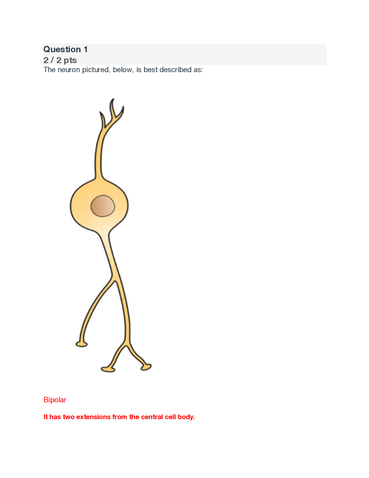
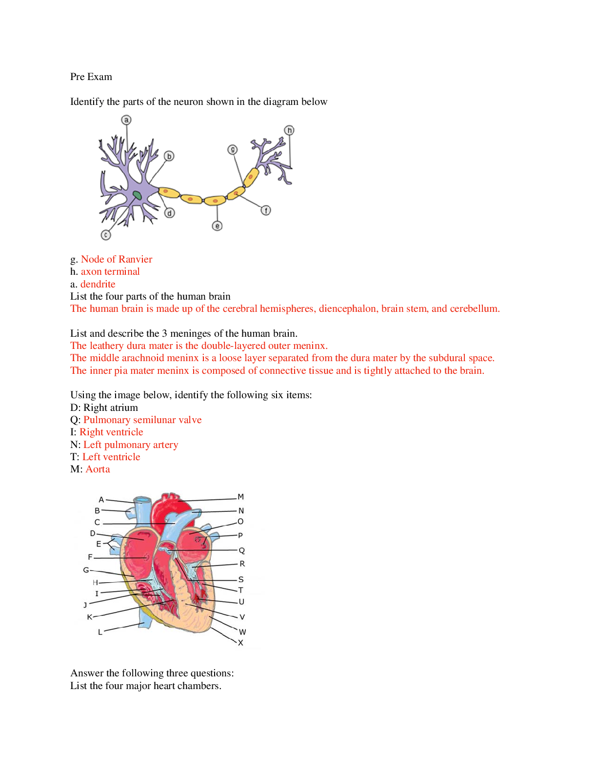
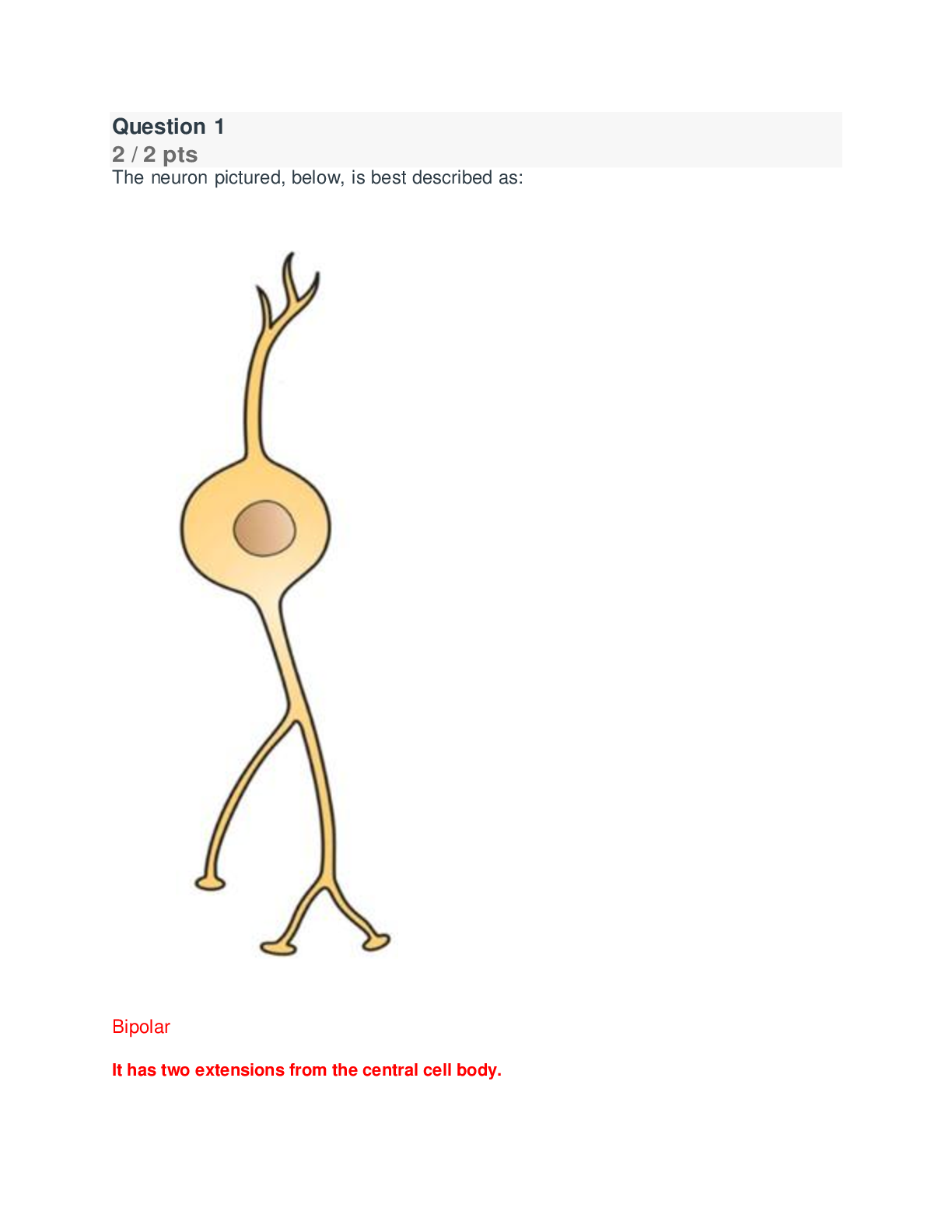
The neuron pictured, below, is best described as: Bipolar It has two extensions from the central cell body. Multipolar Unipolar Pseudounipolar Name and describe what is occurring in the neuron cell me... mbrane in section 4 of the diagram. Include the charge of the membrane during this phase. Your Answer: • Afterpolarization (Hyperpolarization) Potassium gates are slow to close and there is an undershoot of the potential. • The charge drops below -70mV and then returns to -70mV once at resting state again. Where is the integration center of a reflex located? CNS (central nervous system) What is true about the flexor withdrawal reflex? A. It does not involve interneurons. B. It involves excitatory interneurons. C. It involves inhibitory interneurons. D. The effect of the reflex is to create a co-contraction of two muscles E. A&D F. B&C Label the nerves (A-C) in the figure below: A: B: C: A- Lateral femoral cutaneous B- Femoral nerve C- Saphenous Which of the following is most likely a symptom of ALS? Impaired ability to swallow Decreased sensation in the hands Shrinkage of cerebral cortex Increased size of brain ventricles All the above What is the purpose of the blood-brain barrier? Describe its maintenance from a cellular level. The blood-brain barrier is a diffusion barrier which prevents most particles from entering the central nervous system tissue, keeping the brain and spinal cord separate from general blood circulation. The blood-brain barrier is formed by the relatively impermeable brain capillaries, due to the glial cells astrocytes. Maintenance of the blood-brain-barrier is important to provide a stable chemical environment for the nervous system. A patient damaged the radial nerve. What action is most likely limited? Elbow flexion Hip extension Wrist flexion Wrist extension What is muscle tone and how is it maintained? Muscle tone is the degree at which muscles remain partially contracted while at rest. Muscle tone is continuously monitored and maintained by the cerebellum to keep bones and joints in place. A patient had a CVA in the area indicated by the red x in the figure, below. What type of blindness is the patient most likely to incur? Explain your reasoning. A. Left eye blindness B. Right eye blindness C. Bilateral left visual field blindness D. Bilateral right visual field blindness. C The right optic tract is damaged. All the sensory information from the left visual fields travels together after the optic chiasm to the right side of the brain. Match the numbers (1-5) in the figure below with the correct terms (A-H). Note: not all terms will be used. 1- A: Cochlear Nerve 2- B: Facial Nerve 3- C: Tympanic Membrane 4- D: Cochlea 5- E: Malleus ......................continued [Show More]
Last updated: 1 year ago
Preview 1 out of 19 pages

Reviews( 0 )
Document information
Connected school, study & course
About the document
Uploaded On
Jan 14, 2022
Number of pages
19
Written in

Additional information
This document has been written for:
Uploaded
Jan 14, 2022
Downloads
0
Views
45


.png)

.png)
.png)
.png)
.png)
