*NURSING > STUDY GUIDE > Rasmussen College: PN 3 Exam 3 Study Guide_ LATEST,100% CORRECT (All)
Rasmussen College: PN 3 Exam 3 Study Guide_ LATEST,100% CORRECT
Document Content and Description Below
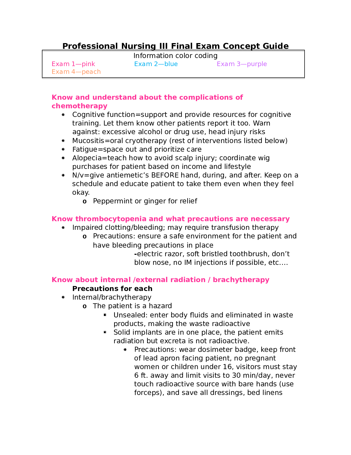
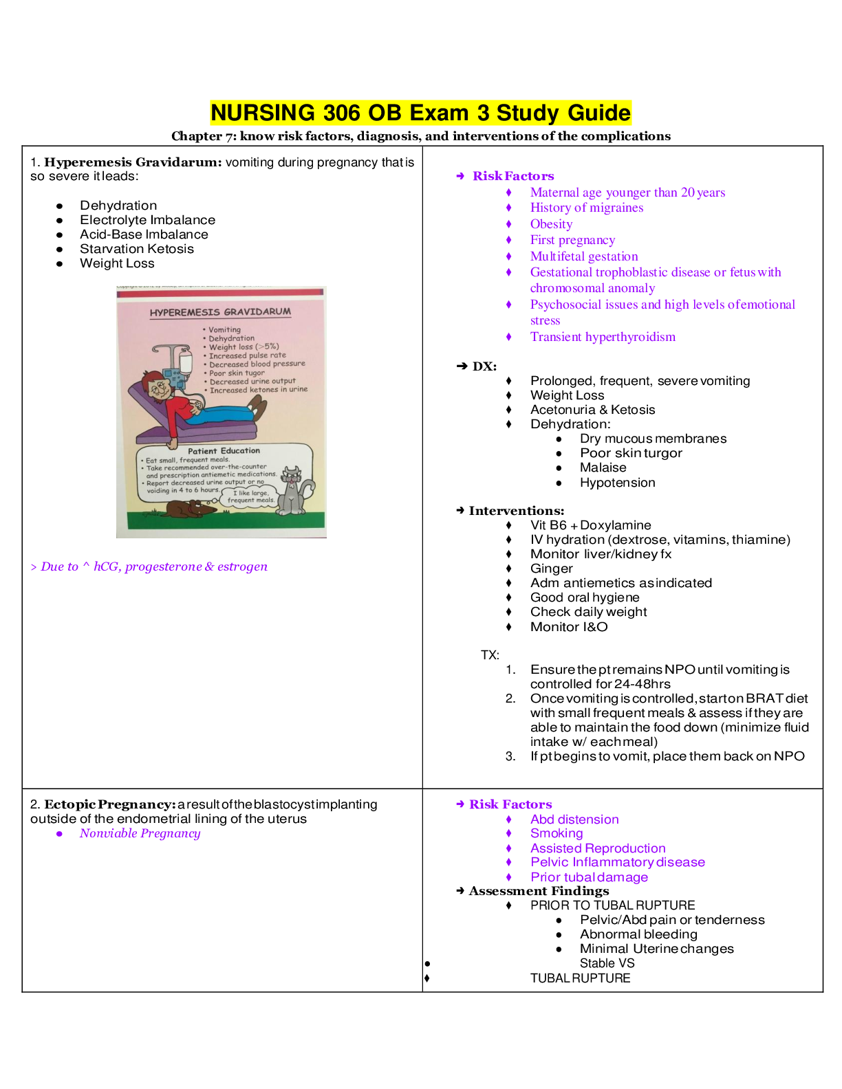
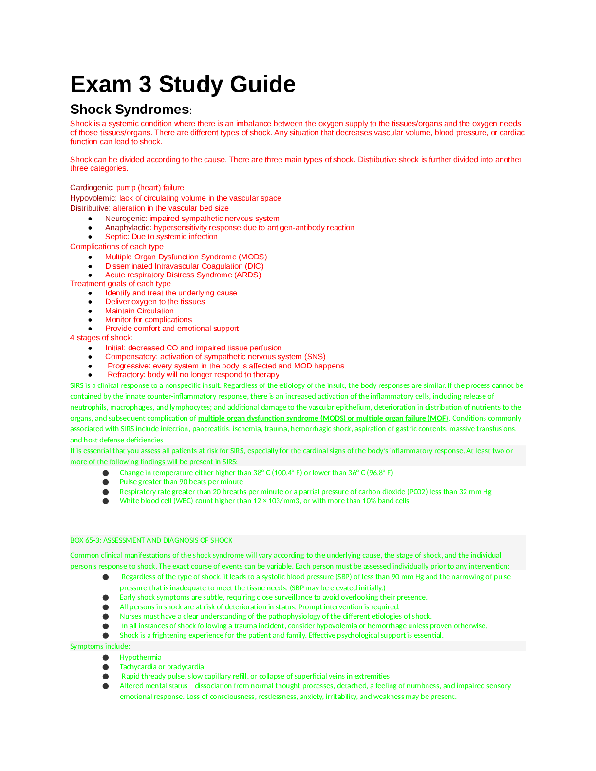
Rasmussen College: PN 3 Exam 3 Study Guide_ LATEST Shock Syndromes: Shock is a systemic condition where there is an imbalance between the oxygen supply to the tissues/organs and the oxygen needs of ... those tissues/organs. There are different types of shock. Any situation that decreases vascular volume, blood pressure, or cardiac function can lead to shock. Shock can be divided according to the cause. There are three main types of shock. Distributive shock is further divided into another three categories. Cardiogenic: pump (heart) failure Hypovolemic: lack of circulating volume in the vascular space Distributive: alteration in the vascular bed size ● Neurogenic: impaired sympathetic nervous system ● Anaphylactic: hypersensitivity response due to antigen-antibody reaction ● Septic: Due to systemic infection Complications of each type ● Multiple Organ Dysfunction Syndrome (MODS) ● Disseminated Intravascular Coagulation (DIC) ● Acute respiratory Distress Syndrome (ARDS) Treatment goals of each type ● Identify and treat the underlying cause ● Deliver oxygen to the tissues ● Maintain Circulation ● Monitor for complications ● Provide comfort and emotional support 4 stages of shock: ● Initial: decreased CO and impaired tissue perfusion ● Compensatory: activation of sympathetic nervous system (SNS) ● Progressive: every system in the body is affected and MOD happens ● Refractory: body will no longer respond to therapy SIRS is a clinical response to a nonspecific insult. Regardless of the etiology of the insult, the body responses are similar. If the process cannot be contained by the innate counter-inflammatory response, there is an increased activation of the inflammatory cells, including release of neutrophils, macrophages, and lymphocytes; and additional damage to the vascular epithelium, deterioration in distribution of nutrients to the organs, and subsequent complication of multiple organ dysfunction syndrome (MODS) or multiple organ failure (MOF). Conditions commonly associated with SIRS include infection, pancreatitis, ischemia, trauma, hemorrhagic shock, aspiration of gastric contents, massive transfusions, and host defense deficiencies ● Change in temperature either higher than 38° C (100.4° F) or lower than 36° C (96.8° F) ● Pulse greater than 90 beats per minute ● Respiratory rate greater than 20 breaths per minute or a partial pressure of carbon dioxide (PC02) less than 32 mm Hg ● White blood cell (WBC) count higher than 12 × 103/mm3, or with more than 10% band cells ● Regardless of the type of shock, it leads to a systolic blood pressure (SBP) of less than 90 mm Hg and the narrowing of pulse pressure that is inadequate to meet the tissue needs. (SBP may be elevated initially.) ● Early shock symptoms are subtle, requiring close surveillance to avoid overlooking their presence. ● All persons in shock are at risk of deterioration in status. Prompt intervention is required. ● Nurses must have a clear understanding of the pathophysiology of the different etiologies of shock. ● In all instances of shock following a trauma incident, consider hypovolemia or hemorrhage unless proven otherwise. ● Shock is a frightening experience for the patient and family. Effective psychological support is essential. Symptoms include: ● Hypothermia ● Tachycardia or bradycardia ● Rapid thready pulse, slow capillary refill, or collapse of superficial veins in extremities ● Altered mental status—dissociation from normal thought processes, detached, a feeling of numbness, and impaired sensory- emotional response. Loss of consciousness, restlessness, anxiety, irritability, and weakness may be present. ● Clinical findings correlated with organs compromised by inadequate oxygen supply and the phases of the shock syndrome. Examples: ○ a. Skin: cold, clammy, cyanotic, poor capillary refill, or warm dry skin due to pooling of blood in extremities. Cyanosis (circumoral, earlobes, finger tips, or toes). ○ b. Kidneys: decreased urine output; anuria, or oliguria. ○ c. Lungs: dyspnea, crackles, or wheezes. ○ d. GI system: thirst, dry mucous membranes; nausea and vomiting; or decreased bowel sounds. Acute Coronary Syndrome: Unstable Angina (VS stable angina) ● Unstable angina: condition in which your heart doesn’t get enough blood flow and oxygen--may lead to a heart attack. DO NOT KNOW THE TRIGGERS ● Stable angina: chest pain or discomfort that most often occurs with activity or emotional stress. KNOW WHAT POTENTIAL TRIGGERS ARE. Usually a chronic condition that causes stable angina. Non ST segment elevation myocardial infarction (NSTEMI)--> Indicates a partial thickness injury to the heart muscle. Less severe than a STEMI. Partial occlusion of a major coronary artery ST segment elevation myocardial infarction (STEMI)--> indicates a full thickness injury of the heart muscle. Assessment of chest pain: Including: Special populations assessment: elderly, women Onset: When did the pain begin? Location: Where is the pain? Duration: How long does the pain last? Characteristics:Describe the pain? Crushing, stabbing, ingestion like, dull, ache for example Associating Factors: Other symptoms associated with the pain such as nausea and/or vomiting, weakness, fatigue, breathlessness, syncope, cold and clammy? Relieving Factors/Radiation: Does the pain radiate such as down the arm, up into the neck for example? Relieving factors: pain stops when activity ceases, relieved by sitting forward or resting? Treatment/Temporal factors: Use of GTN, pain was relieved by rest or decrease in physical activity. Pain non comparable to previous ischemic chest pain Severity (Intensity): A numerical scale (1 no pain- 10 worse pain experienced ) is used to gauge pain severity Older adults: Typical chest pain→ Usually intense and unremitting for 30-60 minutes. Is retrosternal and often radiates up to the neck, shoulder, and jaws, and down to the left arm. The chest pain is usually described as a pressure sensation that can be perceived as squeezing, aching, burning, or even sharp. Anxiety, lightheadedness, cough, nausea, profuse sweating, shortness of breath, wheezing, rapid or irregular heart rate, fullness/indigestion/choking feeling Women may or may not experience chest pain but may experience any of the symptoms above. Treatments/Surgical Procedures ● Meds: Anticoagulatns, antiplatelets, ACE inhibitors, beta blockers, Calcium Channel Blocker, Statins, diuretics, vasodilators, pain relievers (morphine), Nitroglycerin ● Surgeries: coronary angioplasty and stenting, coronary artery bypass surgery Patient Education for Anginal Episodes ● Stop activity, sit or lie down. ● Place one nitroglycerin tablet under the tongue and allow to dissolve. (Do not chew or swallow.) ● Tablet will cause a tingling sensation, heart pounding, flushing, and headache. ● Stay in resting position for 15 to 20 minutes and get up slowly after taking nitroglycerin to prevent fainting from postural hypotension. ● If angina is not relieved in five minutes, the dose may be repeated two times at five-minute intervals for a total of three doses. ● If angina is not relieved after two doses, seek immediate medical attention. ● Report angina that increases in frequency, lasts longer, limits previous level of activity, and occurs at rest. ● Carry tablets at all times in the original prescription container. Pharmacology→(what Jenni told us to remember) MONA (Morphine, Oxygen, Nitro, Ativan) Antiplatelet agents-Aspirin, Varopaxar Nitrates-relax smooth muscles Analgesics-Morphine-decreases sympathetic stress, in addition to providing some preload reduction. Beta-adrenergic blocker-Esmolol, Metoprolol-Beta blockers have antiarrhythmic and antihypertensive properties, as well as the ability to reduce ischemia. They minimize the imbalance between myocardial supply and demand by reducing afterload and wall stress. Glycoprotein IIB/IIIA inhibitors-Glycoprotein IIb/IIIa receptor antagonists include abciximab, eptifibatide, and tirofiban. Glycoprotein IIb/IIIa antagonists prevent the binding of fibrinogen, thereby blocking platelet aggregation. Anticoagulants-Heparin augments the activity of antithrombin III and prevents the conversion of fibrinogen to fibrin. It does not actively lyse but is able to inhibit further thrombogenesis. This agent prevents recurrence of a clot after spontaneous fibrinolysis. Low molecular weight heparins-Lovenox Adenosine diphosphate receptor antagonists-Thienopyridine adenosine 5'-diphosphate (ADP) antagonists approved for antiplatelet activity in the United States include clopidogrel (Plavix), ticlopidine, prasugrel, and ticagrelor. Modifiable Risk factors→ The major modifiable risk factors are hyperlipidemia, hypertension (blood pressure greater than 140/90 mm Hg), cigarette smoking, diabetes mellitus, obesity, and sedentary lifestyle. In addition, the remaining modifiable risk factors include low daily fruit and vegetable intake, alcohol consumption, and psychosocial index Cholesterol: purpose of each type→ Total cholesterol–a measure of the total amount of cholesterol in your blood, including low-density lipoprotein (LDL) cholesterol and high-density lipoprotein (HDL) cholesterol. LDL (bad) cholesterol–the main source of cholesterol buildup and blockage in the arteries HDL (good) cholesterol–HDL helps remove cholesterol from your arteries Triglycerides–another form of fat in your blood that can raise your risk for heart disease Total Cholesterol Level Less than 200mg/dL Desirable 200-239 mg/dL Borderline high 240mg/dL and above High LDL (Bad) Cholesterol Level Less than 100mg/dL Optimal 100-129mg/dL Near optimal/above optimal 130-159 mg/dL Borderline high 160-189 mg/dL High 190 mg/dL and above Very High HDL (Good) Cholesterol Level Less than 40 mg/dL A major risk factor for heart disease 40—59 mg/dL The higher, the better 60 mg/dL and higher Considered protective against heart disease What Affects Cholesterol Levels: Diet. Saturated fat and cholesterol in the food you eat make your blood cholesterol level rise. Saturated fat is the main problem, but cholesterol in foods also matters. Reducing the amount of saturated fat and cholesterol in your diet helps lower your blood cholesterol level. Weight. Being overweight is a risk factor for heart disease. It also tends to increase your cholesterol. Losing weight can help lower your LDL and total cholesterol levels, as well as raise your HDL and lower your triglyceride levels. Physical Activity. Not being physically active is a risk factor for heart disease. Regular physical activity can help lower LDL (bad) cholesterol and raise HDL (good) cholesterol levels. It also helps you lose weight. You should aim to be physically active for 30 minutes on most, if not all, days. Things outside of your control that also can affect cholesterol levels include: Age and Gender. As women and men get older, their cholesterol levels rise. Before the age of menopause, women have lower total cholesterol levels than men of the same age. After the age of menopause, women's LDL levels tend to rise. Heredity. Your genes partly determine how much cholesterol your body makes. High blood cholesterol can run in families. HTN: values in each category→ TABLE 24-2: Blood Pressure Classification BP CLASSIFICATION SBP mm Hg DBP mm Hg Normal Less than or equal to 120 or less than or equal to 80 Prehypertension 120–139 or 80–90 Stage I hypertension 140–159 or 90–99 Stage II hypertension Less than or equal to 160 or less than or equal to 100 Diet: including specific food examples→ TLC diet: Total fat 25–30%, saturated fats to less than or equal to 7%, complex carbohydrates 50–60%, protein 15% of total daily calories, total cholesterol less than or equal to 200 mg/day, fiber 25 g/day Steam, bake, broil, grill, or stir fry foods Avoid high fat foods such as meat, avocados, olives, nuts, butter, salad dressing, organ meats and shrimp, egg yolks, palm and coconut oils, high fat dairy products, fried foods Choose fresh fruits and vegetables, egg substitutes or egg whites, lean meats, fish, vegetable oils, low fat dairy products Buy a heart-healthy cookbook Achieve and maintain a healthy weight After approval by the health care provider, exercise regularly Non-modifiable risk factors→ Primary causes of hyperlipidemia include dietary sources and genetic disorders. Secondary causes of hyperlipidemia are due to hormone imbalances, liver and kidney dysfunction, alcohol consumption, and certain drugs such as beta blockers and cyclosporine Renal Dysfunctions: Types: RENAL ASSESSMENT SUBJECTIVE You can ask the following questions when obtaining a renal assessment from the patient: ● What is the general state of health? ● Is there a history of neurological deficit, diabetes, or other debilitating disease that could lead to urinary stasis, damage to the nephron, or impaired healing? ● Have there been recent hospitalizations and drug therapies? ● Is there a history of urinary calculi or prostate gland hypertrophy? ● Is there a recent history of catheterization? ● Have you experienced burning on urination, cloudy urine, suprapubic pain, colicky abdominal pain, or flank or back pain? ● How long have the symptoms persisted, and how were they treated in the past? OBJECTIVE The physical examination must include: ● Palpation of the abdomen for masses and tenderness. ● Palpation of costovertebral angle with a gentle tap of the fist over the kidney to elicit diagnostic discomfort in the presence of an inflammatory process. ● Urine inspection for clarity with sample collection for laboratory analysis. Vital point: A catheter specimen will be necessary for females during menses or for those patients who lack the capacity to produce a reliable, noncontaminated specimen. Pyelonephritis→ acute or chronic infection of one or both kidneys. Acute pyelonephritis is a sudden and severe kidney infection. It causes the kidneys to swell and may permanently damage them. Pyelonephritis can be life-threatening. When repeated or persistent attacks occur, the condition is called chronic pyelonephritis. The chronic form is rare, but it happens more often in children or people with urinary obstructions. 85 % of cases is related to E.coli. Risk factors… pregnancy, urinary obstruction, congenital malformation, urinary tract trauma, calculi, and diabetes. Asymptomatic bacteriuria or cystitis may lead to acute pyelonephritis. Symptoms usually appear within two days of infection. Common symptoms include: ● a fever greater than 102°F (38.9°C) ● pain in the abdomen, back, side, or groin ● painful or burning urination ● cloudy urine ● pus or blood in the urine ● urgent or frequent urination ● fishy-smelling urine ● Tachypnea ● Flank pain ● disuria Other symptoms can include: ● shaking or chills ● nausea ● vomiting ● general aching or ill feeling ● fatigue ● moist skin ● mental confusion Maintain bedrest. Encourage increase in fluid intake to maintain urine output 1500ml per day. I& O. urine culture. Monitor electrolytes. Monitor kidney function. Monitor for edema and signs of renal failure. Medication therapy… antimicrobials, urinary antiseptics and analgesics for pain. Diet is high calorie and low protein diet if oliguria is present, hygiene to prevent further infection, The collaborative management for pyelonephritis includes: ● Monitor vital signs and fluid balance. ● Ensure adequate hydration via an accurate intake and output record, encourage and provide at least 2 liters a day of fluid intake or maintain a patent intravenous (IV) access. ● Monitor electrolytes, white blood count (WBC), blood urea nitrogen (BUN), and creatinine. ● Provide adequate pain management via analgesics and urinary antiseptics, such as phenazopyridine (Pyridium) or a combination urinary antiseptic or antispasmodic agent, such as urised (Urisedon or Urisedamine) or urisept (Uriseptic). ● NIC: Assist with hygiene. NURSING DIAGNOSES Based on the information gathered, examples of nursing diagnoses in the patient with pyelonephritis may include the following: ● Risk for imbalanced body temperature ● Pain related to ureteral colic or bladder spasm ● Fear in response to the diagnosis of pyelonephritis ● Deficient knowledge related to completion of drug therapy, optimal fluid intake, or need to empty bladder every two hours to reduce bacterial count ● Ineffective coping related to anxiety, malaise (body discomfort and fatigue), and lowered activity level Polycystic Kidney Disease→ P (PKD) is an inherited disorder in which clusters of cysts develop primarily within your kidneys. Cysts are noncancerous round sacs containing water-like fluid. The cysts vary in size and, as they accumulate more fluid, they can grow very large. S/S: High blood pressure, Back or side pain, Headache, Increase in the size of your abdomen, Blood in your urine • Frequent urination • Kidney stones • Kidney failure • Urinary tract or kidney infections Abnormal genes cause polycystic kidney disease, and the genetic defects mean the disease runs in families. Rarely, a genetic mutation can be the cause of polycystic kidney disease. There are two types of polycystic kidney disease, caused by different genetic flaws: ● Autosomal dominant polycystic kidney disease (ADPKD). Mutation on chromosomes 4 &16...Signs and symptoms of ADPKD often develop between the ages of 30 and 40. In the past, this type was called adult polycystic kidney disease, but children can develop the disorder. Only one parent needs to have the disease in order for it to pass along to the children. If one parent has ADPKD, each child has a 50 percent chance of getting the disease. This form accounts for about 90 percent of cases of polycystic kidney disease. Polycystic liver disease is the most common expression of the disease outside the kidney. Nearly 75% of those with ADPKD will develop cystic livers by their seventh decade. Nearly 8% will develop cranial aneurysms. Cysts may develop in other organs systems, such as the pancreas, arachnoid membrane, spleen, ovaries, testicles, or seminal vessels ● Autosomal recessive polycystic kidney disease (ARPKD). This type is far less common than is ADPKD. The signs and symptoms often appear shortly after birth. Sometimes, symptoms don't appear until later in childhood or during adolescence. Both parents must have abnormal genes to pass on this form of the disease. If both parents carry a gene for this disorder, each child has a 25 percent chance of getting the disease ● Type 1 arpkd→ more aggressive and usually causes patient death by age 15. ● Type 2 milder… will have an onset of hypertension and develop renal fialure at older age than type 1. ***Pain, the most common presenting symptom in ADPKD, may be unilateral or bilateral and range from dull and achy to knife-like, stabbing pain. Pain is caused by blood vessel rupture with bleeding into the cyst or perinephric tissues. As the hemorrhagic cyst empties, the patient may present a sudden onset of hematuria (blood in the urine) ranging from mild to severe and obstructive (Torra, 2009). Gross hematuria is another source of pain. The collaborative management for polycystic kidney disease includes: ● Pain management to prevent disability from chronic pain or analgesics. ○ Notify health care provider if measures are unsuccessful or if current complaint is a significant change from patient’s past experience of pain. ○ Teach patients that nonsteroidal anti-inflammatory drugs (NSAIDs) pose a hazard to remaining kidney function via further compromise to intrarenal blood flow. ○ Prepare the patient for guided ultrasound drainage of troublesome cysts or for surgical intervention, as appropriate. ● Blood pressure control to prevent complications. ○ Monitor blood pressure after patient has taken medication, if possible. ○ Early diagnosis and management of infected cysts or parenchymal infection as evidenced by fever, deep tenderness over kidney, diaphoresis, bacteremia, leukocytosis, and bacteriuria (Torra, 2009). ○ Notify health care provider if measures are unsuccessful or if current complaint is a significant change from patient’s past experience of pain. ○ Monitor WBC, hemoglobin, and hematocrit levels. ● Antibiotic therapy for infected cysts. First line antibiotics for management of cysts in polycystic kidney disease: ○ Cephalosporins ○ Penicillin derivatives ○ Aminoglycosides ● Resistant infection following use of the previously mentioned antibiotics may call for a lipid-soluble antibiotic to penetrate the less permeable cysts: ○ Clindamycin and newer derivatives ○ Gentamicin ○ Promote adequate fluid and nutritional intake; administer antipyretic medications, as appropriate. ● Provide counseling to consider both the benefits and consequences of genetic testing in asymptomatic adult patients. ● Facilitate social work referral, as appropriate. Complications: high blood pressure, pregnancy complications, growth of cysts in the liver, development of an aneurysm in the brain, heart valve abnormality, colon problems, chronic pain.First, the nurse must assess the subjective components of the patient by obtaining a complete medical and family history that focuses on renal disease, family history of polycystic kidney disease, pain, hematuria, increase in abdominal girth, and symptoms of UTI. Next, the nurse assesses the objective aspects of the patient with a complete physical examination, which must include careful palpation of the abdomen and costovertebral angle. Possible findings include enlarged, irregular shaped kidneys and an enlarged liver with palpable cysts. The urine may be positive for blood. Proteinuria indicates an advanced stage of the disease Nursing diagnoses in the patient with polycystic kidney disease may include the following: ● Anxiety related to potential for renal failure and to the possibility for genetic transfer of the disease to offspring ● Acute pain related to blood vessel rupture with bleeding into the cyst or perinephric tissue ● Potential for injury, related to fluid, electrolyte, and metabolic acid-base balance ● Knowledge deficit related to genetics, disease process, and treatment regimen For polycystic kidney disease, certain tests can detect the size and number of kidney cysts you have and evaluate the amount of healthy kidney tissue, including: ● Ultrasound exam. During an ultrasound, a wand-like device called a transducer is placed on your body. It emits inaudible sound waves that are reflected back to the transducer — like sonar. A computer translates the reflected sound waves into images of your kidneys.Ultrasound is the most useful diagnostic tool in combination with a family history or gene linkage to ADPKD. Renal ultrasound is the most reliable and least expensive test for identifying ADPKD. Advantages include avoidance of contrast media in the patient with renal disease or pregnancy. ● Computerized tomography (CT) scan. As you lie on a movable table, you're guided into a big doughnut-shaped device that projects very thin X-ray beams through your body. Your doctor is able to see cross-sectional images of your kidneys. ● Magnetic resonance imaging (MRI) scan. As you lie inside a large cylinder, magnetic fields and radio waves generate cross-sectional views of your kidneys. ● Surgery→ Hemorrhagic cyst and cyst dome removal may be necessary for those individuals with frequent or obstructive episodes of bleeding. Nephrectomy may be necessary following repeated resistant infections, cysts creating uncontrollable pain, or uncontrollable hypertension with significant loss of renal function Treating this includes treating the signs and symptoms involved with it including high blood pressure, pain, Immunological and Vascular Diseases→ Alport syndrome→ is a complex genetic disorder. The genetic anomaly varies; thus, patients have differing presenting syndromes. Renal dysfunction, deafness, or visual disturbance may occur depending on genetic mutation. Encompasses a group of heterogeneous inherited disorders involving the basement membranes of the kidney and frequently involving the cochlea and the eye. These disorders are the result of mutations in type IV collagen genes. The mode of inheritance is X-linked in 80% of cases, autosomal recessive in 15% of cases, and autosomal dominant in about 5% of individuals with Alport syndrome. The form most affecting the renal system is X-linked Alport syndrome. Collagen anomaly in the glomerular and tubular basement membranes results in a characteristic thickening and thinning of the membrane. Focal ruptures develop in the basement membrane, allowing spillage of red blood cells and protein leakage. Hematuria is a common finding. There are three genetic forms of Alport syndrome. The X-linked dominant form, the autosomal recessive form, and the autosomal dominant form each have differing molecular genetics. X-linked Alport syndrome accounts for approximately 80% of the cases. The primary methods for diagnosing Alport syndrome are by renal biopsy and a complete family history. Wegener’s granulomatosis is a multisystem disease. It presents with respiratory disease in 75% of the cases with classic necrotizing and cavitating lesions of the pulmonary parenchyma (functional elements of an organ), trachea, and subglottal region . Shortness of breath and hemoptysis may be the presenting symptoms. Wegener’s granulomatosis may cause nasal crusting or septal defect, hearing loss, chronic sinusitis, or ophthalmological disorders. Fever, weakness, malaise, and cough with hemoptysis are common presenting symptoms. Renal involvement develops in up to 95% of all cases and is varied in the clinical presentation. Evidence of renal involvement includes microscopic hematuria, proteinuria (protein in the urine), decreased glomerular filtration, and hypertension.Incidence of Wegener’s granulomatosis is only slightly higher in males than in females. Peak incidence is in the fourth and fifth decades. Testing… CT of the lungs, bronchoscopy with lung biopsy, and renal biopsy. In addition, the laboratory tests are antineutrophil cytoplasmic antibodies (ANCA), electrolytes, CBC, and urine studies. Treatment...Wegener’s granulomatosis was formerly a lethal disease. Advances in diagnosis and treatment have decreased mortality rates but have not yet effected a cure. The primary treatment for Wegener’s granulomatosis is pharmacological, which uses glucocorticoids and cyclophosphamide Anti-GBM→ Goodpasture’s Syndrome Pathophysiology: Anti GBM is an autoimmune disorder that attacks the lungs and kidneys, leading to bleeding from lungs and kidney failure. Antigens from antibodies attack the basement cell membrane of the glomerulus, causing rapidly progressive glomerulonephritis. Risk Factors: Strong genetic link, smoking or cocaine inhalation, infections, exposure to inhaled solvents, hydrocarbons (in petroleum and natural gas), metal dusts. Diagnosis: Urinalysis: shows hematuria and foamy urine, blood tests to check for Antibodies, Chest X-ray to identify lung damage. Most accurate diagnosis is kidney or lung biopsy. Clinical Manifestations: Shortness of breath, pulmonary hemorrhage, flu-like symptoms, weakness, malaise, pallor, anemia, hematuria, hemoptysis, decreased urine output, edema, hypertension, fatigue, nausea/vomiting, swelling in the legs, difficulty urinating, back pain, swelling in the hands and feet. Complications: Vascular membrane destruction of capillary beds in the lungs and kidneys. Kidney failure is the most common complication of anti-GMB. Pulmonary hemorrhage may occur. Respiratory failure and dialysis dependence. Relapse may occur due to the circulating antibodies that normally clear within 8 weeks, but an early relapse (ie, within the first 2 mo) may occur when circulating antibodies are still present. This typically manifests as alveolar hemorrhage. There is an increased risk for infection while on immunosuppressive therapy. Medications: Treatment usually includes oral immunosuppressive drugs such as cyclophosphamide and corticosteroids. These drugs decrease the immune system's production of Goodpasture syndrome antibodies. In some cases, intravenous corticosteroids may be needed to control bleeding in the lungs. Current treatment includes high dose oral or IV corticosteroid therapy to control the inflammatory process in conjunction with plasmapheresis to reduce the circulating immune complexes. Interventions: Anti-GBM disease carries a 50% mortality rate without early recognition and intervention. Immunosuppressive therapy is required to inhibit antibody production and rebound hyper-synthesis, which may occur following discontinuation of plasma exchange. Plasmapheresis has been shown to be beneficial in the treatment of Goodpasture syndrome by removal of anti-GBM antibodies. Renal transplantation has been used for end-stage renal disease secondary to Goodpasture syndrome. Monitor BUN/ creatinine clearance, GFR. Rhabdomyolysis Acute Renal Failure/Acute Kidney Failure Medications Chronic Renal failure→ The common underlying cause of progression to CRF is glomerulosclerosis. Regardless of the initial insult, glomerulosclerosis is the end result. As the level of glomerular function declines, need for intervention increases. Failure of the kidneys to maintain homeostasis has significant health implications. Cardiovascular disease is the major cause of death in the patient with ESRD TABLE 54-4: Stages of Chronic Kidney Disease STAGE DESCRIPTION GFR (ML/MIN/1.73M2) 1 Kidney damage with normal or increased GFR Greater than or equal to 90 2 Kidney damage with mildly decreased GFR 60–89 3 Moderately decreased GFR 30–59 4 Severely decreased GFR 15–29 5 Kidney failure Less than 15 (or dialysis) TABLE 54-5: Clinical Manifestations of Chronic Kidney Failure SYSTEM MANIFESTATION Cardiovascular Volume overload, hypertension, anemia-related increased cardiac output with left ventricular dysfunction, and dyslipidemia. Endocrine Anemia secondary to decreased secretion of erythropoietin in low oxygen states. Decreased production of 1,25-dihydroxyvitamin D (calcitriol). Skeletal Renal bone disease related to high levels of phosphorus competing with calcium and to the kidney’s decreased production of 1,25-dihydroxyvitamin D (calcitriol) and increased levels of parathyroid hormone (PTH). Dermatological Dry scaly skin with brown pigmentation with brownish discoloration of the nails. Pruritus is a consequence of hyperphosphatemia and thought also to be caused by histamine release. GI Occult blood loss, anorexia, nausea, and vomiting. Constipation is common and secondary to dietary restrictions, fluid restrictions, decreased mobility, and phosphate binders. Hematological Metabolic acidosis, hyperkalemia, hyponatremia secondary to dilution, hypermagnesemia, hypocalcemia, and hyperphosphatemia. Genitourinary Hormonal changes lead to sexual dysfunction in males and infertility in females. Menses ranges from severe and erratic to amenorrhea. Neurological Uremic encephalopathy manifested as cognitive impairment in its mildest form and ranging to uremic coma. Myoclonus (restless legs) ranging from to uncontrollable mild twitching contractions. Mixed sensorimotor polyneuropathy in ESRD manifested primarily as a prickly, burning sensation. Autonomic neuropathy causing diminished cardiovascular reflexes and dialysis fluid removal- related hypotension. Immunological Immunosuppression with increased risk of infection; the second cause of death in ESRD. Psychological Anxiety and depression related to disease process, lack of knowledge, changes in lifestyle, sexual dysfunction, rising health care costs, or loss of income. Medications→ BOX 54-6: PHARMACOLOGICAL THERAPY IN CRF ● Phosphate binders (to be taken with each meal), for example, calcium carbonate (TUMS), calcium acetate (PhosLo), sevelamer HCl (Renagel) ● Recombinant erythropoietin (Epogen or Procrit) for CRF-related anemia; dose adjusted for target hematocrit ● 1,25 Dihydroxyvitamin D3 (e.g., Calcitriol, Rocaltrol) ● Water-soluble replacement because of dialysis loss; includes folate, vitamins C and B (e.g., Berocca and Nephrocap) ● Avoid magnesium-containing antacids and cathartics ● Opioids: Monitor closely for respiratory depression and adjust dose and interval, as appropriate ● Drug dose or interval adjustment is necessary for most drugs dependent on renal clearance ● Schedule daily drugs for after dialysis to prevent dialysis loss; alternatively, check with a pharmacist for information on drugs used during dialysis. Causes: Dietary Considerations→ Patients should be taught to avoid the following foods that are high in potassium: fresh fruits and vegetables, particularly citrus, tomatoes, and potatoes. They can reduce the potassium content of potatoes by boiling and replacing the water during the cooking process. They should also avoid dried fruits and vegetables, legumes and nuts, chocolate, and protein-rich foods The nutritional care for ARF/AKI involves caloric intake adequate to prevent protein catabolism and starvation ketoacidosis; protein intake restriction, except in highly catabolic patients of 0.8 to 1 g/kg/day; and phosphate restriction, as appropriate. This may require limiting dairy foods, although a reduced protein diet provides phosphate restriction. In addition, there should be potassium restriction in patients at risk for hyperkalemia at 2 g/day. Daily dietary management includes: ● Protein intake: HD, 1.2 g/kg/day with 50% of high biological value; PD, 1.2 to 1.3 g/kg/day with 50% biological value and increases to 1.5 g/kg/day with malnutrition or peritonitis (Krause, 2010). ● Sodium is restricted in ESRD to minimize thirst. Daily recommendations are: HD, 1000 to 3000 mg/day; PD, 2000 to 4000 mg/day. ● Potassium: HD, 40 to 70 mEq/day; PD, 75 to 100 mEq/day. ● Phosphate: HD and PD, less than 17 mg/kg/day. ● Fluid intake: HD, 1000 mL/day plus volume to replace any urinary output. PD: may not have a fluid restriction or may be individualized based on current clinical presentation. The goal is to limit weight gain between dialysis treatments to 2 to 5 percent of the established dry weight Genetic Considerations Urine output: WNL values: >30 mL/hr Oliguria: 0.5mL/kg/hr Dialysis Types Advantages/Disadvantages of each Hemodialysis Hemodialysis (HD) is the mainstay of RRT. The patient’s blood is pumped through semipermeable capillaries in a hemodialyzer. Dialysate fluid containing a premixed concentrate of electrolytes flows countercurrent to bloodflow through the intercapillary spaces of the dialyzer. Solute clearance is directly related to the concentration gradient and flow rate. Prescribed fluid removal is facilitated by transmembrane pressure and the ultrafiltration capacity of the hemodialyzer. Typical HD treatment duration is three to four hours on a three times per week schedule. Outpatients are assigned a regular time for their HD procedure at an outpatient facility (usually three times a week) or taught to do HD in their home with a dedicated partner. Peritoneal Dialysis The capillaries of the peritoneal membrane allow solute clearance down a concentration gradient between the instilled dialysate and the plasma. Fluid removal is by osmotic gradient with dialysate dextrose concentration providing the higher osmotic pressure. Capillary pore size will allow some protein loss but is not large enough to allow phosphate clearance. Phosphate binding is required. PD requires filling the peritoneal cavity with a prescribed volume of peritoneal dialysate, allowing it to dwell for a prescribed period of time, then draining and discarding the effluent (waste materials). Continuous ambulatory peritoneal dialysis (CAPD) requires manual dialysate instillation and removal on a predetermined schedule. Continuous cyclic peritoneal dialysis is delivered by a computerized cycler. The dialysis cycler is programmed for length of therapy and desired fill volumes. The computer tracks volumes filled and drained for accurate ultrafiltration information. CAPD is usually a nighttime procedure that allows the patient to sleep through the process. CAPD requires manual fills and drains. Determining ultrafiltration volume requires weighing effluent bags. Advantages to PD include portability (the patient can perform the PD relatively easily at home or when traveling), higher level of self-efficacy and independence, daily maintenance dialysis with constant lower levels of azotemia, and moderate dietary and fluid restrictions. Disadvantages include protein loss and potential for life-threatening peritoneal infection. Kidney Transplantation→ Though 70 has been considered the maximum age for eligibility, a good state of health will make most any patient a reasonable candidate. Recurrent and persistent noncompliance, acute or chronic infection, malignancy, substance abuse, unstable psychosis that would impair consent and compliance, ABO incompatibility, and a positive cross-match will render the patient ineligible for transplantation Patients with diabetic nephropathy who are in reasonable health are prime candidates for a kidney transplant and may be considered for a pancreas or kidney transplant. Cross-match and tissue typing by blood sample are utilized to align the recipient with a histocompatible organ. Two primary considerations are ABO blood group antigens and human leukocyte antigen (HLA) molecules. LIVE DONOR EXCLUSION CRITERIA Live donor exclusion criteria include: ● Age younger than 18 years ● Severe hypertension (HTN) or end-organ damage secondary to HTN ● Diabetes mellitus or a strong family history of diabetes mellitus ● Impaired renal function including proteinuria and hematuria ● Family history of polycystic kidney disease or hereditary nephritis ● History of nephrolithiasis or hypercoagulability ● Morbid obesity ● Uncontrolled psychiatric disorder ● Any infection, HIV, hepatitis B, or hepatitis C Medications→ Agents of immunosuppression and their targets are: ● Glucocorticoids: Block cytokines and migration of phagocytes indirectly block T cell activation ● Azathioprine: Blocks DNA to prevent lymphocyte proliferation following antigenic stimulation ● Mycophenolate: Selective inhibition of T and B lymphocyte proliferation ● Cyclosporine: Inhibits calcineurin, blocks interleukins and TNF ● Tacrolimus (FK506): Inhibits calcineurin and T cell activation ● Rapamycin (Sirolimus): Inhibits T cell activation Drug Therapy for Patients after a Renal Transplant IMMUNOSUPPRESSIVE AGENTS IMPORTANT CONSIDERATIONS SIDE EFFECTS Corticosteroids: IV: Methylprednisolone Oral: Prednisolone or prednisone Be alert to tapering doses Cataracts, infection, osteoporosis, and avascular necrosis of the head of the femur Calcineurin inhibitors Diabetes mellitus, hypertension, hyperlipidemia, nephrotoxicity, seizures and thrombocytopenia, hemolytic uremic syndrome, and gingival hyperplasia Cyclosporin (Neoral) Tacrolimus (FK506, Prograf) Schedule phlebotomy to facilitate accurate serum level samples Sirolimus (Rapamune) Adhere strictly to 12-hour dosing Lymphoma, malignancy, lymphocele, thrombocytopenia, and leukopenia Azathioprine (Imuran and AZA San) Adhere strictly to 12-hour dosing Encourage use of sun block Bone marrow suppression, leukopenia, thrombocytopenia, and anemia; increased risk of malignancy, hepatotoxicity, and hair loss Mycophenolate mofetil (CellCept) Adhere strictly to 12-hour dosing Diarrhea, increased cytomegalovirus (CMV) infection, leukopenia, and mild anemia Thymoglobulin Premedicate with acetaminophen, Benadryl, and corticosteroids to minimize or prevent flulike response Thrombocytopenia, neutropenia, secondary infection, secondary malignancy, lymphoproliferative disorders, and sepsis OKT3 Premedicate as per thymoglobulin Pulmonary edema and acute renal failure The nurse must perform the following as part of the nursing management of a patient experiencing a renal transplant: ● Maintain fluid balance by closely monitoring urinary output and providing expedient replacement. ● Monitor laboratory studies for graft function and electrolyte balance. ● Monitor for abrupt decrease in urine output or abrupt increase in pain or swelling at the graft site, as this may indicate renal vascular thrombosis or acute rejection. ● Monitor wound drainage for any signs of ureteral anastomosis failure. ● Adhere to the immunosuppression schedule and facilitate accurate timing of phlebotomy blood drawing of samples for related drug levels. ● Monitor blood glucose and anticipate changes related to high-dose glucocorticoids. ● Arrange for a supply of medications prior to discharge to prevent any disruption in immunosuppression. ● Teach the patient medication management, follow-up protocols, signs of transplant rejection or infection and appropriate response, importance of adequate hydration, and infection prevention. ● Offer the patient support during periods of delayed graft function or graft failure. ● Arrange postdischarge follow-up appointments. Endocrine Dysfunctions Pituitary Gland Dysfunctions: -HYPERsecretion of the (anterior) Pituitary Gland: s/sx of ALL Hyperprolactinemia: Elevated serum prolactin. Prolactin is an amino acid produced in the pituitary gland. Primary function is to enhance breast development during pregnancy and to induce lactation. It is pulsatile, which means it increases with sleep, stress, pregnancy, and chest wall trauma. Must be drawn after fasting. Normal fasting values are less than 25- 30 ng/mL. Normal levels are generally higher in women. Abnormal levels are usually caused by a tumor on the pituitary gland, or by certain prescriptions (calcium channel blockers, SSRI and tricyclic antidepressants, opiates, antipsychotics, and estrogen). ***Etiology of hyperprolactinemia includes pituitary adenoma, primary hypothyroidism, traumatic injury to or surgery on the pituitary stalk, breast disease, estrogen therapy, pregnancy, or drugs. Renal and liver disease and hypothyroidism should also be considered potential causes. Hyperprolactinemia is also seen in multiple sclerosis, spinal cord lesions, systemic lupus erythematosus, and other diseases. The most common cause of hyperprolactinemia is ingestion of drugs, including dopamine antagonists, haloperidol, risperidone, metoclopramide, opioids, cimetidine, and verapamil S/SX: Both men and women with hyperprolactinemia have infertility, decreased sex drive, bone loss. Women may also have: vaginal dryness, pain during intercourse, no periods or irregular periods, production of breast milk when NOT pregnant or nursing. Men may also have erectile dysfunction, gynecomastia, decreased muscle mass and decreased body hair. Treatment: prescription medications to decrease prolactin production, surgery to remove tumor, and radiation of surgery is not effective. ***Patients who present with symptoms of hyperprolactinemia should have a basal prolactin level, thyroid panel, and gonadal function test done. Tests to rule out renal and liver disease are advised. In addition, women with amenorrhea should have a pregnancy test. In most incidences, a PRL level of greater than 200 ng/mL is diagnostic of a prolactinoma. A prolactin level of less than 200 ng/mL does not rule out a pituitary tumor . A newer modality for treating pituitary tumors is stereotactic radiosurgery (gamma knife). MEDICATION→ Dopaminergic medications, which stimulate dopamine receptors, are used to medically manage prolactinomas. They are capable of controlling hyperprolactinemia, shrinking the tumor, and restoring menses and fertility. These drugs include bromocriptine (Parlodel) and cabergoline (Dostinex). Historically, bromocriptine, the first available dopamine agonist, has effects at both the hypothalamic and pituitary levels. Dosage is 2.5–10 mg a day orally in divided dosages. Side effects include postural hypotension and gastrointestinal (GI) problems. Acromegaly: Hormonal disorder when pituitary gland produces too much growth hormone during childhood. Bones increase in size, including those of your hands, feet, and face. Usually affects middle-aged adults. In children who are still growing, too much growth hormone can cause gigantism. Acromegaly is uncommon and occurs gradually, so often goes undiagnosed. Can be life-threatening. Usually caused by a tumor on the pituitary gland. Treatment: surgery and radiation to remove tumor, medications to lower hormone levels or block effect of growth hormone on body tissues. S/SX: Enlarged hands and feet, changes in shape of face (protruding lower jaw and brow, enlarged nose, thickened lips, wider spacing between teeth), coarse, oily, and thickened skin, excessive sweating and body odor, skin tags, fatigue and muscle weakness, deepened husky voice, severe snoring, impaired vision, headaches, enlarged tongue, women may have menstrual cycle irregularities, men may have erectile dysfunction, barrel chest, enlarged organs. ***Excess GH also causes an alteration in fat and carbohydrate metabolism. The multiple effects of GH on carbohydrate metabolism include hyperglycemia, which occurs due to a decreased glucose uptake by peripheral tissues and an increased production of glucose by the liver. Rather than normal use of carbohydrates for energy, with excess GH the creation of ketones is boosted and free fatty acids are used for energy. These events lead to glucose intolerance, hyperinsulinemia, and the probability of developing diabetes. Testing→ Increased values of hGH may be associated with certain medications (e.g., glucagon, levodopa, insulin, estrogens, or oral contraceptives). In acromegaly, the hGH level is greater than 10 ng/mL (Aron et al., 2007b). Prior to the hGH test the patient should fast, and the test should be drawn at 8.00 a.m. because of the circadian cycle. The glucose suppression test involves measuring GH following the administration of 100 g of glucose. Normally, GH secretion is lowered to less than 2 ng/mL. A result greater than 2 ng/mL is considered conclusive of a diagnosis of acromegaly. Hypercortisolism (Cushings): Serious condition of excess cortisol in blood, caused by pituitary secreting too much ACTH. ACTH stimulates adrenals to produce cortisol. Cortisol is commonly referred to as the stress hormone. S/SX: fullness and rounding of the face, added fat hump on the back of the neck, easy bruising, abdominal striae, excessive weight gain usually in abdominal region while arms and legs remain thin, red cheeks, excess hair growth on the face/neck/chest/abdomen/thighs, weakness and fatigue, menstrual disorders, decreased fertility/sex drive, high blood pressure, diabetes, mood and behavior disorders. Easy bruising, poor wound healing, excess hair growth in females, voice changes, emotional liability, Treatment: Surgery and radiation, medications can inhibit the adrenal glands production of cortisol, but there aren’t any drugs that can lower ACTH production. The treatment of choice is selective transsphenoidal resection with tumor removal to correct the hypercortisolism. Radiotherapy may be prescribed. In extreme cases, bilateral adrenalectomy has been done when all other therapies have failed. Lifelong hormone replacement therapy is required following the surgery. Tests… serum ACTH (elevated in hyperfunction of the pituitary; decreased in hypofunction of the pituitary), plasma cortisol, dexamethasone, ACTH suppression test, urine free cortisol (UFC) (requires 24 hour urine collection), salivary cortisol, CT and MRI of the pituitary and adrenal glands, and inferior petrosal sinus sampling (IPSS) The compromised immune system makes the patient with hypercortisolism susceptible to opportunistic or bacterial infections. Even minor wounds and infections heal slowly. The patient may not respond normally to an infection with increased body temperature and pulse rate. Even a low-grade temperature may be a red flag for the patient with hypercortisolism. Nursing diagnoses in the patient with hypersecretion of the adrenal glands may include the following: ● Activity intolerance related to heart failure, and weakness due to hypercortisolism, which causes proximal muscle weakness, decreased muscle mass, and hypokalemia. ● Disturbed body image related to appearance changes. ● Risk for infection related to altered resistance to infection due to compromised immune system, altered production of cytokines, hyperglycemia, and negative nitrogen balance. ● Risk for injury related to fractures from osteoporosis. ● Impaired skin integrity related to loss of tissue strength with increased capillary fragility. ● Ineffective sexuality patterns related to loss of libido in females secondary to elevated androgens; impotence and decreased libido in males secondary to excess cortisol. PITUITARY Hormones most often affected are growth hormone (GH) and gonatropic hormones ( luteinizing hormone, follicle, stimulating hormone) TSH, ACTH or ADH may also be envolved. … mild to moderate obesity (GH,TSH) … reduced CO (GH,ADH) … infertility, sexual dysfunction (gonadotropins, ACTH) ... fatigue, low blood pressure (TSH, ADH, ACTH, GH) … Tumors of pituitary may also cause headaches and visual defects (pituitary located near optic nerve) -HYPERsecretion of the (posterior) Pituitary Gland: SIADH SIADH: Excess ADH is released, but not in response to the bodys need for it. Causes include: trauma, stroke, malignancies, medication, and stress. Syndrome results in water intoxification and hyponatremia. S/S: signs of fluid volume overload, changes of levels of consciousness, mental status changes, hypertension, weight gain, tachycardia, anorexia, N/V, and hyponatremia. The primary forms of diagnostic tests for SIADH are serum and urine studies. Hyponatremia and low serum osmolality (less than 280 mosm/kg) confirm the condition. The urine is inappropriately concentrated (greater than 100 mOsm/kg; urinary sodium is greater than 20 mmoL/d) Interventions: Monitor VS and cardiac and neurological status, provide safe environment, monitor I/O, daily weights, monitor fluid and electrolyte status, monitor serum and urine osmolality, restrict fluid intake as prescribed, administer diuretics and IV (usually NS or hypertonic saline), monitor IV fluids carefully, and medications that inhibit ADH-induced water reabsorption and produce water diuresis may be prescribed. -HYPOsecretion of the (anterior) Pituitary Gland: s/sx Hypopituitarism: Hyposecretion of one or more of the pituitary hormones. Causes:Tumors, trauma, encephalitis, autoimmunity, and stroke. S/S: mild to moderate obesity, reduced cardiac output, infertility, sexual dysfunction, fatigue, low blood pressure, headaches, visual defects. Interventions: Emotional support, hormone replacement. -HYPOsecretion of the (posterior) Pituitary Gland: s/sx Diabetes insipidus: Hyposecretion of ADH caused by stroke or trauma, or may be idiopathic. Kidney tubules fail to reabsorb water. S/S: Excretion of large amounts of dilute urine, polydipsia, dehydration, inability to concentrate urine, low urinary specific gravity, fatigue, muscle pain, weakness, headache, postural hypotention, and tachycardia. Intervention: monitor VS, neurological status, and cardiovascular status. Provide safe environment. Monitor electrolyte values and for signs of dehydration. Maintain client intake of adequate fluids. Monitor I and O, weight, serum osmolality, and specific gravity of urine. Avoid foods or liquids that produce diuresis. Prescribed- vasopressin tannate, desmopressin. Wear a medical alert bracelet. Adrenal Gland Dysfunctions: HYPOsecretion of the adrenal gland: s/sx Addisons: Addison's disease is a disorder that occurs when your body produces insufficient amounts of certain hormones produced by your adrenal glands. In Addison's disease, your adrenal glands produce too little cortisol and often insufficient levels of aldosterone as well. Also called adrenal insufficiency, Addison's disease occurs in all age groups and affects both sexes. Addison's disease can be life- threatening. Treatment for Addison's disease involves taking hormones to replace the insufficient amounts being made by your adrenal glands, in order to mimic the beneficial effects produced by your naturally made hormones. Symptoms Addison's disease symptoms usually develop slowly, often over several months, and may include: ● Extreme fatigue ● Weight loss and decreased appetite ● Darkening of your skin (hyperpigmentation) ● Low blood pressure, even fainting ● Salt craving ● Low blood sugar (hypoglycemia) ● Nausea, diarrhea or vomiting ● Abdominal pain ● Muscle or joint pains ● Irritability ● Depression ● Body hair loss or sexual dysfunction in women Acute adrenal failure (addisonian crisis) Sometimes, however, the signs and symptoms of Addison's disease may appear suddenly. In acute adrenal failure (addisonian crisis), the signs and symptoms may also include: ● Pain in your lower back, abdomen or legs ● Severe vomiting and diarrhea, leading to dehydration ● Low blood pressure ● Loss of consciousness ● High potassium (hyperkalemia) and low sodium (hyponatremia) ● Severe headace ● shock When to see a doctor See your doctor if you have signs and symptoms that commonly occur in people with Addison's disease, such as: ● Darkening areas of skin (hyperpigmentation) ● Severe fatigue ● Unintentional weight loss ● Gastrointestinal problems, such as nausea, vomiting and abdominal pain ● Lightheadedness or fainting ● Salt cravings ● Muscle or joint pains Your doctor can help determine whether Addison's disease or some other medical condition may be causing these problems. Causes ● ● Adrenal glands Addison's disease results when your adrenal glands are damaged, producing insufficient amounts of the hormone cortisol and often aldosterone as well. These glands are located just above your kidneys. As part of your endocrine system, they produce hormones that give instructions to virtually every organ and tissue in your body. Your adrenal glands are composed of two sections. The interior (medulla) produces adrenaline-like hormones. The outer layer (cortex) produces a group of hormones called corticosteroids, which include glucocorticoids, mineralocorticoids and male sex hormones (androgens). Some of the hormones the cortex produces are essential for life — the glucocorticoids and the mineralocorticoids. ● Glucocorticoids. These hormones, which include cortisol, influence your body's ability to convert food fuels into energy, play a role in your immune system's inflammatory response and help your body respond to stress. ● Mineralocorticoids. These hormones, which include aldosterone, maintain your body's balance of sodium and potassium to keep your blood pressure normal. ● Androgens. These male sex hormones are produced in small amounts by the adrenal glands in both men and women. They cause sexual development in men, and influence muscle mass, libido and a sense of well-being in both men and women. Primary adrenal insufficiency Addison's disease occurs when the cortex is damaged and doesn't produce its hormones in adequate quantities. Doctors refer to the condition involving damage to the adrenal glands as primary adrenal insufficiency. The failure of your adrenal glands to produce adrenocortical hormones is most commonly the result of the body attacking itself (autoimmune disease). For unknown reasons, your immune system views the adrenal cortex as foreign, something to attack and destroy. Other causes of adrenal gland failure may include: ● Tuberculosis ● Other infections of the adrenal glands ● Spread of cancer to the adrenal glands ● Bleeding into the adrenal glands, which may present as adrenal crisis without any preceding symptoms. Secondary adrenal insufficiency Adrenal insufficiency can also occur if your pituitary gland is diseased. The pituitary gland makes a hormone called adrenocorticotropic hormone (ACTH), which stimulates the adrenal cortex to produce its hormones. Inadequate production of ACTH can lead to insufficient production of hormones normally produced by your adrenal glands, even though your adrenal glands aren't damaged. Doctors call this condition secondary adrenal insufficiency. Another more common cause of secondary adrenal insufficiency occurs when people who take corticosteroids for treatment of chronic conditions, such as asthma or arthritis, abruptly stop taking the corticosteroids. Addisonian crisis If you have untreated Addison's disease, an addisonian crisis may be provoked by physical stress, such as an injury, infection or illness Intervention… IV glucocorticoids as ordered, following resolution of the crisis administer glucocorticoid and mineral corticoid orally as directed. Monitor vitals, neurological check (noting irritability and confusion), I &O, Labs(Na, K, and glucose levels, IV fluids as prescribed to restore electrolyte balance. Protect from infection.. Bedrest and quiet environment. Thyroid Gland Dysfunctions HYPERsecretion of the thyroid gland: s/sx Graves→ Enlarged thyroid gland, palpitations, cardiac dysrhythmias, such as tachycardia or afib. Protruding eyeballs (exophthalmos). Hypertension, heat intolerance, diaphoresis, weight loss, diarrhea, smooth soft skin and hair, nervousness and fine tremors of the hands. Irritability, agitation and mood swings. Interventions… rest, sedatives as prescribed, cool and quiet environment, daily weight, high calorie diet, avoid stimulants, adminisiter PTU, propranolol for tachycardia, possible radioactive iodine therapy or thyroidectomy Thyroid Crisis/Storm→ elevated temp, tachycardia, systolic hypertension, nausea, vomiting and diarrhea, agitation, tremors, anxiety, irritability, agitation, restlessness, confusion and seizures as progress, delirium and coma Interventions… patent airway and adequate ventilation. Administer antithyroid meds, sodium iodide solution, propranolol and glucocorticoids. Monitor vitals, monitor continuously for cardiac dysrhythmias. Nonsalicylate antipyretics as prescriibed, cooling blanket HYPOsecretion of the thyroid gland: s/sx Myxedema Coma→ This is rare but serious disorder results for persistently low thyroid production. Hypotension, bradycardia, hypothermia, hyponatremia, hypoglycemia, generalized edema, respiratory failure,, coma. Intervention...patent airway, aspiration precautions, IV fluids( normal or hypertonic saline) as prescribed. IV glucose as ordered. Corticosteriods, temp hourly, blood pressure checks, keep warm, monitor changes in mental status, monitor electrolytes and glucose levels. HYPERparathyroidism: s/sx→ hypercalcemia & hypophosphatemia. Fatigue and muscle weakness. Skeletal pain and tenderness bone deformities that result in pathological fractures, anorexia, nausea, vomiting epigastric pain. Weight loss. Constipation, hypertension, cardiac dysrhythmias, renal stones Intervention… vital signs (bp), monitor for cardiac dysrhythmias, I and O, check for renal stones, monitor for skeletal pain, move pt slowly, encourage fluid intake, Lasix as ordered, phosphate as prescribed , monitor calcium and phosphorus levels, prepare client for parathyroidectomy. HYPOparathyroidism: s/sx→ hypocalcemia & hyperphosphatemia. Numbness and tingling in face, muscle cramps (abd or extremities) + trousseau’s sign or chvostek’s sign. BRONCHOSPASM, LARYNGOSPASM, CARPOPEDAL SPASM, DYSPHAGIA, PHOTOPHOBIA, CARDIAC DYSRHYTHMIAS, SEIZURES. Hypotension, anxiety, irritability, depression. Interventions... monitor vital signs, monitor for hypocalcemia and tetany, initiate seizure precautions, place a tracheostomy set, oxygen and suction at bedside, prepare to give calcium gluconate iv for hypocalcemia, high calcium, low phosphorus diet. Vitamin d supplements as prescribed. Phosphate binders as ordered. Medic alert bracelet. [Show More]
Last updated: 1 year ago
Preview 1 out of 46 pages
Instant download
Buy this document to get the full access instantly
Instant Download Access after purchase
Add to cartInstant download
Reviews( 0 )
Document information
Connected school, study & course
About the document
Uploaded On
Feb 04, 2022
Number of pages
46
Written in

Additional information
This document has been written for:
Uploaded
Feb 04, 2022
Downloads
0
Views
35