Cell Division Gizmo _ ExploreLearning.pdf.. 99% proven pass rate
Document Content and Description Below
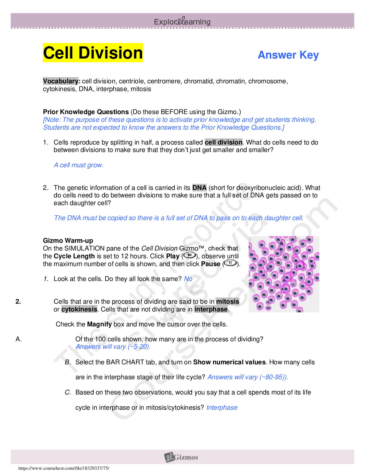
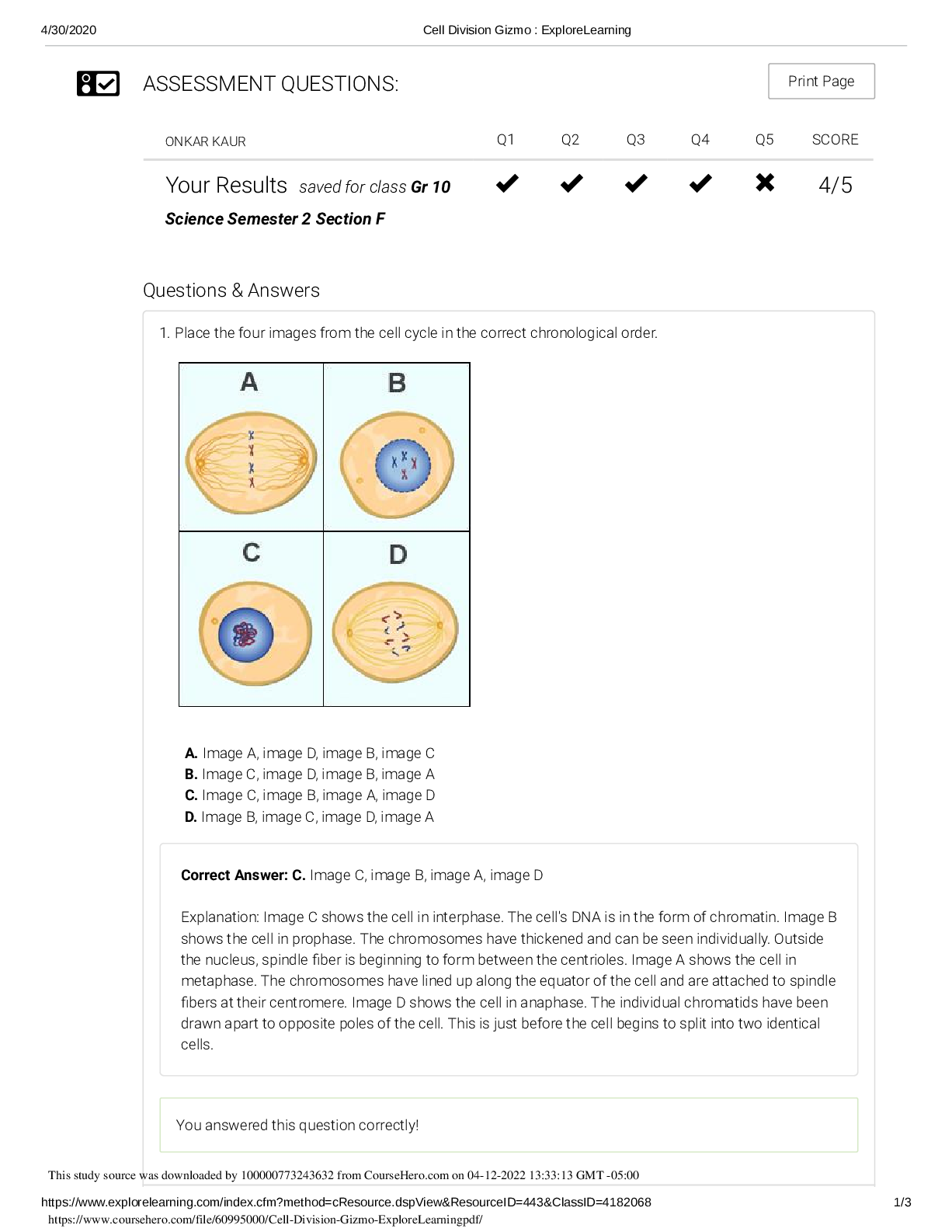
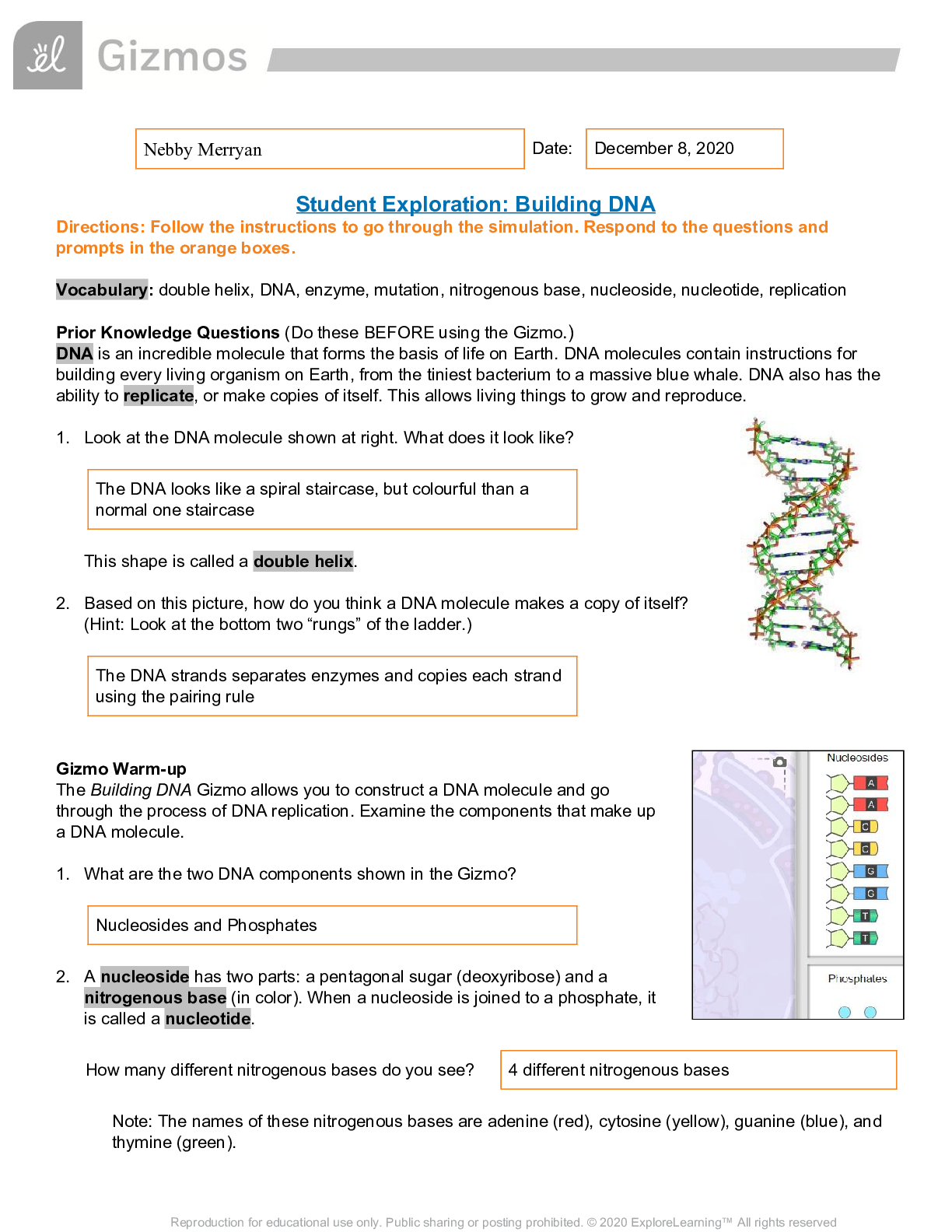
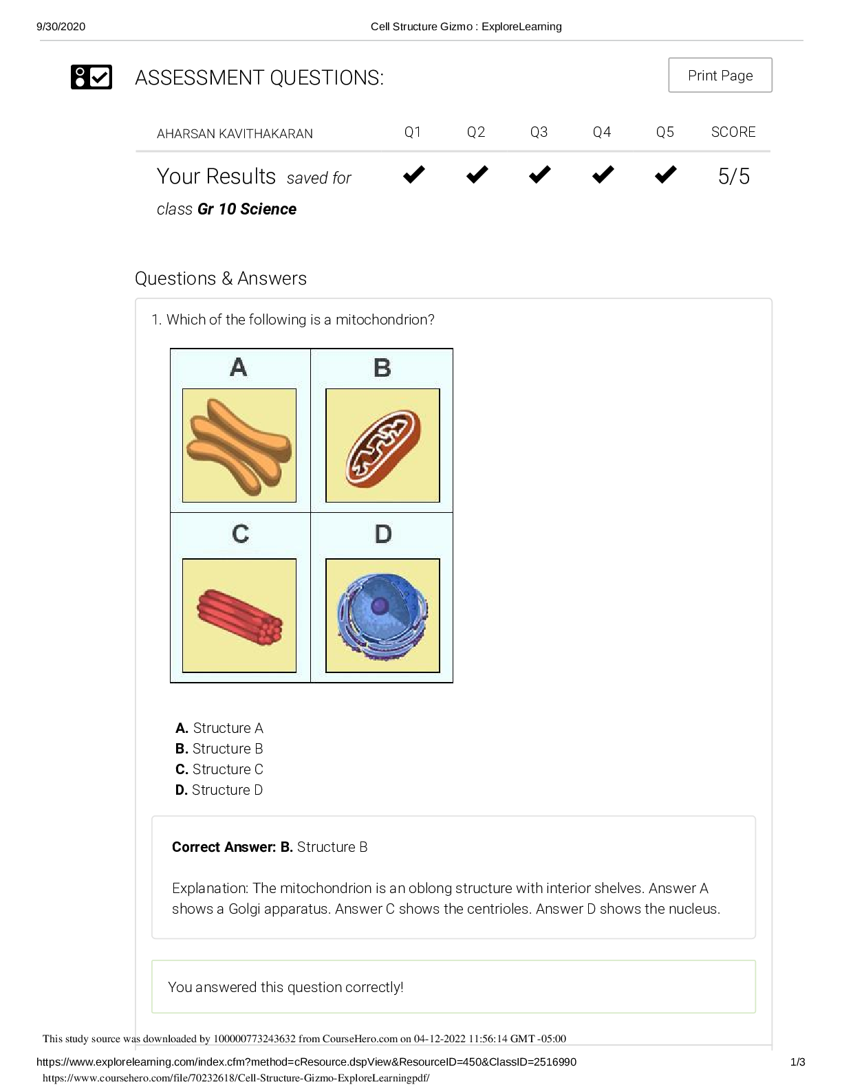
1. Place the four images from the cell cycle in the correct chronological order. A. Image A, image D, image B, image C B. Image C, image D, image B, image A C. Image C, image B, image A, image D... D. Image B, image C, image D, image A Correct Answer: C. Image C, image B, image A, image D Explanation: Image C shows the cell in interphase. The cell's DNA is in the form of chromatin. Image B shows the cell in prophase. The chromosomes have thickened and can be seen individually. Outside the nucleus, spindle fiber is beginning to form between the centrioles. Image A shows the cell in metaphase. The chromosomes have lined up along the equator of the cell and are attached to spindle fibers at their centromere. Image D shows the cell in anaphase. The individual chromatids have been drawn apart to opposite poles of the cell. This is just before the cell begins to split into two identical [Show More]
Last updated: 1 year ago
Preview 1 out of 3 pages
Reviews( 0 )
Document information
Connected school, study & course
About the document
Uploaded On
Apr 12, 2022
Number of pages
3
Written in

Additional information
This document has been written for:
Uploaded
Apr 12, 2022
Downloads
0
Views
51

.png)
.png)