PaEasy Neuro Test bank question and answers, graded A+. 2022
Document Content and Description Below
PaEasy Neuro Test bank question and answers, graded A+. 2022 A 75-year-old man is involved in a motor vehicle accident and strikes his forehead on the windshield. He complains of neck pain and ... severe burning in his shoulders and arms. His physical examination reveals weakness of his upper extremities. What type of spinal cord injury does this patient have? A anterior cord syndrome B central cord syndrome C Brown-Séquard syndrome D complete cord transection E cauda equina syndrome - ✔✔Central Cord Syndrome the central cord syndrome involves loss of motor function that is more severe in the upper extremities than in the lower extremities, and is more severe in the hands. There is typically hyperesthesia over the shoulders and arms. Anterior cord syndrome presents with paraplegia or quadriplegia, loss of lateral spinothalamic function with preservation of posterior column function. Brown-Séquard syndrome consists of weakness and loss of posterior column function on one side of the body distal to the lesion with contralateral loss of lateral spinothalamic function one to two levels below the lesion. Complete cord transection would affect motor and sensory function distal to the lesion. Cauda equina syndrome typically presents as low back pain with radiculopathy. A 17-year-old boy develops progressively abnormal muscle fatigability. He is diagnosed as suffering from myasthenia gravis and is admitted into a hospital. In the course of his treatment with pyridostigmine, he develops increased weakness, nausea, vomiting, sweating, and bradycardia. What is the best management for this patient? Answer Choices 1 Thymectomy 2 Corticosteroids 3 Plasmapheresis 4 Withdrawal of pyridostigmine 5 Addition of neostigmine - ✔✔Withdrawal of pyridostigmine Explanation The patient is having a cholinergic crisis. The over-dosage of cholinesterase inhibitors, such as pyridostigmine and neostigmine, results in cholinergic crisis; it is characterized by salivation, lacrimation, urinary incontinence, emesis, and other symptoms like colic, diarrhea, and miosis. These symptoms are similar to myasthenic crisis, and because treatment differs in both, the 2 conditions need to be differentiated. After stabilizing the patient, edrophonium testing should be done in order to distinguish between a myasthenic and a cholinergic crisis. The management of patients with cholinergic crisis involves prompt withdrawal of cholinesterase inhibitors. The immediate administration of atropine is also recommended. Thymectomy is indicated in severe cases of myasthenia gravis. It is especially effective in young women. Steroids sometimes help if immunosuppression is needed for thymoma, which may be associated with myasthenia gravis. Plasmapheresis is a treatment for severe cases of myasthenia gravis with or without thymectomy. Neostigmine is an alternative cholinesterase inhibitor. There is no rationale in switching from pyridostigmine if a cholinergic crisis has already occurred. A 33-year-old African-American woman presents with a new onset of left facial droop. The droop was present when she awoke this morning, and it has worsened since then. She reports that she had been feeling fine. She denies headaches, visual changes, and weakness or tingling in the extremities. Exam reveals an anxious, overweight woman. She is afebrile. Cranial nerve exam reveals weakness of the entire left side of the face, including the forehead. The left eye does not close entirely, and there is a widened palpebral fissure. Sensation is intact. All cranial nerves, other than VII, appear intact. Her tympanic membranes are clear, and there are no skin lesions around the face or head. The rest of her exam is unremarkable. Labs reveal normal CBC, serum electrolytes, thyroid function tests, and liver transaminases. A chest X-ray is clear. Question What is the most likely cause of this patient's facial droop? Answer Choices 1 Bell's palsy 2 Sarcoidosis 3 Ramsay-Hunt syndrome 4 Lyme disease 5 Cerebrovascular accident (CVA) - ✔✔Bell's Palsy Explanation This patient has a classic presentation of Bell's palsy. Bell's palsy is a sudden, unilateral facial weakness and affects about 1 out of every 60 or 70 persons in their lifetime. The etiology is unknown at this time, but herpes simplex virus type 1 has been associated with some cases. The presumed pathogenesis of the palsy is that inflammation of the facial nerve causes swelling then secondary compression, and ischemia occurs where the nerve courses through the temporal bone. Pain behind the ear may precede the paralysis by a day or 2. Hyperacusis and loss of taste may occur, if the lesion is proximal enough; lacrimation may be affected as well. Sensation is not affected, although patients may experience numbness or heaviness in the face. In some cases, there is a mild lymphocytosis of the CSF. MRI may reveal swelling and uniform enhancement of the geniculate ganglion and facial nerve. Also, in some cases, MRI may reveal a swollen facial nerve entrapped within the temporal bone. Prognosis depends upon the degree of paralysis. Approximately 80% of patients will recover within a few weeks to a month. Of those with an incomplete paralysis in the 1st week, nearly all will experience complete recovery within several months. Of those with a more complete paralysis, like this patient, nerve conduction studies and electromyography can indicate prognosis. The likelihood of completely recovery is 90% if the nerve branches in the face retain normal excitability to supramaximal electrical stimulation; however, the likelihood is only about 20% if electrical excitability is absent. Treatment includes symptomatic care, such as taping the upper eyelid closed during sleep and using lubricating agents in the eye In order to prevent corneal desiccation. Some studies have shown improvement in patients treated with glucocorticoids; other studies showed improvement with prednisone and acyclovir together. Sarcoidosis can cause a form of bilateral facial paralysis (facial diplegia) called uveoparotid fever (Heerfordt's syndrome). Given this patient's African American background, sarcoidosis should be considered as a possible etiology of her symptoms. However, the unilateral nature of the paralysis, combined with a negative chest X-ray, makes sarcoidosis unlikely. A serum angiotensin converting enzyme (ACE) could be drawn if sarcoidosis is a concern. Ramsay-Hunt syndrome is a facial palsy associated with a vesicular eruption in the pharynx, external auditory canal, and other surrounding areas of skin. This is thought to be secondary to herpes zoster infection of the geniculate ganglion; often the 8th cranial nerve is affected, as well. This patient did not have any skin lesions around the ear, thus making this diagnosis very unlikely. However, this demonstrates the importance of a thorough examination in facial palsy patients, including careful examination inside the ears. Lyme disease is a frequent cause of facial paralysis in areas where Lyme infection is endemic. However, the majority of patients who have facial palsy caused by Lyme disease note an antecedent rash adjacent to the site of a tick bite. Blood tests for Lyme disease can help identify that as an etiology. However, this patient denied any risk factors for tick bites such as camping/ travel, making this diagnosis less likely. The family history of CVA and the fact that she is currently taking oral contraceptives do confer some increased risk for thrombotic events in this patient. However, with a stroke, the forehead is usually spared from facial palsy, since the upper facial muscles (the frontalis and orbicularis oculi muscles) are innervated by corticobulbar pathways from both motor cortices. The lower facial muscles are innervated only by the contra-lateral hemisphere. This patient's facial palsy is complete and includes the forehead because the facial nerve itself is affected in Bell's palsy. A 13-year-old boy presents with dysmorphic features (e.g., a long face, prominent jaw, and large ears), large testes, and intellectual disability (intellectual developmental disorder). History reveals that 2 other family members also have intellectual disability. Question What is the most likely clinical diagnosis in this child? Answer Choices 1 Klinefelter syndrome 2 Noonan syndrome 3 Patau syndrome 4 Fragile X syndrome 5 Prader-Willi syndrome - ✔✔Explanation The most likely clinical diagnosis is fragile X syndrome; it accounts for 3% of cases of intellectual disability (intellectual developmental disorder). Macroorchidism and hereditary intellectual disability are 2 specific features of this syndrome. Fragile sites are regions of chromosomes that have a tendency for separation, breakage, or attenuation under particular growth conditions. 1 fragile site associated with fragile X syndrome is on the distal long arm of chromosome Xq27.3. In Klinefelter syndrome, most children (80%) have a male karyotype with an extra X,47,XXY. The remaining 20% have a higher grade of sex chromosome aneuploidy, (i.e., 48,XXXY; 49XXXXY; 48,XXYY). Klinefelter syndrome is the most common cause of hypogonadism and infertility in boys/men; puberty occurs at the normal time, but the testes and penis remain small. Noonan syndrome is an autosomal dominant disorder. Clinical features are similar to Turner syndrome, but Noonan syndrome affects both sexes, while Turner syndrome occurs only in girls/women. Clinical features include a short stature, a low posterior hairline, a shield-like chest, and a short webbed neck. Pulmonary stenosis due to valve dysplasia is the most common cardiac anomaly in Noonan syndrome. In Turner syndrome, the common cardiac defects are bicuspid aortic valve, coarctation of aorta, aortic stenosis, and mitral valve prolapse. Patau syndrome (trisomy 13) is characterized by intellectual disability (intellectual developmental disorder), cleft lip and palate, microcephaly, low-set ears, central nervous system malformations (e.g., holoprosencephaly), and microphthalmia. Prader-Willi syndrome manifests with hypotonia, intellectual disability (intellectual developmental disorder), hypogonadism, and hyperphagia (leading to obesity). Some children with Prader-Willi syndrome have a partial deletion of the paternally derived chromosome 15 and loss of paternally-expressed genes. A 54-year-old man presents after having a generalized seizure. The patient is HIV positive, but he has been unable to afford antiretroviral therapy since losing his job 2 years ago. Other than cachexia, the physical exam is unremarkable. Upon further inquiry, the patient also notes that he has become short-tempered and hypercritical; at times he seems confused. An MRI of the brain is performed, and it reveals several cortical ring-enhancing lesions. Question What is the most likely diagnosis? Answer Choices 1 AIDS dementia complex 2 Cryptococcal meningitis 3 Cytomegalovirus encephalitis 4 Progressive multifocal leukoencephalopathy 5 Toxoplasma encephalitis - ✔✔Toxoplasma encephalitis Explanation The patient's symptoms and MRI findings are most consistent with the diagnosis of toxoplasma encephalitis. Toxoplasmosis is the most common cerebral mass lesion among HIV-positive patients. Infection with the Toxoplasma gondii parasite is relatively common and usually asymptomatic. Reactivation occurs in HIV-positive patients due to failing cellular immunity, and it causes a multifocal necrotizing encephalitis. Seizures may be the initial manifestation of central nervous system (CNS) infection; other common clinical manifestations include focal neurologic deficits (e.g., impaired speech and hemiparesis). Personality change, lethargy, headache, and confusion are also observed. The MRI in patients with toxoplasma encephalitis characteristically reveals multiple, ring-enhancing lesions with surrounding edema; these lesions usually occur bilaterally in the frontal and parietal cortices. AIDS dementia complex describes a constellation of cognitive symptoms seen among HIV-positive patients. The condition occurs when the HIV virus disseminates to the CNS. Within the CNS, the virus tends to concentrate in the basal ganglia and subcortical regions. Symptoms include a constellation of cognitive, behavioral, and motor disturbances that cause varying degrees of functional impairment. Characteristic MRI findings include non-enhancing white matter, cerebral atrophy, and ventricular enlargement. The diagnosis requires that other central nervous system infections, carcinoma, general medical conditions, and substance abuse have been excluded. Cryptococcal meningitis is caused by the encapsulated fungus Cryptococcus neoformans. Among HIV-positive patients, the illness may be the result of new infection or reactivation of latent infection. Presenting signs are often nonspecific; they include headache, fever, change in mental status, and nausea or vomiting. Nuchal rigidity and photophobia may also be present, and elevated intracranial pressure is not uncommon. MRI findings vary, but they include lesions in the basal ganglia; meningeal enhancement, cerebral edema, and shrunken ventricles may also be seen. Cytomegalovirus (CMV) infection causing encephalitis is usually observed in patients with evidence of CMV infection at other sites. MRI findings vary, but they often show areas of focal necrosis within the brain parenchyma, meninges, or periventricular regions. Symptoms typically reflect progressive dementia, with episodes of confusion, apathy, and focal neurologic deficits. Progressive multifocal leukoencephalopathy is often a fatal disorder; it is caused by reactivation of a latent JC viral infection. Focal neurologic deficits (e.g., hemiparesis and gait disturbance) are often the initial presenting symptoms; they are followed by progressive cognitive decline, coma, and death. The MRI commonly reveals multiple, non-contrast enhancing foci in the cerebral white matter. A 48-year-old Caucasian woman with a past medical history of hypertension and hypercholesterolemia was diagnosed recently with a cerebral aneurysm. The treatment plan for the aneurysm is endovascular coiling. Among other complications, this patient has an increased risk of thromboembolism postoperatively. Question What is the initial preventive medication that will be used to help minimize this risk in this patient? Answer Choices 1 Abciximab 2 Heparin 3 Coumadin 4 Eptifibatide 5 Tirofiban - ✔✔Heparin Explanation Complications of endovascular coiling can include thromboembolism and intra-procedural aneurysm rupture. A study found the incidence of thromboembolism in up to 12.5 % of patients in their patient population group. For this reason, many providers will choose to administer some type of combination of heparin and/or antiplatelet therapy to help minimize this potential complication. Coumadin is not indicated as an appropriate choice for this acute procedure. The medications abciximab, eptifibatide, and tirofiban are all anti-platelet/glycoprotein IIb/IIIa inhibitors. These all have been shown to be safe, but they are currently only recommended for use in percutaneous coronary intervention and the prevention of complications in patients with unstable angina/non-ST-elevation myocardial infarction. A 70-year-old woman is brought to your attention by her family because of the slowly progressive gait disorder, the impairment of mental function, and urinary incontinence. About 1 year ago, she started having weakness and tiredness in her legs, followed by unsteadiness; her steps became shorter and shorter, and she also experienced unexplained backward falls. She is becoming emotionally indifferent, inattentive, and her actions and thinking have became "dull". Over the past month, she has started having urinary urgency and involuntary leaking of urine. Besides multivitamins and local application of the Timolol for glaucoma, she takes no other medications; there are no other symptoms. Question What is most likely the best method of treating the patient's urinary problems? Answer Choices 1 Antimuscarinic drug (Tolterodine) 2 Antibiotic (Sulfamethoxazole/trimethoprim) 3 Acetylcholinesterase inhibitor (Donepezil) 4 Ventriculoperitoneal shunt 5 Kegel exercises - ✔✔Ventriculoperitoneal shunt Explanation Clinical triad of slowly progressive gait disorder, followed by impairment of mental function and then sphincteric incontinence strongly suggests the presence of normal-pressure hydrocephalus. Ventricular expansion is the cause of symptoms, and surgical CSF shunting is the main treatment modality. The potential benefit from surgery is usually evaluated by testing gait, cognition, and micturition before and after CSF drainage. Antimuscarinic Tolterodine is an antispasmodic that is used for symptomatic treatment of urinary incontinence in patients with an overactive bladder (urge incontinence). Antimuscarinic drugs are contraindicated in patients with glaucoma. A urinary tract infection will probably manifest with a strong, persistent urge to urinate, burning sensation when urinating, passing frequent, small amounts of urine that has unusual smell and the appearance. Your patient has no such signs and symptoms; therefore, in this case, antibiotics are not indicated. Donepezil is used to treat dementia, but in the case of normal-pressure hydrocephalus, the problem is anatomic (the distortion of the periventricular limbic system and frontal lobes), and the best treatment is probably surgical. Kegel exercises can prevent or control urinary incontinence and other pelvic floor problems in cases of pelvic sphincter weakness. However, pelvic sphincter weakness will probably manifest as stress incontinence. What is the treatment of choice for primary generalized seizures? Answer Choices 1 Phenytoin 2 Phenobarbital 3 Valproic acid 4 Carbamazepine 5 Gabapentin - ✔✔Valproic acid Explanation Epilepsy is defined as a syndrome characterized by recurrent, unprovoked seizures. A seizure is best defined as a discrete neurologic event caused by abnormal, excessive, spontaneous discharge of cerebral cortical neurons. The phenomenology of a single seizure is dependent upon the origin and subsequent spread of neuronal discharge. Epileptic seizures, as opposed to symptomatic seizures provoked by acute brain disturbances (e.g. hyponatremia, meningitis, cocaine use), are treated medically with anticonvulsants. Specific anti-epileptic drugs are efficacious for different types of seizure. Primary generalized epilepsy is commonly associated with developmental brain disorders of childhood (congenital and acquired). Types of primary generalized seizures include tonic-clonic, absence, tonic, clonic, myoclonic, and atonic. Anticonvulsants indicated for primary generalized seizures include valproic acid, clonazepam, and ethosuximide. Localization-related epilepsy results in partial (focal) seizures with or without secondary generalization. These are best treated with phenytoin, carbamazepine, phenobarbital, and valproic acid. The newer anticonvulsants, gabapentin, felbamate, and lamotrigine are also effective for partial seizures. A 64-year-old man presents with difficulty speaking, trouble swallowing, and right arm weakness. Physical examination reveals dysarthria, tongue deviation to the right, paralysis of the left soft palate, left facial droop, left facial sensory loss, and right arm weakness. Question These findings suggest involvement in what area? Answer Choices 1 Medulla 2 Left brain stem 3 Left cerebral hemisphere 4 Pons 5 Right midbrain - ✔✔Left Brain stem Explanation Although precise localization of neurologic deficits is often difficult, certain patterns are important to recognize. Brain stem involvement results in 'crossed findings' (e.g., right facial weakness and left arm weakness) because the lesions affect the brain stem nuclei directly (uncrossed) and the corticospinal tract as it is crossing to the opposite side of the body. Because of the unique anatomic arrangement of the brainstem, a unilateral lesion within the structure can cause 'crossed findings' in which ipsilateral dysfunction of 1 or more cranial nerves is associated with hemiplegia and hemisensory loss on the contralateral side of the body, as described in this case. Medullary syndromes, such as lateral medullary syndrome, are associated with vertigo, nausea, vomiting, nystagmus as well as hiccups, and diplopia; they were not observed in this patient. Medial medullary syndrome is associated with tongue deviation toward the lesion, which is opposite to what is described in this case. Cerebral (cortical) lesions produce contralateral motor and sensory findings in the limbs and contralateral cranial nerve deficits (e.g., right arm and right facial paralysis). Pontine lesions result in coma, miosis, gaze paresis, and altered respiratory patterns. Cerebellar lesions produce nystagmus, dizziness, nausea and vomiting, and the inability to stand or walk if midline cerebellar areas are involved. What is the therapeutic window for using recombinant tissue plasminogen activator in acute ischemic stroke? Answer Choices 1 1 hour 2 2 hours 3 3 hours 4 4 hours 5 6 hours - ✔✔3 hours Explanation Tissue plasminogen activator (TPA) catalyzes the conversion of plasminogen to plasmin, which promotes fibrinolysis. Ischemic stroke is caused by sudden occlusion of a cerebral artery by thrombus or embolus, producing a focal neurologic deficit. Timely reperfusion of ischemic brain tissue has been shown to limit irreversible neuronal injury in both humans and animals. The benefit of TPA in acute ischemic stroke was demonstrated in a large, multicentered national study. Patients whose symptoms began within 3 hours of treatment with TPA had significantly less disability at the end of 3 months than those given placebo. Although treated patients had higher incidence of secondary brain hemorrhage (6.4% vs. 0.8 %), there was no difference in mortality between the 2 groups. TPA must be given no later than 3 hours of symptom onset in patients with acute ischemic stroke. Prior to treatment, it is essential to exclude cerebral hemorrhage on a head CT scan. The presence of a bleeding diathesis, uncontrolled hypertension, recent prior stroke, and past history of cerebral hemorrhage are important contraindications for using TPA in acute ischemic stroke. TPA given later than 6 hours of stroke onset is associated with unacceptable risk of cerebral hemorrhage; it is not known whether fibrinolytic therapy can be given between 3 and 6 hours of stroke onset. A 19-year-old woman presents to the Accident and Emergency Department following a road traffic accident 20 minutes prior. She states her car was just lightly bumped from behind, but she is concerned she might have whiplash, as her neck is stiff and sore. She also says she's experiencing tingling in her left arm down to her thumb. On physical examination, her bicep strength is +3/5 on the left and +5/5 on the right. Her bicep tendon reflex is 2+ on the left and 2+ on the right. In addition, her brachioradialis tendon reflex is 1+ on the left and 2+ on the right. The remainder of the physical examination is normal. Question Based on the above presentation, what cervical nerve root is most likely affected? Answer Choices 1 C4 2 C5 3 C6 4 C7 5 C8 - ✔✔C6 Explanation The most likely nerve affected in this case is the C6 nerve root. Impingement of this nerve can cause numbness and tingling down the arm into the thumb, with weakness in the bicep muscle and diminished brachioradialis tendon reflex in the affected extremity. Impingement of the C4 nerve root may cause neck and upper shoulder numbness and pain. Impingement of C5 nerve root may cause deltoid and shoulder numbness and pain, and bicep tendon reflex may be diminished. Impingement of C7 also causes numbness and pain down the affected arm, but reflex may be diminished into the middle finger and the triceps,. Impingement of C8 may cause numbness and tingling primarily on the lateral surface of the arm and into the lateral hand into the fourth and fifth digits. It may also cause dysfunction of the hand as it innervates the small hand muscles. You are on duty in the ER when a young man is brought in by ambulance. The paramedics report that they were called by this man's roommate who reported the patient was confused and dazed, and obviously very ill. The patient is acutely febrile and delirious; there is evidence of meningeal involvement. Subsequent diagnosis reveals viral encephalitis. What is the most common cause of viral encephalitis worldwide? Answer Choices 1 La Crosse 2 Herpes simplex 3 Japanese B 4 Eastern equine 5 Western equine - ✔✔Japanese B Explanation Viral encephalitis is secondary to viral infection of the brain tissue itself, causing widespread death of nerve cells and glia. Approximately 1,000-5,000 cases per year are reported in the U.S. Worldwide, Japanese B encephalitis is the most common cause of epidemic encephalitis. However, herpes simplex is the most common cause of sporadic encephalitis in the U.S. Arthropod-borne viral encephalitides are transmitted through mosquitoes (most often), and ticks and are named for the region of the US affected. These included LaCrosse, St. Louis, Venezuelan equine, Western equine, and Eastern equine encephalitides. LaCrosse, a type of California encephalitis, is the most common arthropod-borne encephalitis in the U.S. Eastern equine encephalitis (EEE) is a summertime disease that affects children in the east, south and on the Gulf Coast. The mortality is 50-75%, with 80% of patients having neurological deficits. Western equine encephalitis (WEE) occurs also in the summer months in states west of the Mississippi River. The mortality is 5-15%, with infants being the most susceptible. Venezuelan equine encephalitis (VEE) occurs in the southwestern U.S. and Central and South America and has very low mortality and morbidity rates. This infection has been linked to epidemics in tens of thousands of cases, however. St. Louis encephalitis occurs in the central, western and southern U.S. and affects older adults in the autumn months. Headaches may be considered primary in origin (migraine, tension-type, and cluster) or due to underlying metabolic or organic disease. Primary headaches are not associated with any structural disease, but it is believed they are caused by biochemical dysfunction. What statement correctly describes cluster headaches? Answer Choices 1 Patients usually have a positive family history for cluster headaches 2 The headache lasts an average of 30 minutes to 3 hours 3 Cluster headaches occur approximately with equal frequency in males and females 4 Cluster headache pain is typically a moderate, bilateral, steady pain 5 Menstruation is an exacerbating factor for cluster headaches - ✔✔The headache lasts an average of 30 minutes to 3 hours Explanation Cluster and migraine headaches are attributable to cerebrovascular imbalances and are both referred to as vascular headaches. The cerebrovascular dysfunction in cluster headaches is likely different from that of migraine headaches. Cluster headaches usually last from 30 minutes to 3 hours; they occur in groups of 1 to 3 daily over a period of several weeks to a few months. Headache-free intervals range from several months to several years. Clusters occur more often in the spring. The pain is described as excruciating and as seeming to bore from one eye back into the head. Accompanying characteristics include ipsilateral tearing, conjunctival congestion, nasal stuffiness, and a partial Horner's syndrome. Aggravating factors are mostly unknown, but alcohol and sleep may be triggers. Physical activity is known to ameliorate cluster headaches. There is rarely a positive family history and cluster headaches occur more frequently in males. Migraine headaches occur more often in females. Alcohol, chocolate, menstruation, weather changes, and physical activity are considered triggering factors. Migraine headaches usually have moderate to severe pain described as a unilateral throbbing, and they can last from several hours to several days. Associated symptoms include nausea, photophobia, phonophobia and, less commonly, aura. Most patients have a family history positive for migraines A 72-year-old man presents with the inability to comprehend spoken language. His speech is fluent and has a normal cadence and rhythm; however, when he talks, it is gibberish. He frequently substitutes one word for another. What is this phenomenon called? Answer Choices 1 Paraphasia 2 Echolalia 3 Alexia 4 Apraxia 5 Agraphia - ✔✔Paraphasia Explanation- Paraphasia is a type of aphasia. The substitution of a similar sounding word for another word is called paraphasia. With paraphasia, the words can also be jumbled. If a patient is able to hear things and repeat them, it is called echolalia. With echolalia, the patient does not understand what he has heard. This is also referred to as echophrasia. Alexia is a type of aphasia. An aphasia in which there is a problem with reading is called alexia. Alexia is word blindness or text blindness. Alexia is also called optical aphasia or visual aphasia. Apraxia refers to the condition in which a patient has difficulty performing motor acts, despite having the muscular capacity and coordination to do so. A patient with apraxia cannot execute the intended movement. A writing disturbance is called agraphia. There are various forms of agraphia. With absolute agraphia, even simple letters cannot be written. This is also referred to as literal agraphia. A 23-year-old woman was diagnosed with multiple sclerosis approximately 2 years ago; she presents to request information regarding her risk of exacerbation should she become pregnant. Question What does current information on the subject indicate? Answer Choices 1 No increased risk of exacerbations during pregnancy or in the post partum period 2 Increased risk during pregnancy but not during the post partum period 3 Decreased risk during pregnancy and during the post partum state 4 Major increased risk during pregnancy and post partum state 5 Decreased risk during pregnancy with increased risk during the post partum state 6 Increased risk such that exacerbations occurring during pregnancy will not remit - ✔✔Decreased risk during pregnancy with increased risk during the post partum state Explanation Confavreux et al have reported the exacerbation rate per year to be 0.7 in non-pregnant state, decreased during pregnancy (0.5 during first trimester, 0.6 during the 2nd trimester, and 0.2 during the 3rd trimester), and increased during the 3-month postpartum state (1.2). However, when calculated over the whole pregnancy and post partum state, the risk of exacerbations is increased very little, if at all. A 37-year-old man fell from a ladder as he finished hanging the Christmas lights on his house. The right side of his head hit the alley cement, and he lost consciousness for about 1 minute; he woke up with a headache, but he had no other complaints. A few hours later, the patient is brought to the emergency room by his neighbor because of an intense headache, confusion, and left hand hemiparesis. On examination, the patient has a bruise located over the right temporal region, mydriasis, and right deviation of the right eye, papilledema, and left extensor plantar response. An emergency CT scan of the head without contrast reveals a lens-shaped hyper-density under the right temporal bone with mass effect and edema. What is the most likely diagnosis? Answer Choices 1 Epidural hematoma 2 Subdural hematoma 3 Subarachnoid hemorrhage 4 Intracerebral parenchymal hemorrhage 5 Acute meningitis - ✔✔Epidural Hematoma Epidural hematoma most often results from a traumatic tear of the middle meningeal artery. Although a lucid interval ranging from minutes to hours followed by altered mental status and focal deficits is typical for epidural hematoma, this clinical picture is only encountered in up to 1/3 of the patients. The collection of blood between the skull and dura mater causes an evident mass effect with ophthalmic nerve palsy and the contralateral hemiparesis. Surgical evacuation of the clot via burr holes is the treatment of choice. Subdural hematoma results from a traumatic rupture of the bridging veins that connect the cerebrum to the venous sinuses within the dura. This venous hemorrhage will result in a gradual increase of the hematoma, with a progressive clinical picture over days or weeks. The CT scan will show a concave, crescent-shaped hyper-density compared to the convex, lens-shaped hyper-density in epidural hematoma. Subarachnoid hemorrhage is the result of an aneurysm rupture; the most common is the congenital berry aneurysm. The clinical picture is of a sudden, severe headache with meningeal irritation. A CT scan will show blood in the subarachnoid space, and a lumbar puncture will reveal xanthochromia CSF. Intracerebral parenchymal hemorrhage is most likely caused by hypertension complicated with Charcot-Bouchard aneurysms. The blood accumulates into the brain substance and most commonly involves the basal ganglia. Acute meningitis is not associated with trauma. Fever and signs of meningeal irritation dominate the clinical picture. Lumbar puncture, indicated if there are no focal neurological signs on clinical examination, will be the diagnostic procedure. The CT scan of the patient presented in this case is characteristic for epidural hematoma, and there is no indication for a lumbar puncture. A 31-year-old woman presents with a purpural rash covering her arms, legs, and abdomen. She also has fever, chills, nausea, abdominal tenderness, tachycardia, and generalized myalgias. Prior to the development of the rash, the patient noted that she had a headache, cough, and sore throat. Laboratory studies were positive for Gram-negative diplococci in the blood, along with thrombocytopenia and an elevation in PMNs. Urinalysis showed blood, protein, and casts. Vital signs are as follows: PB 92/66, P 96, RR 14, T 39. The patient denies any foreign travel and does not have any sick contacts. However, she does work part time as a nurse in a local hospital. Question The patient is diagnosed with Meningococcemia; she is admitted to the hospital and placed in respiratory isolation. What major course of therapy should this patient receive? Answer Choices 1 Steroids 2 Supportive care 3 Antibiotics 4 Transfusion 5 Bactericidal/permeability-increasing protein - ✔✔Antibiotics Antibiotics are the treatment of choice for meningococcemia. The preferred drug for active infection is penicillin G. For those allergic to penicillin, chloramphenicol and cephalosporins (ie, cefotaxime, cefuroxime) may be used as alternatives. Patients will also receive supportive care, but antibiotic therapy must be initiated quickly if the patient is to survive. Intensive care placement may be necessary if organ failure is imminent. Ventilatory support, inotropic support, and IV fluids are necessary in some. If adrenal insufficiency occurs, corticosteroid replacement may be considered. A central venous line helps to provide large amounts of volume expanders and inotropic medications for adequate tissue perfusion. Steroids have not been shown to play a major role in the treatment of meningococcemia. However, they have been used in addition to antibiotic therapy. In the case of adrenal insufficiency, for example, steroid replacement has been shown to be beneficial. Transfusion does not generally play a major role in treatment. If the patient suffers from a devastating coagulopathy, blood or blood products may be replaced as necessary. Bactericidal/permeability-increasing protein is a protein stored in the granules of neutrophils. It binds to endotoxin in vitro and neutralizes it. This technique is experimental, and it is not used in everyday treatment of meningococcemia. In myasthenia gravis, weakness is a result of insufficient acetylcholine transmission at the neuromuscular junction; however, weakness can also occur with overdosing of the cholinergic medications used to treat myasthenia. What symptom helps differentiate a myasthenic crisis from a cholinergic crisis? Answer Choices 1 Respiratory failure 2 Bilateral ptosis 3 Muscle fasciculations 4 Diplopia 5 Normal muscle stretch reflexes - ✔✔Muscle Fasiculations Signs of cholinergic overdosage include muscle fasciculation, rhinorrhea, lacrimation, salivation, increased bronchial secretions, nausea, or diarrhea. The presence of any of these suggests that the patient's weakness may be due to cholinergic crisis. The other signs are due to weakness and can occur in either condition. A 54-year-old man presents after having a generalized seizure. The patient is HIV positive, but he has been unable to afford antiretroviral therapy since losing his job 2 years ago. Other than cachexia, the physical exam is unremarkable. Upon further inquiry, the patient also notes that he has become short-tempered and hypercritical; at times, he seems confused. An MRI of the brain is performed, and it reveals several cortical ring-enhancing lesions. Question What is the most likely diagnosis? Answer Choices 1 AIDS dementia complex 2 Cryptococcal meningitis 3 Cytomegalovirus encephalitis 4 Progressive multifocal leukoencephalopathy 5 Toxoplasma encephalitis - ✔✔Toxoplasma encephalitis The patient's symptoms and MRI findings are most consistent with the diagnosis of toxoplasma encephalitis. Toxoplasmosis is the most common cerebral mass lesion among HIV-positive patients. Infection with the Toxoplasma gondii parasite is relatively common and usually asymptomatic. Reactivation occurs in HIV positive patients due to failing cellular immunity, and it causes a multifocal necrotizing encephalitis. Seizures may be the initial manifestation of central nervous system (CNS) infection; other common clinical manifestations include focal neurologic deficits, such as impaired speech and hemiparesis. Personality change, lethargy, headache, and confusion are also observed. The MRI in patients with toxoplasma encephalitis characteristically reveals multiple, ring-enhancing lesions with surrounding edema; these lesions usually occur bilaterally in the frontal and parietal cortices. AIDS dementia complex describes a constellation of cognitive symptoms seen among HIV positive patients. The condition occurs when HIV virus disseminates to the CNS. Within the CNS, the virus tends to concentrate in the basal ganglia and subcortical regions. Symptoms include a constellation of cognitive, behavioral, and motor disturbances that cause varying degrees of functional impairment. Characteristic MRI findings include non-enhancing white matter, cerebral atrophy, and ventricular enlargement. The diagnosis requires that other central nervous system infections, carcinoma, as well as general medical conditions and substance abuse have been excluded. Cryptococcal meningitis is caused by the encapsulated fungus Cryptococcus neoformans. Among HIV positive patients, the illness may be the result of new infection or reactivation of latent infection. Presenting signs are often nonspecific; they include headache, fever, change in mental status, and nausea or vomiting. Nuchal rigidity and photophobia may also be present, and elevated intracranial pressure is not uncommon. MRI findings vary, but they include lesions in the basal ganglia; meningeal enhancement, cerebral edema, and shrunken ventricles may also be seen. Cytomegalovirus (CMV) infection causing encephalitis is usually observed in patients with evidence of CMV infection at other sites. MRI findings vary, but they often show areas of focal necrosis within the brain parenchyma, meninges, or periventricular regions. Symptoms typically reflect progressive dementia, with episodes of confusion, apathy, and focal neurologic deficits. Progressive multifocal leukoencephalopathy is often a fatal disorder; it is caused by reactivation of a latent JC viral infection. Focal neurologic deficits such as hemiparesis and gait disturbance are often the initial presenting symptoms; they are followed by progressive cognitive decline, coma, and death. The MRI commonly reveals multiple, non-contrast enhancing foci in cerebral white matter. A 1-year-old boy presents with increasing lethargy. He is barely responsive, and his parents deny any trauma or injury. What is the most common cause of nontraumatic altered levels of consciousness? Answer Choices 1 Seizure disorder 2 Diabetic ketoacidosis 3 Inborn errors of metabolism 4 Toxic ingestion 5 Infection - ✔✔infection Awareness of self and the surrounding environment or consciousness may be altered into different abnormal states of consciousness. Consciousness can shift from loss of clear thinking or confusion, usually accompanied by disorientation, to delirium, a succession of confused and unconnected ideas manifested in children as extreme mental and motor excitement, to lethargy, a profound type of slumber where movement or speech is limited, to stupor or deep sleep where arousal is achieved only by repeated vigorous stimuli, finally to coma, unresponsiveness to even painful stimuli. Non-traumatic coma is most common in infants and toddlers with another smaller peak of occurrence in adolescence. The most common cause of non-traumatic altered level of consciousness in children is infection of either the brain (encephalitis), meninges (meningitis), or both; infections account for more than 1/3 of cases. Prolonged seizures, anticonvulsive therapy, and postictal states can also lead to altered levels of consciousness. The most common metabolic cause of alteration of consciousness is diabetic ketoacidosis, which can occur at any age, but is most common in adolescence. Caused by severe insulin deficiency, hyperglycemia and ketogenesis lead initially to polyuria, polydipsia, hyperpnea, vomiting, and abdominal pain. As the process progresses, hyperosmolar dehydration and acid/base and electrolyte disturbances occur. Advanced stages alter level of consciousness and can lead to coma. Alterations of consciousness due to inborn errors of metabolism that present with electrolyte and glucose abnormalities typically present in infancy. The availability of gluconeogenic precursors or the functions of the enzymes required for production of hepatic glucose are affected. Metabolic defects causing hypoglycemia include glycogen storage disease, galactosemia, fatty acid oxidation defects, carnitine deficiency, several of the amino acidemias, hereditary fructose intolerance, and defects of other gluconeogenic enzymes. Toxic ingestion and exposure are very common in toddlers and adolescents, with a toddler's ability to explore his environment filled with often brightly colored medications and intentional ingestion by adolescents typically involving over-the-counter medications or psychotropic drugs such as antidepressants. A 44-year-old man starts to notice that his eyelids are drooping. Some time afterwards, his jaw becomes weak. He has difficulty swallowing and also experiences weakness in his limbs. He is quite embarrassed when he eats because he must use his hand to help support his jaw. His weakness gets progressively worse. Finally, he seeks medical attention. His physical examination demonstrates the weakness in his limbs; however, no sensory defects are present. A Tensilon test is done and is positive. His doctor is concerned about an associated malignancy. What is the underlying pathology of this disease? Answer Choices 1 Inhibition of acetylcholine release 2 Blockage of the sodium channels 3 Demyelination 4 Subacute combined degeneration of the spinal cord 5 Antibodies to the acetylcholine receptor - ✔✔5 Antibodies to the acetylcholine receptor Antibodies directed towards the acetylcholine receptor at the neuromuscular junction are seen with myasthenia gravis. This man has myasthenia gravis. Ocular muscle weakness, ptosis, dysphagia, and limb weakness can all be seen with myasthenia gravis. When the initial symptom is ocular weakness, Eaton Lambert Syndrome is extremely unlikely. Eaton Lambert Syndrome tends to not involve the extra-ocular muscles or the muscles involving chewing, swallowing, or speech. The Tensilon test is used in the diagnosis of myasthenia gravis. The Tensilon test consists of the administration of edrophonium. Edrophonium is a quick acting anticholinesterase. Thymic tumors are associated with myasthenia gravis. Thymic tumors are also referred to as thymomas. Approximately 10 - 15% of patients with myasthenia gravis have an associated thymoma. The majority of patients with myasthenia gravis have hyperplasia of their thymus. Botulinum toxin inhibits acetylcholine release. The site of action is at the neuromuscular junction. Botulinum toxin is an enterotoxin produced by Clostridium botulinum. Botulism can result from incorrectly canned foods. Tetrodotoxin is a toxin produced by puffer fish. The sodium channels are blocked by tetrodotoxin. The blockage of the sodium channels interferes with the inflow of sodium. As a result, the propagation of nerve and muscle action potentials is affected. Demyelination refers to the loss of myelin around the axon. Several disorders result in demyelination. An example of a demyelinating disease is multiple sclerosis. Subacute combined degeneration of the spinal cord is also called combined systems disease. Subacute combined degeneration of the spinal cord is a neuropathy secondary to B12 deficiency. It is seen in patients with pernicious anemia, especially pernicious anemia that has been present for quite some time. Symptoms include paresthesias and a loss of proprioception. A 74-year-old man presents after his wife witnessed him grab his head in pain and fall to the floor. He has not regained consciousness. His current blood pressure is 150/96 mm Hg, and his heart rate is 65 bpm. Emergent head CT shows a subarachnoid hemorrhage. Question In addition to life saving interventions, what prescription medication will most benefit this patient at this time? Answer Choices 1 Furosemide (loop diuretic) 2 Prednisone (glucocorticoid) 3 Tranexamic acid (antifibrinolytic agent) 4 Labetolol (beta blocker) 5 Nimodipine (calcium channel blocker) - ✔✔Nimodipine (CCB) Nimodipine, a calcium channel blocker, has been shown to improve outcomes in patients following aneurysmal SAH. The mechanism of action is thought to be prevention of ischemia. Glucocorticoids (e.g., prednisone) are often utilized in patients with SAH because they offer symptomatic relief of headache and neck pain, but they have not been proved to decrease cerebral edema. Antifibrinolytic agents (e.g., Tranexamic acid) can be utilized in patients with a diagnosed aneurysm who cannot undergo directed treatment, but they are not routinely used following aneurysmal rupture. Labetalol (a beta blocker) may be utilized to treat elevated BP, but it must be used with caution because it can also decrease cerebral perfusion. Diuretics (e.g., furosemide) have no identified role in the treatment of aneurysmal SAH. A 37-year-old woman presents to her GP surgery with a history of right-sided facial weakness and peri-auricular discomfort since she awoke this morning. She is afebrile. Question What is the most likely diagnosis? Answer Choices 1 Trigeminal neuralgia 2 Bell's Palsy 3 Multiple sclerosis 4 Myasthenia gravis 5 Primary lateral sclerosis - ✔✔Bell's Palsy Explanation The correct answer is Bell's palsy, which is a condition typically with sudden onset that affects the facial nerve, causing unilateral facial weakness. Trigeminal neuralgia presents with sharp pain on one side of the mouth that radiates to the ipsilateral ear, eye, or nostril. Multiple sclerosis is a demyelinating disorder, causing a multitude of symptoms that typically include diplopia or blurred vision early on, then an insidious onset of progressive weakness, numbness, and/or tingling in the extremities. Myasthenia gravis commonly presents with ptosis and diplopia, as well as difficulty swallowing, fatigue, and muscle weakness. Primary lateral sclerosis is an upper motor neuron disease that causes limb weakness, stiffness, and fasciculations. A 62-year-old man presents with vision problems and difficulty swallowing. Over the last week, he has had a constellation of symptoms; they began with numbness and tingling in his feet and progressed to weakness that now affects both lower and upper extremities. Within the last day, he has started to notice difficulty swallowing and double vision. He also feels it is difficult for him to take a big breath. His past medical history is noncontributory, and he takes no medications. Exam reveals bilateral absence of patellar and ulnar reflexes. A lumbar puncture is performed to confirm the diagnosis. Question What cerebrospinal fluid (CSF) finding is most likely? Answer Choices 1 Decreased CSF glucose content 2 Decreased CSF protein content 3 Elevated CSF polymorphonuclear cell count 4 Elevated CSF protein content 5 Elevated CSF lymphocyte count - ✔✔Elevated CSF proteins Correct response is elevated CSF protein content. Symmetrical ascending paralysis as described in this patient is indicative of Guillain-Barré syndrome (GBS). The cause of GBS is unknown, but it is generally thought to be an inflammatory autoimmune process. More than half of patients with GBS report an antecedent illness. The antibodies produced in response to antigens present in the infectious agent are thought to cross-react with components of human neurons, prompting an acute postinfectious demyelinating process. Lumbar puncture characteristically reveals elevated CSF protein content. Other results are normal, although the white blood cell count may be somewhat elevated. Decreased CSF glucose and increased polymorphonuclear cell counts are seen in acute bacterial meningitis. Decreased CSF glucose and elevated CSF lymphocyte counts are commonly seen with meningitis caused by fungi. Viral meningitides usually presents with elevated lymphocyte counts, normal CSF glucose, and normal or slightly elevated CSF protein levels. A 70-year-old woman is brought to your attention by her family because of the slowly progressive gait disorder, the impairment of mental function, and urinary incontinence. About 1 year ago, she started having weakness and tiredness in her legs, followed by unsteadiness; her steps became shorter and shorter, and she also experienced unexplained backward falls. She is becoming emotionally indifferent, inattentive, and her actions and thinking have became "dull". Over the past month, she has started having urinary urgency and involuntary leaking of urine. Besides multivitamins and local application of the Timolol for glaucoma, she takes no other medications; there are no other symptoms. Question What is most likely the best method of treating the patient's urinary problems? Answer Choices 1 Antimuscarinic drug (Tolterodine) 2 Antibiotic (Sulfamethoxazole/trimethoprim) 3 Acetylcholinesterase inhibitor (Donepezil) 4 Ventriculoperitoneal shunt 5 Kegel exercises - ✔✔Ventriculoperitoneal shunt Clinical triad of slowly progressive gait disorder, followed by impairment of mental function and then sphincteric incontinence strongly suggests the presence of normal-pressure hydrocephalus. Ventricular expansion is the cause of symptoms, and surgical CSF shunting is the main treatment modality. The potential benefit from surgery is usually evaluated by testing gait, cognition, and micturition before and after CSF drainage. Antimuscarinic Tolterodine is an antispasmodic that is used for symptomatic treatment of urinary incontinence in patients with an overactive bladder (urge incontinence). Antimuscarinic drugs are contraindicated in patients with glaucoma. A urinary tract infection will probably manifest with a strong, persistent urge to urinate, burning sensation when urinating, passing frequent, small amounts of urine that has unusual smell and the appearance. Your patient has no such signs and symptoms; therefore, in this case, antibiotics are not indicated. Donepezil is used to treat dementia, but in the case of normal-pressure hydrocephalus, the problem is anatomic (the distortion of the periventricular limbic system and frontal lobes), and the best treatment is probably surgical. Kegel exercises can prevent or control urinary incontinence and other pelvic floor problems in cases of pelvic sphincter weakness. However, pelvic sphincter weakness will probably manifest as stress incontinence. A 5-month-old male infant presents after a seizure involving all 4 limbs. His mother tells you that he was born full term without any complications, and he was well until 2 days ago when he developed a fever. He vomited multiple times yesterday and was irritable. He has not had diarrhea or a cough. He was given antipyretic medication for his fever. He has no known allergies. His immunizations are up-to-date. His developmental milestones have been in accordance with his age. On physical exam, his temperature is 102.7 F, and his pulse is 154/min; BP is 90/50 mmHg, and RR is 20/min. He is lethargic, pale, and focal neurological deficits are present. His anterior fontanel is bulging. You suspect that he has bacterial meningitis. Question After drawing blood samples for investigations, what is the most appropriate next step? Answer Choices 1 Intravenous phenytoin 2 Intravenous empirical antibiotics 3 MRI of the head 4 Lumbar puncture 5 Intravenous glucose - ✔✔intravenous emiprical antibiotics The infant in the vignette appears to have bacterial meningitis. The initial approach to the patient should be the "ABCs." After assessing and stabilizing the patient's airway and obtaining IV access, intravenous antibiotics should be given immediately. As bacterial meningitis is associated with high morbidity and mortality, prompt initiation of empirical antibiotics is crucial for better prognosis. The choice of antibiotics is dependent on the patient's age and specific predisposing conditions. Use of broad-spectrum cephalosporins, such as ceftriaxone or cefotaxime with vancomycin, may be used in infants more than 1 month old. Ideally, serum glucose, blood culture, complete blood count, and serum chemistries should be drawn when IV access is obtained; however, drawing labs should not delay beginning antibiotics. Intravenous glucose is necessary if the patient is found to be hypoglycemic; bedside serum glucose is mandatory in any patient that presents with a seizure. Intravenous phenytoin and an MRI of the head might also be necessary for a patient such as the one in the vignette, but would not emergently precede antibiotics. The diagnosis of bacterial meningitis rests on CSF examination performed after lumbar puncture. However, LP is deferred in patients with evidence of increased intracranial pressure, new onset seizure, cardiorespiratory compromise, or focal neurological deficits. Antibiotics should be given, and CT scan of the head should be performed. If CT scan is negative, LP can be performed. A 12-year-old girl presents with a 3-day history of progressive dysarthria, dysphagia, and weakness. The patient was well until 3 days prior to admission to the hospital; at that time, she developed the onset and subsequent gradual worsening of dysarthria. She attributed the dysarthria to a sore throat that she had had about 2 weeks earlier. 3 days prior to admission, she also had the onset of mild dysphagia; it mostly occurred with liquids. 24 hours prior to admission, she developed weakness in both upper extremities, which increased and began to involve the lower extremities. This limb weakness was neither worsened by activity nor improved by rest. She also developed tingling in her toes 24 hours prior to presentation. When she became unable to walk without assistance on the day of admission, she decided to seek medical attention and was admitted to the hospital. Past medical history is significant for measles and mumps. Because of family religious beliefs, she has not had any immunizations. She is very athletic, and frequently plays soccer with friends and siblings in the fields on her grandfather's horse farm. Physical examination reveals a well-developed, well-nourished girl. She is awake, alert, cooperative, and in no acute distress. Temperature is 98.7 F by mouth, blood pressure of 140/80 mm Hg, heart rate is 84/min and regular, and respirations are 22/min and unlabored. There are multiple scratches and abrasions in varying stages of healing over most of her extremities. Her speech is moderately dysarthric. She experiences some mild choking when she tries to drink a glass of water. She can smile weakly, but she cannot raise her eyebrows against resistance. She shows mild bilateral weakness of eye adduction. Pupillary responses are normal. There is mild to moderate upper extremity and mild lower extremity weakness, greater distally than proximally. Her motor strength is sustained over at least 30 seconds without fatigue. Her gait is ataxic, and she cannot walk without assistance. Reflexes are hypoactive to absent, and the response to plantar stimulation is downgoing bilaterally. Sensation is intact, except for mildly impaired position and vibratory sensation in both feet. A complete blood count, chemistry profile, chest X-ray, and EKG are all normal. Computed tomography of the brain, with and without contrast, is negative. A nerve conduction study reveals a moderate degree of mostly motor demyelinating peripheral neuropathy, highly suggestive of Guillain-Barre. Question What statement best describes the patient's prognosis? Answer Choices 1 With a predominantly demyelinating rather than axonal neuropathy, her prognosis is especially bad 2 Mortality is 25-30% 3 Full functional recovery is expected in 45% in several months to a year 4 Whether recovery is full or partial, relapses do not occur during the recovery phase 5 Her rapidly evolving clinical course indicates a poor prognosis - ✔✔5 Her rapidly evolving clinical course indicates a poor prognosis This patient must be watched very closely for the very real possibility of respiratory failure and the need for ventilatory support (2). Mortality is expected to be less than 5% with good medical support (1). With a demyelinating pattern on EMG, her prognosis is better. A consistent indicator of residual muscle weakness is an EMG pattern of axonal damage, with the more severe degrees of damage suggesting the worse prognosis (1). About 85% of patients with GBS have a full functional recovery within a year; however, some may be left with minor residuals such as areflexia on exam (2). Between 5-10% of patients with Guillain-Barre have 1 or more relapses; these cases are referred to as chronic inflammatory demyelinating peripheral neuropathy (CIDP) (1). A 45-year-old African-American man with no significant past medical history presents with a 1-hour history of left retroorbital headache. The headache was of a sudden onset and began upon waking that morning. It is described as excruciating, stabbing, sharp, and lancinating; it is rated as severe in intensity. He denies any preceding infections, nausea, vomiting, photophobia, or osmophobia; he also denies fever, chills, stiff neck, focal weakness, numbness, tingling, vision, hearing, gait, or speech changes. He recalls a similar episode several months ago; it lasted about a week, and it dissipated without complications. His physical exam is remarkable for painful distress, lacrimation with conjunctival injection, nasal congestion, rhinorrhea, left ocular miosis, and left forehead diaphoretic flushing. Question What pharmacologic agent is the most beneficial for this patient at this time? Answer Choices 1 Sumatriptan 2 Verapamil 3 Lithium 4 Topiramate 5 Prednisone - ✔✔Sumatriptan The correct response is sumatriptan. This patient's most likely diagnosis is most likely a cluster headache. Pharmacologic management of cluster headache may be divided into abortive/symptomatic and preventive/prophylactic strategies. Abortive agents are used to stop or reduce the severity of an acute attack, and include oxygen, triptans, ergot alkaloids, and anesthetics. Inhalation of high-flow concentrated oxygen is extremely effective for aborting attacks. 5-Hydroxytryptamine-1 (5-HT1) receptor agonists, such as triptans or ergot alkaloids with metoclopramide, are often the first line of treatment. Stimulation of 5-HT1 receptors produces a direct vasoconstrictive effect and may abort the attack. Subcutaneous injection of sumatriptan can be effective, in large part because of the rapidity of onset. Studies have indicated that intranasal administration is more effective than placebo but not as effective as injections. Prophylactic agents are used to reduce the frequency and intensity of individual headache exacerbations. Preventive and prophylactic medications include calcium channel blockers, mood stabilizers, and anticonvulsants. Verpamil is the most effective calcium channel blocker for prophylaxis. It inhibits calcium ions from entering slow channels, select voltage-sensitive areas, or vascular smooth muscle, thereby producing vasodilation and preventing the initial vasoconstrictive phase of cluster headaches. It can be combined with ergotamine or lithium. Preliminary evidence suggests that prophylactic lithium may interfere with substance P and vasoactive intestinal peptide (VIP)-induced arterial relaxation. Anticonvulsants such as Divalproex and Topiramate are preventative medications whose mechanism of action may involve regulation of central sensitization. Prednisone is very effective for aborting the cluster headache cycle or providing intermediate prophylaxis as bridging therapy between acute and prophylactic agents. It is effective for treatment that does not respond to lithium. A 48-year-old woman presents after a seizure. Prior to the seizure, she experienced confusion and disorientation preceded by nausea, vomiting, and blurred vision. Symptoms appeared after working for several hours in the garden under the sun. Her medical history is significant for the presence of schizophrenia, for which she takes chlorpromazine at bedtime. Her temperature is 41 C; BUN and creatinine are elevated; and there is neutrophilia, hemoconcentration, and lactic acidosis. You think that the event is possibly drug-related. Question What is the most likely diagnosis? Answer Choices 1 Heat cramps 2 Neuroleptic malignant syndrome 3 Heat stroke 4 Malignant hyperthermia 5 Heat exhaustion - ✔✔Heat stroke Heat disorders can be exertional and nonexertional. Both can be drug-related. Neuroleptics (e.g., phenothiazines, thioxanthenes) may impair thermoregulation due to both anticholinergic and antidopaminergics effects. Anticholinergics inhibit sweating, therefore disturbing thermoregulation during exercise or under conditions of environmental heat stress. Antidopaminergics elevate the set point of the temperature regulation center in hypothalamus. Your patient most probably suffered heat stroke. It is a life-threatening condition characterized by elevated body temperature with nausea, blurred vision, confusion, disorientation, and seizures. Hemoconcentration, anuria, rhabdomyolysis, kidney dysfunction, lactic acidosis, and even disseminated intravascular coagulation may result. Heat cramps are a mild disorder characterized by painful muscle contractions due to temporary fluids and electrolytes depletion. There are no signs and symptoms of neurological dysfunction, and body temperature is normal. Neuroleptic malignant syndrome is an idiosyncratic reaction to neuroleptics, most commonly phenothiazines and butyrophenones, and it is characterized by rigidity, fever, and autonomic instability. It is not connected with the exposure to heat and exertion. Men under 40 are at greatest risk. Malignant hyperthermia is a nonexertional idiosyncratic reaction to the anesthetic. Heat exhaustion is a condition with a severity that lies between heat cramps and heat stroke. Body temperature might be slightly elevated, and there may be neurological signs like headache, but there will be neither severe confusion nor seizures. A 17-year-old adolescent male presents with unexplained neurological symptoms. His liver is enlarged on palpation, and he has other symptoms of hepatitis. Blood work reveals depressed ceruloplasmin levels. An ophthalmological examination reveals Kayser-Fleischer rings. What is the most likely diagnosis? Answer Choices 1 Krabbe disease 2 Lesch-Nyan syndrome 3 Marfan syndrome 4 Wilson disease 5 Hunter syndrome - ✔✔Wilson Disease This young man is suffering from Wilson's disease, a genetic disorder of copper metabolism. It is inherited as an autosomal recessive mutation in the ATP7B genes located on chromosome 13. The protein of the ATP7B gene is a copper-transporting ATPase The frequency of the heterozygous carriers is relatively high (1/90). The incidence of homozygous recessive affected individuals is about 1/30,000. It affects all ethnic groups and both sexes equally. Neurological symptoms, hepatitis, and Kayser-Fleisher rings, greenish-brown deposits of copper in the corneal endothelial (Descemet membrane) basement membrane near the peripheral cornea where it meets the iris (limbus), are characteristic of Wilson's disease. About 40% of the dietary copper is absorbed in the gastrointestinal tract, and makes its way to the liver bound to albumin. In the liver, it is complexed with ceruloplasmin, a blood protein that carries most of the copper. Ceruloplasmin levels are abnormally low in patients with Wilson disease, although the disease is not, per se, a mutation in the ceruloplasmin gene. Ceruloplasmin is recycled in the liver by the usual lysosomal degradation pathway, and the unused copper is excreted in bile. When excessive copper is absorbed in the gut, it accumulates in the brain (producing neurological symptoms), the liver (producing hepatitis and hepatomegaly), and the cornea. Penicillamine can be used to treat this disease. It chelates copper and provides some symptomatic relief. Unfortunately, when used appropriately in pregnant women to treat potentially life-threatening Wilson disease, it is harmful to the fetus and can produce cutis laxa in the newborns of penicillamine-treated patients (see Figure H6.3). A 62-year-old woman presents with excruciating pain, burning, and swelling in her left forearm and wrist. She reports that symptoms initially began with a fracture 4 months ago. The fracture was casted, and the patient was told it had healed well with cast removal at 8 weeks. She is frustrated because her symptoms have persisted and worsened, rather than improving, as she was told they would. The patient has continued to use a sling and limit use of the left arm so she doesn't worsen her condition. She is unable to wear a jacket or long sleeves, as even the fabric touching her skin causes pain. She denies fevers, pain in other areas, and new trauma. She denies polyuria and polydipsia. Her past medical history is unremarkable; she is a menopausal woman, with no known medical conditions, no history of surgeries, no regular medications and no allergies. She lives with her husband, and she is a homemaker. She denies drug, alcohol, and tobacco use. On physical exam, she is a small, thin, pleasant woman, and she is fully oriented. Vitals are normal. No gait or balance abnormalities are noted when she walks or gets onto the exam table. Her left forearm has some mild edema and erythema, as well as tenderness with even light touch. Distribution of findings includes the region from the elbow to wrist, both the anterior and posterior surfaces. Left wrist strength and range of motion are decreased compared to the right. Distal pulses, capillary refill, and reflexes are normal. The remainder of the exam, including mental status, is normal. Question What medication would be most appropriate for this patient's likely diagnosis? Answer Choices 1 Gabapentin 2 Heparin 3 Methadone 4 Probenecid 5 Vancomycin - ✔✔Gabapentin This patient's diagnosis is most likely a complex regional pain syndrome (CRPS). CRPS most often develops after a minor trauma and classic characteristics include pain out of proportion with findings and history, allodynia (pain sensation with normally non-painful stimuli) and motor and sensory disturbances in the affected extremity. The mechanism for CRPS development is not well understood. Prolonged immobilization following injury is a risk factor for development of CRPS. Diagnosis is clinical, and testing is done to rule out other disorders. Treatment is multi-modal and primarily consists of physical therapy, focusing on mobilization and desensitization. Other treatments are often off-label and targeted at chronic pain relief; they include corticosteroids, bisphosphonates, tricyclic antidepressants, anticonvulsants, and topical anesthetics. Gabapentin would be a reasonable choice for this patient. Heparin would be used if this patient's symptoms were attributed to a deep venous thrombosis (DVT). DVT is rare in the upper extremities; it is not associated with the hyperesthesia and allodynia shown in this patient. Methadone, a longer-acting opioid medication, can be used for chronic pain. However, for several reasons, this is not the most appropriate medication to use in CRPS. Opiates carry addiction potential and increase fall risk in the elderly. CRPS is often categorized as early (<6 months' duration) versus late (>6 months). For early CRPS, opiates should be avoided and even with late CRPS, other medications options should be utilized before initiating opiates. Probenecid is a medication which inhibits urate resorption. It is used for gout, a painful inflammatory condition which may cause acute pain, erythema and swelling in the affected joint(s). Gout tends to be more acute and affect joints (not the forearm, as with this patient). Vancomycin is an IV glycopeptide antibiotic; it is used for severe bacterial infections, including cellulitis. Cellulitis, which commonly presents with erythema, edema and tenderness in an extremity, may be considered on the differential for this patient. Cellulitis tends to occur more acutely. This patient had a normal 8 week exam with symptoms of CRPS, but no findings of cellulitis at that time. A 46-year-old man recently recovered from a bout of influenza, and he now presents due to an 8-hour history of right-sided facial paralysis. He is having trouble closing his right eye, cannot raise his right eyebrow, and cannot smile with the right side of his mouth. Question What medication(s) should this patient be started on? Answer Choices 1 Ibuprofen 800mg PO q 6 hours plus acyclovir 400mg PO 5 times daily 2 Prednisone 60mg PO daily alone 3 Prednisone 60 mg PO daily plus acyclovir 400mg PO 5 times daily 4 Acyclovir 400mg PO 5 times daily alone 5 Prednisone 60mg PO daily plus augmentin 500mg PO 2 times daily - ✔✔3 Prednisone 60 mg PO daily plus acyclovir 400mg PO 5 times daily The correct answer is prednisone 60mg PO daily plus acyclovir 400mg PO 5 times daily. Either medication alone can be used with fairly good results, but the most recent research has shown that patients treated within 3 days of onset with both a glucocorticoid and an antiviral have a better outcome than patients treated with either prednisone or acyclovir alone. Ibuprofen 800mg PO q 6 hours plus acyclovir 400mg PO 5 times daily is not the correct answer. Acyclovir is an appropriate choice for treatment, as it is an antiviral, and herpes simplex virus is the likely etiologic agent in the pathogenesis of Bell's palsy. However, a nonsteroidal anti-inflammatory is not the correct type of anti-inflammatory to treat Bell's palsy, so ibuprofen is an incorrect choice. Prednisone 60mg PO daily alone is not the correct answer. Prednisone is an appropriate treatment choice for Bell's palsy. It is typically given 1mg/kg for 5-7 days and then tapered over the next 5-7 days. This appears to shorten the duration of symptoms and improve the functional outcome. It should be started as early as possible, but not at all if it has been 7 days or more since the onset of symptoms. However, outcomes are even better if this treatment is used in conjunction with an antiviral, such as acyclovir. Acyclovir 400mg PO 5 times daily alone is not the correct answer. Acyclovir 400mg PO 5 times per day for 10 days is a good treatment option for Bell's palsy. It is best if started within 72 hours of symptoms, but can be started up to 7 days after the onset. However, outcomes are even better if this treatment is used in conjunction with a glucocorticoid, such as prednisone. Prednisone 60mg PO daily plus augmentin 500mg PO 2 times daily is not the correct answer. Prednisone is an appropriate treatment choice for Bell's palsy. It is typically given 1mg/kg for 5-7 days and then tapered over the next 5-7 days. This appears to shorten the duration of symptoms and improve the functional outcome. However, because it is an antibiotic used to treat bacterial infections, augmentin would not be used to treat Bell's palsy. Bell's palsy is caused by a virus, not a bacterium; therefore, an antibiotic would not help the outcome. A 4-month-old febrile infant presents with loss of appetite, irritability, seizures, focal sensory and motor deficits, and an acute petechial rash. On physical examination, a bulging fontanelle is noted; rectal temperature is 102.8°F. What study would be most important in this child's evaluation? Answer Choices 1 CBC 2 Urinalysis 3 Chemistry panel 4 Serum glucose 5 CSF analysis - ✔✔CSF analysis The clinical picture is suggestive of meningitis, which is diagnosed after obtaining and evaluating the cerebrospinal fluid (CSF). Although a bulging fontanelle is an indication of increased intracranial pressure (ICP), it is not an automatic contraindication for a lumbar puncture (LP) unless the patient displays additional signs of increased ICP. Tonic seizures would be an example of an indication of increased ICP. Even though this child has had seizures, an LP to run a CSF evaluation is still the best answer. This will be the most important study and diagnostic tool for this child. It is likely that a CT or MRI would be done to look at the level of swelling in the brain, and possibly determine the level of ICP. It would be likely for either of these tests to be done prior to a lumbar puncture. Normally, meningitis causes fever, inactivity, and mental status changes; however, these symptoms are often hard to detect in young children. In infants, signs and symptoms may include appearing to be slow or inactive (lack of alertness), irritable, vomiting, or feeding poorly. When CSF infections are suspected, blood should be obtained for a CBC, general chemistry panel, and culture. However, obtaining CSF analysis is the most important. A 46-year-old man presents with severe insomnia and anxiety. While hospitalized, an overnight polysomnogram was performed over 2 consecutive nights. Sleep latency was 60 minutes; REM latency was 45 minutes. He reports feeling paresthesias deep within his legs while lying in bed, especially while falling asleep. He denies recent illness or illicit drug use. His physical exam and lab work were within normal limits. Question What is the most likely cause of the patient's symptoms? Answer Choices 1 Deep vein thrombosis 2 Peripheral neuropathy 3 Restless legs syndrome 4 Alcoholic peripheral neuropathy 5 Periodic limb movements in sleep - ✔✔Restless leg syndrome Restless legs syndrome (RLS) is a neurological disorder with symptoms of an unpleasant sensations in the legs, such as insects crawling inside the legs, burning, tugging, or creeping. There is an uncontrollable urge to move the limb when at rest (lying down and trying to relax activates the symptoms). Most people with RLS have difficulty falling asleep and staying asleep. Women may be slightly more affected than men. Symptoms may begin at any stage of life, although the disorder is more common with increasing age. The severity of the disorder appears to increase with age. In some patients, symptoms will improve over a period of weeks or months. In most cases, the cause of RLS is idiopathic. A family history of the condition is seen in many cases, suggesting a genetic component. People with familial RLS tend to be younger when symptoms start, and they have a slower progression of the condition. Hypoglycemia can worsen the condition. The disorder is diagnosed clinically by evaluating the patient's history and symptoms. Needle electromyography and nerve conduction studies should be considered if polyneuropathy is suspected on clinical grounds, even if results of neurologic examination are apparently normal. Ropinirole is approved for the treatment of moderate-to-severe RLS. Benzodiazepines may be prescribed for patients who have mild or intermittent symptoms. These drugs help patients obtain a more restful sleep, but they do not fully alleviate the symptoms and can cause daytime drowsiness. For more severe symptoms, opioids may be prescribed since they can cause relaxation and decrease pain. Anticonvulsants, such as carbamazepine and gabapentin, are also useful for some patients; they can decrease the creeping and crawling sensations. There may be no symptoms associated with deep vein thrombosis (DVT), but the classical symptoms of DVT include pain, swelling, redness of the leg, and dilation of the surface veins. Peripheral neuropathy, of which alcoholic peripheral neuropathy is a type, causes tingling or burning pain in the feet. At times, it may be so severe that it interferes with walking, which is a result of injury to sensory fibers. As the condition worsens, the pain typically decreases and numbness increases. Periodic limb movements in sleep are associated with periodic episodes of highly repetitive limb movements during sleep. These repetitive episodes of muscle contractions are usually grouped into series. A 32-year-old man presents with a severe headache; he has had 2 similar headaches within the past week. He describes a burning, 'hot poker'-type of pain located primarily behind his right eye. He notes that his eye waters profusely with the headache; in addition, his nose is initially congested, then it starts running. Only his right side is affected. The headache is so severe that he cannot work or sleep through it, and he is unable to concentrate on anything else. The headaches have been unresponsive to over-the-counter pain medications. The episodes seem to last about 1 hour. He denies any other symptoms. This patient has no chronic medical conditions, and he takes no regular medications. Question What is the most likely underlying pathophysiology of this patient's condition? Answer Choices 1 Antigen binds to IgE, triggering the release of histamines and other inflammatory substances 2 Growth of an intracranial tumor near the base of skull, leading to pressure on the cerebellum 3 Increased emotional stress, leading to contractions of head and neck musculature 4 Increased trigeminal nerve and parasympathetic activity, leading to vasodilation 5 Reactivation of varicella zoster virus, leading to inflammation in a ganglion - ✔✔4 Increased trigeminal nerve and parasympathetic activity, leading to vasodilation This patient presents with a history indicating cluster headaches. While the exact mechanism is unclear, it is known that there is increased trigeminal nerve and parasympathetic activity, leading to vasodilation in the intracranial vasculature. Key features of cluster headaches include unilateral pain, involvement of the eye and nose (autonomic/parasympathetic system), an episodic pattern of attacks, and excruciating pain. Antigen binding to IgE, leading to release of histamines, leukotrienes, and prostaglandins is the typical type I hypersensitivity reaction of allergies. Seasonal allergies could cause a headache, along with nasal and ophthalmic symptoms; however, this patient's severity of headaches exceeds a usual allergic sinus headache. If this patient's symptoms had been caused by an allergic response he would have presented with a history of allergies, chronic lower-grade headaches, and sinus symptoms; he would not have acute, episodic attacks of high intensity. While the growth of an intracranial tumor could present with headaches, this patient's history is not suggestive of cerebellar dysfunction. If there were a tumor in this patient's cerebellar region, some motor, and possibly language, dysfunction would be expected. Headaches with lacrimation and rhinitis are not typically seen from a tumor. Increased emotional stress and tight neck musculature is associated with tension-type headaches, which are the most common form of headaches. Typically, these headaches are more chronic; they are associated with psychosocial stressors and respond to analgesics. Nasal and ophthalmic symptoms are not seen with tension-type headaches. The reactivation of the varicella zoster virus (shingles) can produce severe throbbing, stinging, or other pain symptoms along the affected dermatome. Shingles would be a disorder to consider on this patient's differential, but the patient's young age, lack of skin lesions, and the episodic nature of the attacks fit much better with a diagnosis of cluster headaches. A 73-year-old man presents after a 15-minute episode of right eye vision loss, which he described as being "like a shade being pulled down". What diagnostic test is most likely to be abnormal? Answer Choices 1 Carotid ultrasound 2 Computed tomography 3 Electrocardiogram 4 Erythrocyte sedimentation rate 5 Electroencephalogram - ✔✔carotid u/s The case described above is a classic example of amaurosis fugax, which is caused by transient occlusion of the ophthalmic artery, a branch of the internal carotid artery. This occurs most often in the setting of internal carotid artery stenosis. Like other transient ischemic attacks, it is often a harbinger of an impending stroke. A 42-year-old man presents to the emergency department with a severe headache. He has been getting several of these headaches recently and has tried all over-the-counter pain relievers and headache medicines with no relief. His current headache started 15 minutes ago. He describes the pain as located next to and behind his left eye and "stabbing/excruciating" in nature. He feels like his left eye tears up profusely with these headaches. He reports he has been healthy otherwise, with no chronic medical conditions, no history of surgery, no medications, and no drug allergies. He denies recent stressors that may have caused his headaches. On physical exam, the patient appears slightly agitated and appears uncomfortable. His left eye's conjunctiva is mildly injected, and lacrimation is noted. His right eye is normal. Cranial nerves II-VII are intact, although the patient expresses discomfort when the light is shown in his left eye. Speech, gait, coordination, and reflexes are all normal. The remainder of his exam is normal. Head MRI is performed and reported as normal. Question Which of the following aspects of patient history would be most consistent with this patient's suspected condition? Answer Choices 1 Head injury prior to onset of headaches 2 Periodic episodes of aphasia and gait disturbances 3 Recent exposure to outdoor pollens 4 Rhinorrhea associated with headaches 5 2 to 3-day duration of each headache - ✔✔Rhinorrhea associated with HA This patient is suffering from cluster headaches. Cluster headaches are classified as trigeminal autonomic cephalalgias and are most common in men. Cluster headaches may be triggered by alcohol, histamine, and nitroglycerin, but often no cause is identified. There may be a genetic component. Classic presentation of cluster headache includes episodic, unilateral, severe headache, usually located in the temporal and/or orbital region, along with associated lacrimation, injection, eyelid edema, miosis or ptosis of the ipsilateral eye, and nasal congestion and rhinorrhea associated with the headaches. Imaging studies, such as head MRI, should be performed to rule out other primary causes of headache, such as vascular malformation, neoplasm, and infection. A head injury prior to the onset of headaches is not associated with cluster headaches. Post-concussion headaches tend to have longer duration, but less severe intensity compared to cluster headaches. Other clues to a post-concussion headache may be memory problems, "fogginess," and nausea, and/or vomiting. The eye symptoms and signs this patient displayed are not consistent with a prior head injury. Periodic episodes of aphasia and gait disturbances would be concerning for a severe disorder, ranging from vascular, infectious, neurodegenerative, and neoplastic in etiology. Patients with cluster headaches would not be expected to have these types of neurologic symptoms. Recent exposure to outdoor pollens would be expected in a patient experiencing allergy-related headaches. A patient with allergy-related headaches would be expected to have much less intense severity and bilateral eye and nasal symptoms. 2 to 3-day duration of each headache can be seen with tension-type headaches and migraines. However, cluster headaches are noted to last 15-180 minutes of duration. A 66-year-old English teacher is in the hospital. Her neurologist gives her a magazine to read, and she is unable to do so. What is this phenomenon called? Answer Choices 1 Paraphasia 2 Echolalia 3 Alexia 4 Apraxia 5 Agraphia - ✔✔Alexia When a patient is able to hear things and repeat them, it is called echolalia. With echolalia, the patient does not understand what he has heard. This [Show More]
Last updated: 1 year ago
Preview 1 out of 190 pages
Instant download

Buy this document to get the full access instantly
Instant Download Access after purchase
Add to cartInstant download
Also available in bundle (1)
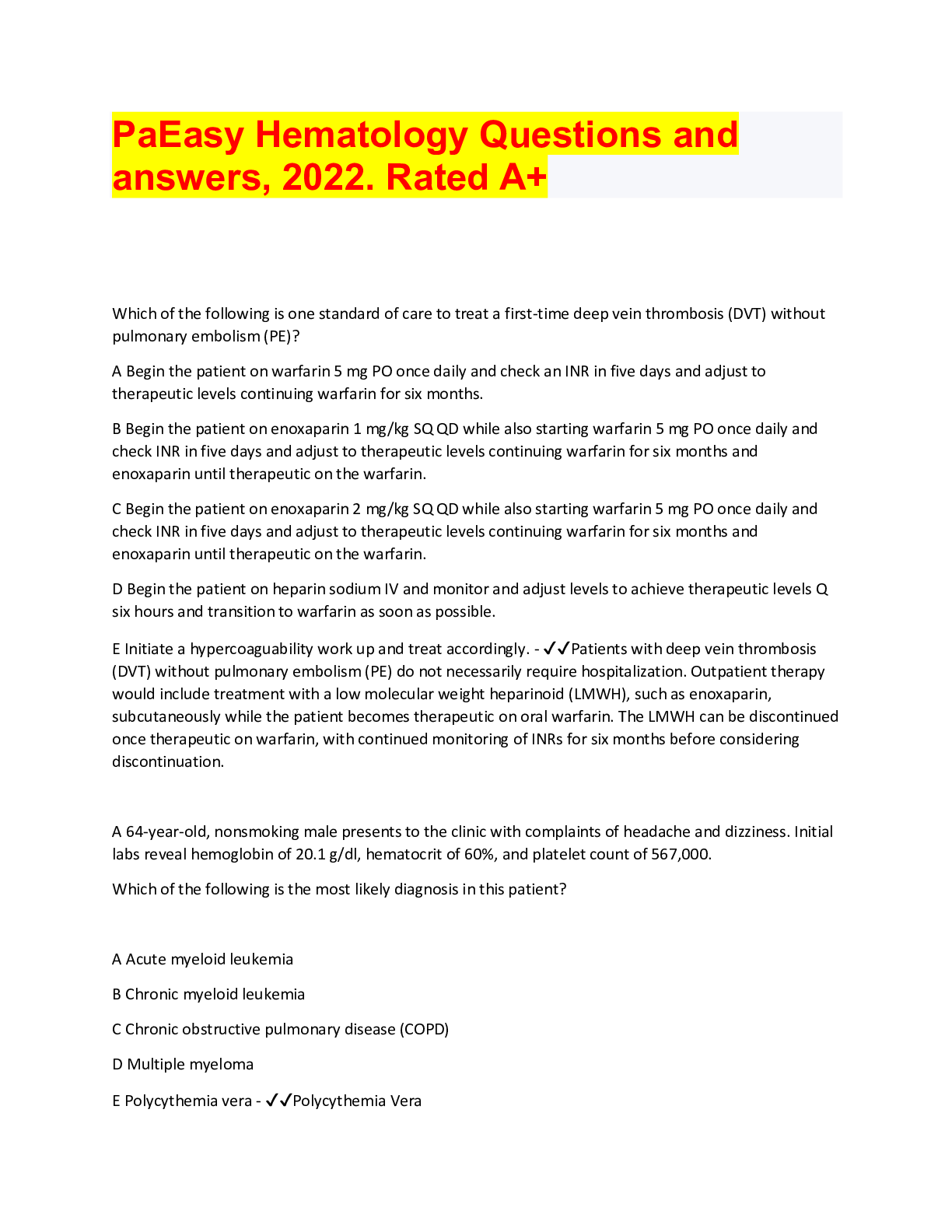
PA easy bundle, Exam Questions and answers, Rated A+
Comprises of exam Questions and answers, latest revisions for exams
By bundleHub Solution guider 1 year ago
$38
11
Reviews( 0 )
Document information
Connected school, study & course
About the document
Uploaded On
Aug 22, 2022
Number of pages
190
Written in

Additional information
This document has been written for:
Uploaded
Aug 22, 2022
Downloads
0
Views
104