NSG 6320 AGNP BOARD EXAM QUESTIONS Dermatoloy Assessment
Document Content and Description Below
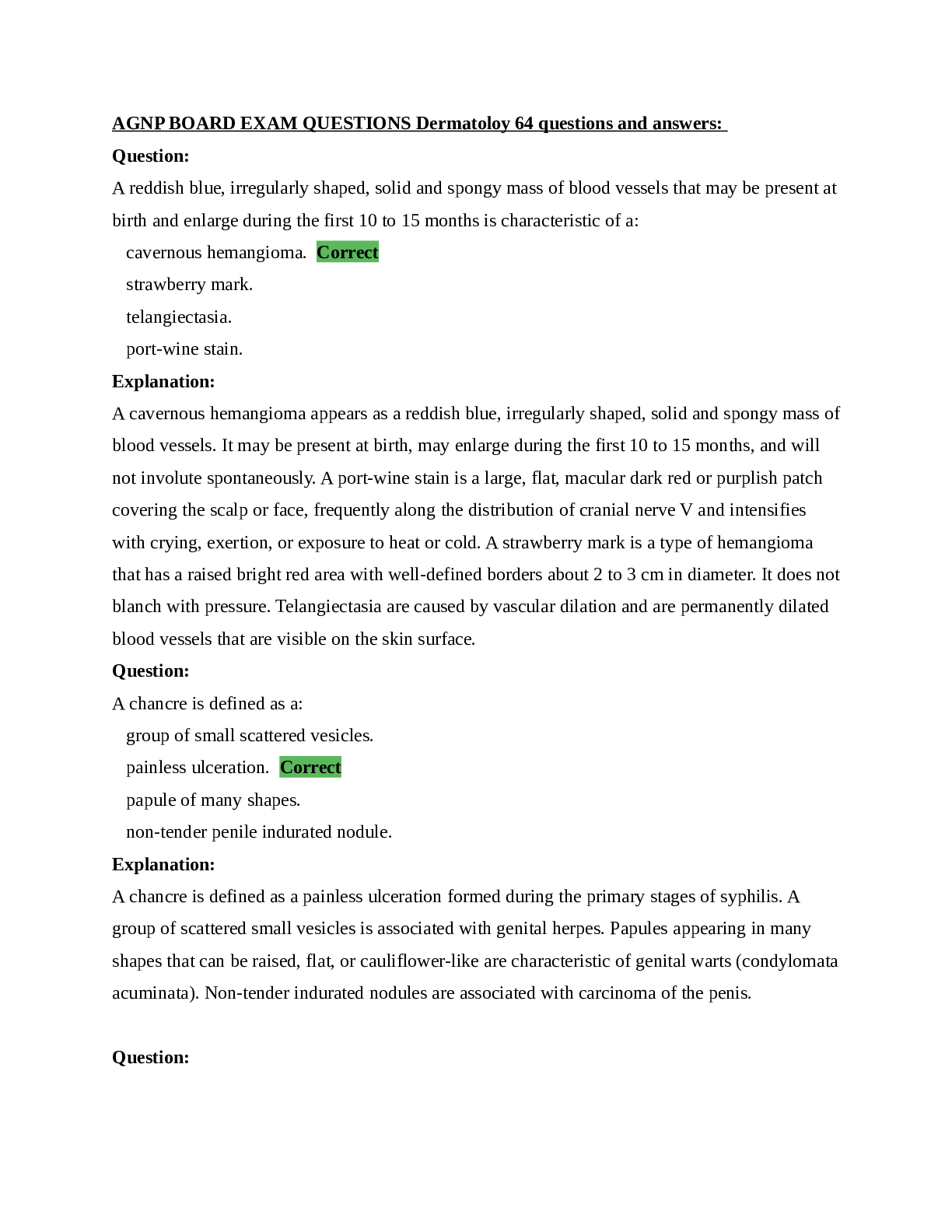
NSG 6320 AGNP BOARD EXAM QUESTIONS Dermatoloy Assessment NSG 6320 AGNP BOARD EXAM QUESTIONS Dermatoloy 64 questions and answers: Question: A reddish blue, irregularly shaped, solid and spongy mass ... of blood vessels that may be present at birth and enlarge during the first 10 to 15 months is characteristic of a: cavernous hemangioma. Correct strawberry mark. telangiectasia. port-wine stain. Explanation: A cavernous hemangioma appears as a reddish blue, irregularly shaped, solid and spongy mass of blood vessels. It may be present at birth, may enlarge during the first 10 to 15 months, and will not involute spontaneously. A port-wine stain is a large, flat, macular dark red or purplish patch covering the scalp or face, frequently along the distribution of cranial nerve V and intensifies with crying, exertion, or exposure to heat or cold. A strawberry mark is a type of hemangioma that has a raised bright red area with well-defined borders about 2 to 3 cm in diameter. It does not blanch with pressure. Telangiectasia are caused by vascular dilation and are permanently dilated blood vessels that are visible on the skin surface. Question: A chancre is defined as a: group of small scattered vesicles. painless ulceration. Correct papule of many shapes. non-tender penile indurated nodule. Explanation: A chancre is defined as a painless ulceration formed during the primary stages of syphilis. A group of scattered small vesicles is associated with genital herpes. Papules appearing in many shapes that can be raised, flat, or cauliflower-like are characteristic of genital warts (condylomata acuminata). Non-tender indurated nodules are associated with carcinoma of the penis. Question: Transverse depressions of the nail plates, usually bilateral, resulting from temporary disruption of proximal nail growth from systemic illness is termed: Terry's nails. Beau's lines. Correct Mees' lines. pitting nails. Explanation: Beau's lines are deep grooved lines that run from side to side on the fingernail. They appear as transverse depressions of the nail plates, usually bilateral, resulting from temporary disruption of proximal nail growth from systemic illness, such as severe illness, cold stress in the presence of Reynaud's disease, and trauma. With Terry's nails, the nail plate turns white with a ground-glass appearance, a distal band of reddish brown, and obliteration of the lunula. Mees' lines present as curving transverse white bands that cross the nail parallel to the lunula. They arise from the disrupted matrix of the proximal nail, vary in width, and move distally as the nail grows out. These lines are seen in arsenic poisoning, heart failure, Hodgkin’s disease, chemotherapy, carbon monoxide poisoning, and leprosy. Pitting nails present as punctate depressions of the nail plate caused by defective layering of the superficial nail plate by the proximal nail matrix. They may be associated with psoriasis, but also seen in Reiter’s syndrome, sarcoidosis, alopecia areata, and localized atopic or chemical dermatitis. Question: A child has a maculopapular, blotchy rash and on examination of his mouth, red eruptions with white centers on the buccal mucosa are visualized. These eruptions are called: rubella spots. aphthous ulcers. Pastia's spots. Koplik spots. Correct Explanation: Koplik spots are seen with measles. They are small, white spots (often on a reddened background) that occur on the inside of the cheeks early in the course of red measles, rubeola. Pastia's spots are pink or red lines that are formed of confluent petechiae found in skin creases and are seen in patients who have scarlet fever. Aphthous ulcers are recurrent small, round, or ovoid ulcers with circumscribed margins, erythematous haloes, and yellow or gray floors occurring in the mouth. Question: When examining the external genitalia of a female patient, excoriations and itchy, small, red maculopapulares were noted. This lesions may be suggestive of: genital herpes. pediculosis pubis. Correct Chlamydia trachomatis. genital warts. Explanation: Excoriations or itchy, small, red maculopapular suggest pediculosis pubis (lice or “crabs”). These symptoms are not consistent with those of genital herpes or, warts, or Chlamydia trachomatis. Question: A 75-year-old man presents with several brown, raised, slightly greasy peeling, velvety appearing lesions on the trunk. These lesions would be classified as: lichenification. seborrheic keratoses. Correct Kaposi's sarcoma. actinic keratoses. Explanation: Seborrheic keratoses are common, benign, yellowish to brown, raised lesions that feel slightly greasy and appear velvety and warty. They usually appear on trunk and face of the elderly. Actinic keratoses are superficial, flattened papules covered by a dry scale and can appear pink, gray, or tan in color. They appear on sun exposed skin of older fair-skinned persons. Lichenification is thickening and roughening of the skin with increased visibility of normal skin furrows. It can be seen in patients with atopic dermatitis. Question: When assessing the skin, it is noted to be thickened, taut, and shiny in appearance. This could be associated with: hypothyroidism. hyperthyroidism. psoriasis. scleroderma. Correct Explanation: Scleroderma appears as skin that is thickened, taut, and shiny in appearance. Psoriasis presents as silvery, scaly papules or plaques, mainly on the extensor surfaces of the skin. In patients who have hyperthyroidism, the skin has a velvety appearance and is usually warm to touch. Skin that appears very dry, rough, and cool to touch can be associated with hypothyroidism. Question: The term asteatosis refers to: skin that appears weather beaten, thickened, yellowed, and deeply furrowed. skin that is dry, flaky, rough, and often itchy. Correct raised yellowish lesions that feel greasy and velvety or warty. painful vesicular lesions in a dermatomal distribution. Explanation: Physiologic changes of aging include loss of elastic turgor, and wrinkling. Skin that appears dry, flaky, rough, and itchy is termed asteatosis. Sun exposure can cause damage to the skin resembling an appearance as weather beaten, thickened, yellowed, and deeply furrowed. Seborrheic keratoses appear as raised, yellowish lesions that feel greasy, velvety, or warty. Painful vesicular lesions in a dermatomal distribution may suggest herpes zoster. Question: The assessment findings of the integumentary system of an 80-year-old that would warrant further evaluation would include: several brown macular spots on the hands and arms. ecchymoses on both forearms. Correct seborrheic keratoses on the scalp. cherry angiomas on the trunk. Explanation: Ecchymoses in the elderly is not a sign of aging and should be evaluated for injury or abuse. Brown macular spots, age spots, on the hands are related to sun exposure. Seborrheic keratoses are thickened lesions on the skin that range from tan to brown to black with a clear border and develop on the trunk but can also occur on the hands, feet, face, and scalp and are not considered harmful. Cherry angiomas are a common type of skin lesion that first occurs in early adulthood and continues with age. These round lesions typically develop on the trunk, hands, and feet. They range from bright to dark red and are asymptomatic with no reported clinical consequences. Question: The patient has recently sustained an injury to the upper thigh. Examination reveals an irregular shaped purplish blue lesion that does not blanch with pressure and does not exhibit pulsatility. This would be considered: petechia. ecchymosis. Correct purpura. a spider vein. Explanation: Ecchymosis is purple or bluish purple area that fades to green, yellow, and brown with time. It is generally larger than petechia and can be round, oval or take on an irregular shape. It does not blanch with pressure and does not pulsate. These lesions are termed bruises and occur secondary to trauma. Petechia and purpura are deep red or reddish purple and fade with time. A spider vein is bluish in color and is mostly seen on the legs near veins. Question: A patient is observed to have pressure related alteration of intact skin along with changes in temperature, consistency, sensation, and color. Which stage of pressure ulcer is consistent with this? Stage I Correct Stage II Stage III Stage IV Explanation: Stage I: Intact skin with non-blanchable redness of a localized area usually over a bony prominence. The area may be painful, firm, soft, warmer or cooler as compared to adjacent tissue. Stage II: Partial thickness loss of dermis presenting as a shallow open ulcer with a red pink wound bed, without slough. May also present as an intact or open/ruptured serum-filled blister. Stage III: Full thickness tissue loss and subcutaneous fat may be visible but bone, tendon or muscle are not exposed. Slough may be present but does not obscure the depth of tissue loss and the depth varies by anatomical location. Stage IV: Full thickness tissue loss with exposed bone, tendon or muscle. Slough or eschar may be present on some parts of the wound bed. The depth varies by anatomical location. Question: Examination of a child who experienced a burn from a curling iron on the forearm appears red without blistering but is painful to touch. This type of burn would be classified as a: superficial thickness burn. Correct superficial partial thickness burn. deep partial thickness burn. full thickness burn. Explanation: Types of burn injuries are chemical, electric, radiation, or thermal and are classified by the depth of damaged skin. Superficial thickness burns appear erythematous without blisters and usually have local pain. Symptoms of superficial partial thickness burns include: moist areas that are red to ivory white in color, blisters forming almost immediately, and painful to touch. Since the pain receptors are intact, pain is perceived. Deep partial thickness burns have a dry waxy, whitish appearance and resemble full thickness burns. Sometimes grafts are needed. Full thickness burns involve the destruction of all skin elements with coagulation of subdermal plexus, muscle, and or tendons. Question: The term used to describe dark-colored adherent crust of dead tissue around an ulcer is: eschar. Correct erythroderma. excoriation. exfoliation. Explanation: Dark-colored adherent crust of dead tissue found on some ulcers is referred to as eschar. Erythroderma occurs when a skin condition affects the whole body or nearly the whole body, resulting in a red appearance all over. An excoriation is a scratch mark or surface injury penetrating the dermis. Exfoliation refers to peeling skin. Question: Skin conditions such as pruritus, hyperpigmentation, and calciphylaxis may be seen in patients who have: diabetes. chronic renal failure. Correct hyperthyroidism. liver disease. Explanation: Pallor, xerosis, pruritus, hyperpigmentation, uremic frost, calciphylaxis (calciphylaxis occurs when calcium accumulates in the skin and small vessels of the skin and fat tissue), “half and half” nails, are skin conditions which may be found in patients with chronic renal disease. These skin conditions are not seen in patients who have diabetes, hyperthyroidism, or liver disease. Question: An adolescent is being examined for acne and findings revealed several closed comedones. These findings are also termed: whiteheads. Correct blackheads. pustules. cysts. Explanation: Whiteheads are closed comedones; blackheads are open comedones; pustules are skin lesions with pus; and cysts are fluid-filled skin lesions. Question: A child was involved in a vehicular accident and sustained burns on the lower extremities. Examination reveals a dry, waxy, whitish appearance of both lower legs and some visualization of the tibialis anterior. This type of burn would be classified as a: superficial thickness burn. superficial partial thickness burn. deep partial thickness burn. full thickness burn. Correct Explanation: Types of burn injuries are chemical, electric, radiation, or thermal and are classified by the depth of damaged skin it caused. Full thickness burns involve the destruction of all skin elements with coagulation of subdermal plexus, muscle, and or tendons. Symptoms of superficial partial thickness burns include: moist areas that are red to ivory white in color, blisters forming almost immediately, and painful to touch. Since the pain receptors are intact, pain is perceived. Superficial thickness burns appear erythematous without blisters and usually have local pain. Deep partial thickness burns have a dry waxy, whitish appearance and resemble full thickness burns. Sometimes grafts are needed. Question: Lesions that develop within hair follicles produce papules that become erythematous, and painful are termed: vesicles. furuncles. Correct nevi. ulcers. Explanation: Boils, also known as furuncles, are painful infections that develop within hair follicles. They usually begin as papules on the skin, then develop a white or soft center. Blisters, also called vesicles, are small lesions filled with clear fluid. A mole (nevus/nevi) is a pigmented area on the epidermis. An ulcer occurs on the skin or a mucous membrane accompanied by the disintegration of tissue. Question: When assessing the skin, it is noted to be very dry and cool to touch. This could be associated with: hypothyroidism. Correct hyperthyroidism. psoriasis. scleroderma. Explanation: Skin that appears very dry, rough and cool to touch can be associated with hypothyroidism. In patients who have hyperthyroidism, the skin has a velvety appearance and is usually warm to touch. Psoriasis can present as silvery, scaly papules or plaques, mainly on the extensor surfaces of the skin. Scleroderma appears as skin that is thickened, taut, and shiny in appearance. Question: A patient presents with a full thickness tissue loss with exposed bone and tendon and eschar of the left hip. Which stage of pressure ulcer development is this? Stage I Stage II Stage III Stage IV Correct Explanation: Stage I: Intact skin with non-blanchable redness of a localized area usually over a bony prominence. The area may be painful, firm, soft, warmer or cooler as compared to adjacent tissue. Stage II: Partial thickness loss of dermis presenting as a shallow open ulcer with a red pink wound bed, without slough. May also present as an intact or open/ruptured serum-filled blister. Stage III: Full thickness tissue loss and subcutaneous fat may be visible but bone, tendon or muscle are not exposed. Slough may be present but does not obscure the depth of tissue loss and the depth varies by anatomical location. Stage IV: Full thickness tissue loss with exposed bone, tendon or muscle. Slough or eschar may be present on some parts of the wound bed. The depth varies by anatomical location. Question: Striae, skin atrophy, and purpura may be associated with: acquired immunodeficiency syndrome (AIDS). Addison's Disease. Cushing's Disease. Correct diabetes. Explanation: Cushing's disease can present with any of the following skin lesions: striae, skin atrophy, purpura, ecchymosis, telangiectasias, acne, moon facies, buffalo hump, or hypertrichosis. Hyperpigmentation of skin and mucous membranes is usually seen in patients who have Addison's Disease. Hairy leukoplakia can be seen in patients who have AIDS. Diabetes may produce any of these skin conditions: necrobiosis lipoidica diabeticorum, diabetic bullae, diabetic dermopathy, granuloma annulare, acanthosis nigricans, candidiasis, neuropathic ulcers, eruptive xanthomas, and peripheral vascular disease. Question: When the examiner lifts a fold of skin and notes how quickly it returns to place, the examiner is assessing skin: mobility. turgor. Correct moisture. temperature. Explanation: When the examiner lifts a fold of skin and notes how quickly it returns to place, the examiner is assessing skin turgor. A good response is for the skin to return to its place immediately. Decreased skin turgor is noted in dehydration and as skin ages. Question: A six-year-old child presents with a few small vesicles that are honey-colored and weeping around the left nare. These lesions are consistent with: impetigo. Correct varicella. Herpes simplex. shingles. Explanation: Impetigo usually appears as red crusty lesions on the face, especially around a child's nose and mouth. The lesions burst and develop honey-colored crusts. Varicella is characteristic of papules, vesicles, and crusted lesions occurring simultaneously. Herpes simplex on the lips and mouth appear as small vesicles or blisters and are termed "fever blisters" or "cold sores". Shingles appear as vesicles or blisters in clusters along the entire path of the nerve or in certain areas supplied by the nerve. Question: To assess skin temperature, the examiner would use the: palmar surface of the hand. dorsal surface of the hand. Correct fingertips. oral thermometer. Explanation: To assess the skin temperature, the examiner should use the backs of the fingers or hand. An oral thermometer measures body temperature. Question: A priority intervention in caring for a child diagnosed with atopic dermatitis should be to: maintain adequate nutrition. keep the child content. keep skin lesions dry. relieve pruritus. Correct Explanation: By relieving pruritus, itching is eased and this helps decrease scratching behaviors which can increase risk of infection. This is the major intervention. The others are helpful but not priority. The key is to keep the lesions moist as dryness will lead to itching and scratching and the potential for infection, especially in the case of impetigo. Question: Which stage of pressure is consistent with findings on dermatologic examination of a full thickness tissue loss and subcutaneous fat visible with mild slough on the right hip? Stage I Stage II Stage III Correct Stage IV Explanation: Stage I: Intact skin with non-blanchable redness of a localized area usually over a bony prominence. The area may be painful, firm, soft, warmer or cooler as compared to adjacent tissue. Stage II: Partial thickness loss of dermis presenting as a shallow open ulcer with a red pink wound bed, without slough. May also present as an intact or open/ruptured serum-filled blister. Stage III: Full thickness tissue loss and subcutaneous fat may be visible but bone, tendon or muscle are not exposed. Slough may be present but does not obscure the depth of tissue loss and the depth varies by anatomical location. Stage IV: Full thickness tissue loss with exposed bone, tendon or muscle. Slough or eschar may be present on some parts of the wound bed. The depth varies by anatomical location. Question: When suspecting pediculosis capitis, the chief complaint is: itching. Correct hives. alopecia. vesicles. Explanation: With pediculosis capitis (lice), itching is the most common symptom and is caused by an allergic reaction. Lice will bite the skin in order to feed on the infected person's blood. Saliva from these bites causes the allergic reaction and itching. The lice lay eggs that eventually hatch which causes more irritation. Vesicles, fluid filled lesions, are not usually seen in this condition. Alopecia may be seen with tinea capitis. Question: Examination of the nail beds revealed pitting in the nail. This is consistent with: leukonychia. psoriasis. Correct paronychia. onychomycosis. Explanation: Small pits in the nails are consistent with early signs of psoriasis. Paronychia is red, swollen, tender inflammation of the nail folds. Onychomycosis is a slow, persistent fungal infection of fingernails and toenails causing change in color, texture, and thickness. The nail may crumble or break with loosening of the nail plate, usually beginning at the distal edge and progressing proximally. Leukonychia is a white discoloration appearing on nails. The most common cause is injury to the base of the nail (the matrix) where the nail is formed. Question: When assessing the skin, it has a velvety appearance and is warm to touch. This could be associated with: hypothyroidism. hyperthyroidism. Correct psoriasis. scleroderma. Explanation: In patients who have hyperthyroidism, the skin has a velvety appearance and is usually warm to touch. Skin that appears very dry, rough and cool to touch can be associated with hypothyroidism. Psoriasis presents as silvery, scaly papules or plaques, mainly on the extensor surfaces of the skin. Scleroderma has a thickened, taut, and shiny skin appearance. Question: A yellowish hue imparted to the skin, mucous membrane, or eye is termed: carotenemia. scleroderma. café-au-lait. jaundice. Correct Explanation: Jaundice appears as a yellow color in the sclera, the palpebral conjunctiva, lips, hard palate, undersurface of the tongue, tympanic membrane, and skin. It is usually seen in patients with liver disease or excessive hemolysis of red blood cells. Carotenemia results in a yellow color of the skin, particularly on the face, palms, and soles from intake of large quantities of carotene (found yellow vegetables). Scleroderma is characterized by sclerosis of the skin or a hardening of the skin with a taunt, shiny appearance. Café-au-lait spots appear as uniform pigmented macules or patches on the skin and never appear in the eyes. Question: A child presents with erythematous papules and vesicles, that are weeping, oozing, and crusty. These lesions are located over the forehead, wrists, elbows, and the backs of the knees. With which of the following conditions are these symptoms associated? An allergic reaction to something Atopic dermatitis Correct Contact dermatitis Psoriasis Explanation: Atopic dermatitis presents with erythematous papules and vesicles, with weeping, oozing, and crusts. Lesions usually appear in characteristic areas: the scalp, forehead, cheeks, forearms and wrists, elbows, and backs of knees. Paroxysmal and severe pruritus can be present. There is usually a family and/or personal history of allergies. An allergic reaction to something usually presents with a generalized rash that resembles hives; erythematous and symmetric rash. Contact dermatitis is associated with a reaction in the area that the object touched. Erythema becomes evident initially, followed by swelling, wheals (or urticaria), or maculopapular vesicles, scales. This is often accompanied by intense pruritus. Psoriasis is scaly, commonly erythematous, and presents with silvery scales, An alternative presentation of psoriasis is plagues. Question: Pressure ulcers, decubitus ulcers, develop over all of the following body parts except the: sacrum. gluteus maximus. Correct ischial tuberosities. greater trochanters. Explanation: Pressure ulcers usually develop over body prominences subject to unrelieved pressure. This results in ischemic damage to underlying tissue form most commonly develop over the sacrum, ischial tuberosities, greater trochanters, and the heels. Question: The term used to describe skin that is red over the entire body is: eschar. erythroderma. Correct excoriation. exfoliation. Explanation: Erythroderma occurs when a skin condition affects the whole body or nearly the whole body, resulting in a red appearance all over. Dark-colored adherent crust of dead tissue found on some ulcers is referred to as eschar. An excoriation is a scratch mark or surface injury penetrating the dermis. Exfoliation refers to peeling skin. Question: A raised, crusted border with central ulceration on the helix is identified. This lesion needs further evaluation because it is most likely: a basal cell carcinoma. a squamous cell carcinoma. Correct cutaneous cyst. chondrodermatitis nodularis helicis. Explanation: A squamous cell carcinoma has a raised, crusted border with central ulceration on the lesion. Biopsy confirms the diagnosis. Basal call carcinoma can present as a red raised nodule with a lustrous surface and telangiectatic vessels with or without ulceration. A cutaneous cyst is a benign, closed sac that lies in the dermis, forming a dome shaped lump. Chondrodermatitis Nodularis Helicus is a condition with painful nodules that develop on the rim of the helix (where there is no subcutaneous tissue). This develops a result of repetitive mechanical pressure or environmental trauma (sunlight). They are small, indurated, dull red, poorly defined, and painful. Question: A patient has a papule with an ulcerated center on the lower lid and medial canthus of the eye. This is consistent with: dacryocystitis. a hordeolum or stye. a chalazion. a basal cell carcinoma. Correct Explanation: Although a basal call carcinoma of the eyelid is uncommon, it does occur most often on the lower lid and medial canthus. It looks like a papule with an ulcerated center. Metastasis is rare but should be referred for removal. Dacryocystitis is an infection and blockage of the lacrimal sac and duct. Hordeolum is often secondary to localized staphylococcal infection of the hair follicles at the lid margin. Chalazion is a beady nodule protruding on the lid. Question: Depigmented macules appearing on the face, hands, feet and other parts of the body due to the lack of melanin are termed: tinea versicolor. vitiligo. Correct cafe-au-lait spots. tinea corporis. Explanation: Vitiligo manifests as depigmented macules appearing on the face, hands, feet and other parts of the body as they lack melanin. This condition is hereditary. Tinea versicolor are hypopigmented macules and are slightly scaly and caused by a fungus. Tinea corporis has annular lesions with central clearing and papules on the borders. This is usually seen in children. Question: An example of a macule is: psoriasis. impetigo. petechia. Correct a nevus. Explanation: A macule is defined as a small flat spot measuring no larger than 1 cm. Examples are a freckle and petechiae. An elevated nevus is an example of a papule. Papules are raised above the level of the skin. Impetigo is an example of a crust which is dried residue of pus, serum, or blood. And psoriasis is an example of a scale, a thin flake of exfoliated epidermis. Dandruff and dry skin also fall under this category. Question: Granuloma annulare, acanthosis nigricans, and candidiasis may be seen in patients who have: acquired immunodeficiency syndrome (AIDS). Addison's Disease. Cushing's Disease. diabetes. Correct Explanation: Hyperpigmentation of skin and mucous membranes is usually seen in patients who have Addison's Disease. Hairy leukoplakia can be seen in patients who have AIDS. Cushing's disease can present with any of the following skin lesions: striae, skin atrophy, purpura, ecchymosis, telangiectasias, acne, moon facies, buffalo hump, or hypertrichosis. Diabetes may produce any of these skin conditions: necrobiosis lipoidica diabeticorum, diabetic bullae, diabetic dermopathy, granuloma annulare, acanthosis nigricans, candidiasis, neuropathic ulcers, eruptive xanthomas, and peripheral vascular disease. Question: A child sustained a "full-thickness" burn injury. This type injury involves tissue destruction down to the: epidermis. dermis. subcutaneous tissue. Correct internal organs. Explanation: A full-thickness burn involves all skin layers, including the epidermis, dermis, and the subcutaneous tissue and fat. Muscles and tendons may be involved. A superficial thickness burn involves the epidermis only. A superficial partial thickness burn involves the epidermis and the dermis. A deep thickness burn involves the entire layer of dermis, and is more severe than a superficial partial thickness burn. Question: When examining the skin, multiple areas of circumscribed elevations of the skin filled with serous fluid measuring approximately 0.5 cm were noted. These types of lesions could be seen in: second degree burns. acne or impetigo. psoriasis or athlete's foot. herpes simplex or varicella. Correct Explanation: A circumscribed superficial elevation of the skin formed by free fluid in a cavity within the skin layers are classified as three different lesions: a vesicle, filled with serous fluid and up to 1 cm.; a bulla, filled with serous fluid and measures greater than 1 cm.; and a pustule, filled with pus. Varicella and herpes simplex are examples of vesicles. Second degree burns are examples of a bulla. Acne and impetigo are examples of pustules. Psoriasis and athlete's foot are examples of a scale type lesion. Question: An infected area of the thigh was edematous, erythematous, tender, and warm to touch. The skin was blistering but free from exudate and the patient's temperature was 102 °F. This is suggestive of: impetigo. cellulitis. Correct a stage II pressure ulcer. erythema multiform. Explanation: Skin that is edematous, erythematous, tender, and warm to touch is consistent with cellulitis. Erythema multiform may appear as a nodule, papule, or macule or may look like hives. They may have blisters or vesicles and have a central sore surrounded by pale red rings, also called a "target", "iris", or "bulls-eye". Impetigo is the dried residue of serum, pus, or blood and can lead to cellulitis if left untreated. A stage II pressure ulcer presents with a partial thickness loss of dermis presenting as a shallow open ulcer with a red pink wound bed, without slough. Question: Ticks can transmit and cause: impetigo, staph infections, and psoriasis. candidiasis, scabies, and tinea capitis. Rocky Mountain spotted fever, Lyme disease, and tularemia. Correct lice, roundworms, and hookworms. Explanation: Ticks cause Rocky Mountain Spotted Fever, Lyme disease, and tularemia. None of the others listed can be transmitted or caused by ticks. Question: When examining the left ear, a raised nodule behind the ear is noted to have a lustrous surface with visible telangiectatic vessels in the center. This lesion needs further evaluation because it is most likely: squamous cell carcinoma. a cutaneous cyst. basal call carcinoma. Correct tophi. Explanation: Basal cell carcinoma can present as a raised, red nodule with a lustrous surface and telangiectatic vessels with or without ulceration. It occurs more frequently in fair -skinned people who have been exposed to large amounts of sun. A cutaneous cyst is a benign, closed sac that lies in the dermis, forming a dome shaped lump. A squamous cell carcinoma can present as a raised, crusted border with central ulceration on the lesion. Biopsy confirms the diagnosis. Tophi are identified as deposits of uric acid crystals secondary to gout and appear as hard nodules in the helix or antihelix. Question: Verruca plana is defined as: small, flat warts located in the superficial surfaces of the skin. Correct dry, rough warts located on the hands. small, tender warts located on the feet. domed-shaped, fleshy lesions located anywhere on the skin. Explanation: Verruca plana are small, flat warts located on superficial surfaces of the skin. Verruca vulgaris are dry, rough warts located on the hands. Small tender warts located on the superficial surfaces of the feet are plantar warts. Molluscum contagiosum are dome-shaped, flashy lesions and are located anywhere on the skin. Question: When assessing the skin, silvery, scaly papules or plaques are noted on the extensor surface of both arms. This could be associated with: hypothyroidism. hyperthyroidism. psoriasis. Correct scleroderma. Explanation: Psoriasis presents as silvery, scaly papules or plaques, mainly on the extensor surfaces of the skin. In patients who have hyperthyroidism, the skin has a velvety appearance and is usually warm to touch. Skin that appears very dry, rough and cool to touch can be associated with hypothyroidism. Scleroderma appears as skin that is thickened, taut, and shiny in appearance. Question: A small child sustained burns to the posterior trunk and posterior surface of both arms. According to the "Rule of Nines" for small children, what percentage of the total body surface area was involved? 9%. 18%. 27%. Correct 32.5%. Explanation: The "Rule of Nines" assigns percentage of body surface area burned based on the location of the burn. The percentages are as follows: head and neck = 18%, anterior and posterior chest = 18% each, arms (anterior and posterior) = 4.5% each, legs (anterior and posterior) = 6.7% each, and perineum = 1%. For this example, 18% for posterior trunk plus 9% for arms = 27%. Question: When the term rubor is used in describing the skin, it means that the appearance is: elastic. dusky red. Correct excoriated. excessively dry. Explanation: Rubor is a term used to describe the response of the skin to inflammation. Skin that appears dusky red is rubor. Question: When examining a patient with atopic dermatitis, there is a thickening and roughening of the skin with increased visibility of the normal skin furrows. This condition is termed: atrophy. excoriation. burrow of scabies. lichenification. Correct Explanation: Lichenification is defined as the thickening and roughening of the skin with increased visibility of the normal skin furrows (numerous grooves of variable depth on the surface of the epidermis). Atrophy is thinning of the skin with loss of normal skin furrows. Excoriation of the skin is an abrasion or scratch mark. The burrow of a scabies lesion includes small papules, pustules, lichenified areas and excoriations. Question: Hairy leukoplakia may be associated with: acquired immunodeficiency syndrome (AIDS). Correct Addison's Disease. Cushing's Disease. diabetes. Explanation: Hyperpigmentation of skin and mucous membranes is usually seen in patients who have Addison's Disease. Hairy leukoplakia can be seen in patients who have AIDS. Cushing's disease can present with any of the following skin lesions: striae, skin atrophy, purpura, ecchymosis, telangiectasias, acne, moon facies, buffalo hump, or hypertrichosis. Diabetes may produce any of these skin conditions: necrobiosis lipoidica diabeticorum, diabetic bullae, diabetic dermopathy, granuloma annulare, acanthosis nigricans, candidiasis, neuropathic ulcers, eruptive xanthomas, and peripheral vascular disease. Question: A child received a burn to the chest from a cup of hot coffee. On examination, the injured area appears moist and red to ivory white in color, blisters are noted, and painful to touch. This burn would be classified as a: superficial thickness burn. superficial partial thickness burn. Correct deep partial thickness burn. full thickness burn. Explanation: Types of burn injuries are chemical, electric, radiation, or thermal and are classified by the depth of damaged skin. Symptoms of superficial partial thickness burns include: moist areas that are red to ivory white in color, blisters forming almost immediately, and painful to touch. Since the pain receptors are intact, pain is perceived. Superficial thickness burns appear erythematous without blisters and usually have local pain. Deep partial thickness burns have a dry waxy, whitish appearance and resemble full thickness burns. Sometimes grafts are needed. Full thickness burns involve the destruction of all skin elements with coagulation of subdermal plexus, muscle, and or tendons. Question: A 45-year-old male presents with a skin lesion that has a smooth, slightly scaly, or pebbly surface, with an irregular edge. Further evaluation reveals a dark brown 2.5 cm lesion. This is most likely a: basal cell carcinoma. melanoma. squamous carcinoma. dysplastic nevus. Correct Explanation: A dysplastic nevus is a type of mole that looks different from a common mole. It may be bigger than a common mole, and its color, surface, and border may be different. It is usually more than 5 millimeters wide and can have a mixture of several colors, from pink to dark brown. Usually, it is flat with a smooth, slightly scaly, or pebbly surface, and it has an irregular edge that may fade into the surrounding skin. Basal cell carcinoma usually arises in sun-exposed areas and classically appears as pearly, erythematous, translucent papules, but may also be subtle red macules or exhibit other morphologies. Squamous cell carcinoma are crusted hyperkeratotic lesions with a rough surface or flat, reddish patches with an inflamed or ulcerated appearance. Melanomas arise from the pigment-producing melanocytes in the epidermis. These melanocytes give skin its color and appear as dark, raised, asymmetric lesions with irregular borders. Question: A forty-year-old man presents with a slow growing lesion on the face. Further examination reveals a lesion with a depressed center, a firm elevated border, and visible telangiectatic vessels. This is most consistent with: squamous cell carcinoma. basal cell carcinoma. Correct Kaposi's sarcoma. malignant melanoma. Explanation: Basal cell carcinoma typically presents with an initial translucent nodule that spreads, leaving a depressed center, a firm elevated border, and visible telangiectatic vessels. They usually appear in adults over forty and seldom metastasize. Squamous cell carcinoma appear on sun exposed skin of fair skinned adults over sixty and look like scaly red patches, open sores, elevated growths with a central depression, or warts; they may crust or bleed. Kaposi's sarcoma is seen in patients with AIDS and appears in many forms and on any part of the body. The abnormal cells form purple, red, or brown blotches or tumors on the skin and can become life threatening when the lesions are in the lungs, liver, or digestive tract. Question: An inflammation of the proximal and lateral nail folds represents: paronychia. Correct onychomycosis. psoriasis. leukonychia. Explanation: Paronychia is red, swollen, tender inflammation of the nail folds. Acute paronychia is usually secondary to a bacterial infection. Chronic paronychia is often secondary to a fungal infection from a break in the cuticle in those who perform “wet” work. Onychomycosis is a slow, persistent fungal infection of fingernails and toenails causing change in color, texture, and thickness. The nail may crumble or break with loosening of the nail plate, usually beginning at the distal edge and progressing proximally. Leukonychia is a white discoloration appearing on nails. The most common cause is injury to the base of the nail (the matrix) where the nail is formed. Psoriasis of the nails may appear as small pits in the nails. Question: A patient presents with a fiery red, slightly raised 2 cm lesion on the upper truck that blanches when pressure is applied. The body of the lesion is surrounded by erythema and radiating legs. This is most likely: a spider angioma. Correct a spider vein. petechiae. a cherry angioma. Explanation: A spider angioma is fiery red, slightly raised and surrounded by erythema and radiating legs that blanch with pressure. These are usually seen in patients with liver disease, pregnancy, vitamin B deficiency, and sometimes patients with no disease. The spider vein is blue. Cherry angiomas are bright or ruby red and increase in size and number as one ages. Petechiae are deep red or reddish purple and are seen when blood is outside the vessels. This suggests a bleeding disorder or injury to the skin. Question: Which model is used to designate risk factors for melanoma? ABCDE HARMM Correct BCRAT NHANES Explanation: The National Melanoma/Skin Cancer Screening Program of the American Academy of Dermatology validates the HARMM model for designating risk factors for melanoma. H - history, A - age, R - regular dermatologist absent, M-mole changing, and M- male gender. ABCDE nomenclature is the method used in recognizing characteristics of possible melanomas. A - asymmetry, B - borders, C - color, D - diameter, and E - evolution. BCRAT is the Breast Cancer Risk Assessment Tool. The National Health and Nutrition Examination Survey (NHANES) is used to predict diabetes prevalence. Question: Skin lesions that appear hypo or hyperpigmented with slightly scaly macules on the trunk, neck, and upper arms are likely: vitiligo. tinea versicolor. Correct actinic keratosis. ichthyosis vulgaris. Explanation: Tinea versicolor is a common superficial fungal infection of the skin, causing hypo or hyperpigmented (“versicolor”), slightly scaly macules on the trunk, neck, and upper arms (short-sleeved shirt distribution). They are easier to see in darker skin and may be more obvious after tanning. In lighter skin, macules may look reddish or tan instead of pale. Patients who have vitiligo may exhibit depigmented macules appear on the face, hands, feet, extensor surfaces, and other regions; and may coalesce into extensive areas that lack melanin. Ichthyosis vulgaris is a skin disorder that causes thin flakes of dead exfoliated epidermis. Question: Which stage of pressure ulcer is consistent with a partial thickness loss of the dermis presenting as a shallow open ulcer with a red pink wound bed, without slough? Stage I Stage II Correct Stage III Stage IV Explanation: Stage I: Intact skin with non-blanchable redness of a localized area usually over a bony prominence. The area may be painful, firm, soft, warmer or cooler as compared to adjacent tissue. Stage II: Partial thickness loss of dermis presenting as a shallow open ulcer with a red pink wound bed, without slough. May also present as an intact or open/ruptured serum-filled blister. Stage III: Full thickness tissue loss and subcutaneous fat may be visible but bone, tendon or muscle are not exposed. Slough may be present but does not obscure the depth of tissue loss and the depth varies by anatomical location. Stage IV: Full thickness tissue loss with exposed bone, tendon or muscle. Slough or eschar may be present on some parts of the wound bed. The depth varies by anatomical location. Question: An infant presents with a rash in the diaper area. Which description likely indicates candidal diaper rash? Red, moist, maculopapular patch with poorly defined borders in diaper area Bright red, moist patches with sharply demarcated borders, some loose scales noted in the diaper area Correct Moist, thin-roofed vesicles with a thin, erythematous base noted in the diaper area Erythematous and symmetric rash noted in the diaper area Explanation: Candidiasis is characteristic of a rash appearing with bright red, moist patches with sharply demarcated borders, with some loose scales noted in the diaper area. Red, moist, maculopapular patch with poorly defined borders in diaper area is diaper dermatitis. Moist, thin-roofed vesicles with a thin, erythematous base noted in the diaper area would be consistent with impetigo. Hives appear as erythematous and symmetric and can be generalized over the body including the diaper area. Question: On examination, a white discoloration appearing on nails was noted. This is consistent with: paronychia. onycholysis. leukonychia. Correct psoriasis. Explanation: Leukonychia is a white discoloration appearing on nails. The most common cause is injury to the base of the nail (the matrix) where the nail is formed. Psoriasis of the nails may appear as small pits in the nail. Paronychia is red, swollen, tender inflammation of the nail folds. Onycholysis refers to separation of the nail plate from the nail bed. The nail may crumble or break with loosening of the nail plate, usually beginning at the distal edge and progressing proximally. Question: If a lesion is described as annular, it appears: circular and begins in center and spreads to periphery. Correct as if the lesions run together. as distinct, individual lesions that remain separate. with concentric rings of color in the lesions. Explanation: Annular lesions are described as circular lesions that begin in the center and spreads to the periphery as with tinea and ringworm lesions. Confluent lesions appear as if the lesions run together and are characteristic of hives. Discrete lesions appear as distinct, individual lesions that remain separate as in acne. Target lesions resemble the iris of the eye and appear with concentric rings of color in the lesions. These are seen in erythema multiforme. Question: A slow, persistent fungal infection of fingernails and toenails is: paronychia. onychomycosis. Correct leukonychia. psoriasis. Explanation: Onychomycosis is a slow, persistent fungal infection of fingernails and toenails causing change in color, texture, and thickness. The nail may crumble or break with loosening of the nail plate, usually beginning at the distal edge and progressing proximally. Leukonychia is a white discoloration appearing on nails. The most common cause is injury to the base of the nail (the matrix) where the nail is formed. Psoriasis of the nails may appear as small pits in the nail. Paronychia is red, swollen, tender inflammation of the nail folds. Question: A dark, raised, asymmetric lesion with irregular borders may be: a comedone. actinic keratosis. a melanoma. Correct actinic purpura. Explanation: A dark, raised, asymmetric lesion with irregular borders may be a melanoma. A comedone is a blackhead. Actinic keratosis is described as superficial, flattened papules covered by a dry scale. Well-demarcated vividly purple macules or patches are called actinic purpura. Question: The term used to refer to skin that is peeling is: eschar. erythroderma. excoriation. exfoliation. Correct Explanation: Dark-colored adherent crust of dead tissue found on some ulcers is referred to as eschar. Erythroderma occurs when a skin condition affects the whole body or nearly the whole body, resulting in a red appearance all over. An excoriation is a scratch mark or surface injury penetrating the dermis. Exfoliation refers to peeling skin. Question: Asymmetry, irregular borders, variation in color, diameter greater than 6 mm, and elevation represent the "ABCDEs" of: benign nevi. basal cell carcinoma. malignant melanoma. Correct skin tumors. Explanation: The American Cancer Society created this tool to assist providers in readily identifying the characteristics of malignant melanoma. With the color variations, the lesions can be mixtures of black, blue, and red. Usually the lesions are raised but can be flat. Benign nevi, basal and squamous cell carcinomas and skin tumors do not follow these characteristics. Question: Purple patches or macules on backs of the hands and forearms of the elderly would be suggestive of: elder abuse. actinic purpura. Correct a bleeding disorder. injury from falls. Explanation: Skin on the backs of the hands and forearms appears thin, fragile, loose, and transparent. There may be purple patches or macules, termed actinic purpura, that fade over time. These spots and patches come from blood that has leaked through poorly supported capillaries and spread within the dermis. [Show More]
Last updated: 1 year ago
Preview 1 out of 29 pages
Instant download
Buy this document to get the full access instantly
Instant Download Access after purchase
Add to cartInstant download
Reviews( 0 )
Document information
Connected school, study & course
About the document
Uploaded On
Dec 30, 2020
Number of pages
29
Written in
Additional information
This document has been written for:
Uploaded
Dec 30, 2020
Downloads
0
Views
25




.png)




 – South University Savannah.png)
 - Prescription (102 Questions) – South University Savannah.png)
 _ Dermatology Prescription (100 Questions and Answers) – South University Savannah.png)
 Assessment question of Endocrinology (48 Questions) – South University Savannah.png)
 Dermatology (64 questions and answers) – South University Savannah.png)
 Prescription Gastroenterology (85 Questions) – South University Savannah.png)
 Prescription of Endocrinology (89 Questions) – South University Savannah.png)
 Assessment Eye, Ear, Nose and Throat (166 Questions) – South University Savannah.png)
 Cardiovascular Assessment (107 Questions and Answers) – South university Savannah.png)

