Anatomy and Physiology - A&P 1 > LAB QUIZ > BIOS-251 Week 6 Lab Assignment: Skeletal System (100% CORRECT ANSWERS) (All)
BIOS-251 Week 6 Lab Assignment: Skeletal System (100% CORRECT ANSWERS)
Document Content and Description Below
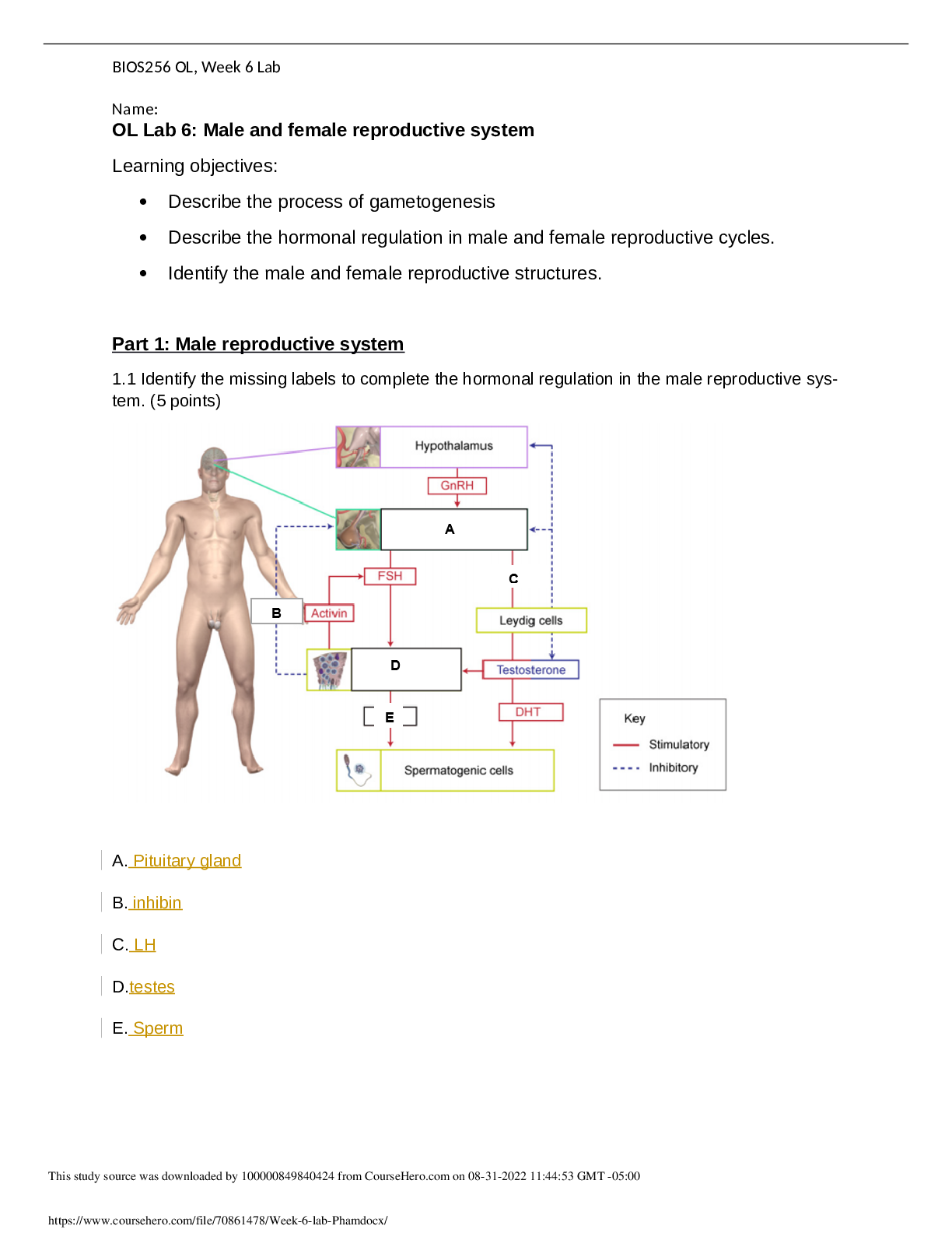
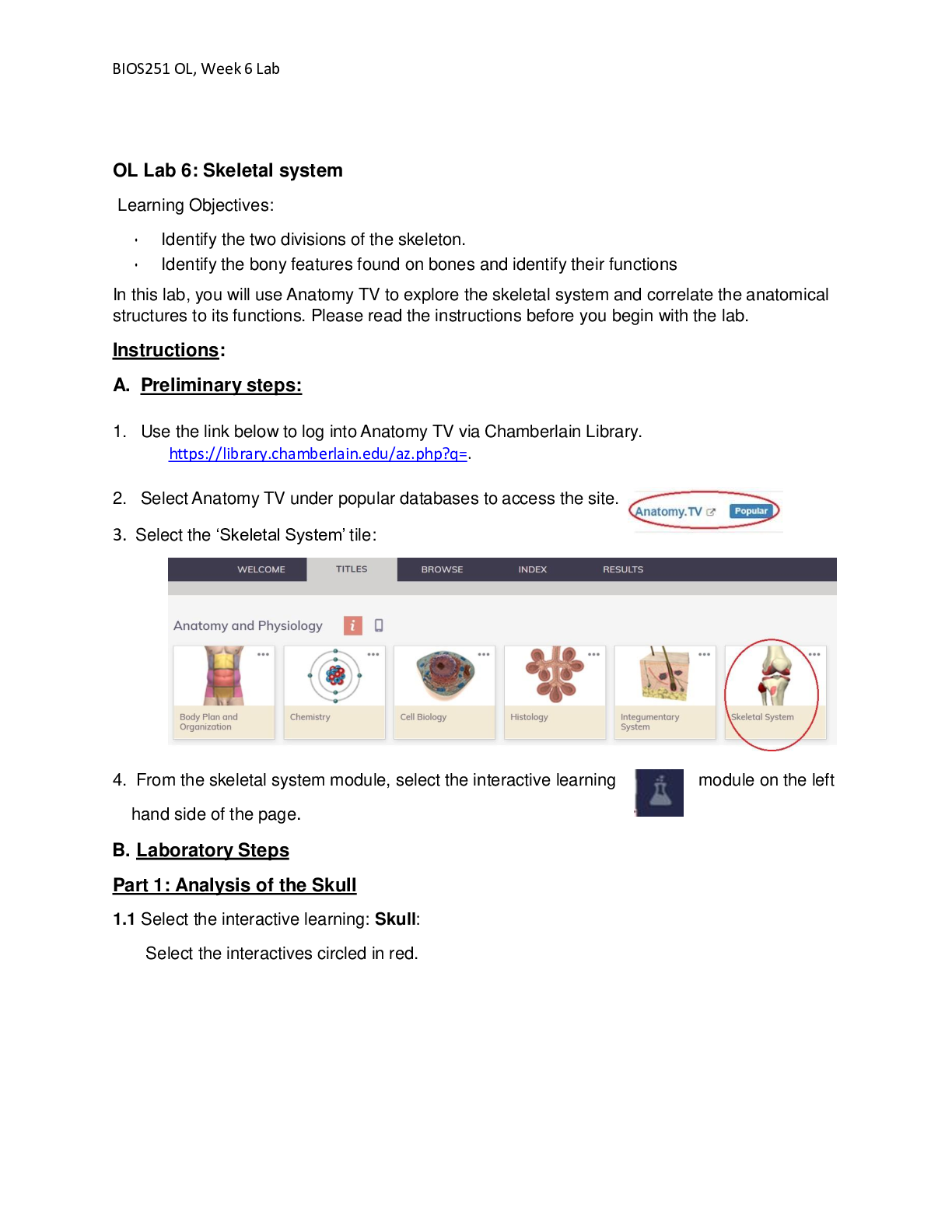
OL Lab 6: Skeletal system Learning Objectives: • Identify the two divisions of the skeleton. • Identify the bony features found on bones and identify their functions In this lab, you will use ... Anatomy TV to explore the skeletal system and correlate the anatomical structures to its functions. Please read the instructions before you begin with the lab. Instructions: A. Preliminary steps: 1. Use the link below to log into Anatomy TV via Chamberlain Library. https://library.chamberlain.edu/az.php?q=. 2. Select Anatomy TV under popular databases to access the site. 3. Select the ‘Skeletal System’ tile: 4. From the skeletal system module, select the interactive learning module on the left hand side of the page. B. Laboratory Steps Part 1: Analysis of the Skull 1.1 Select the interactive learning: Skull: Select the interactives circled in red. 1.2- On the image identify and select a one anatomical structure at a time. The region selected will appear highlighted. 1.3- Use the controls at the bottom of the page as required. Follow the recommendations provided for each structure. 1.4- Take a screen shot of the image or save the image using the download icon on the right hand side tab. 1.5- Create a word document titled “lastname_Lab 6”. Import the image into the document. Use the functions ‘Insert’ and ‘tool’ (shapes) on word to label the images. 1.6- In complete sentence write the functional role of the selected anatomical structure under the image. 1.7- Save the file before you proceed to the next structure. 1.8- Follow these steps to analyze all the structures listed below. Facial Bones: a. Bone: compact bone - Compact bone provides the bone with protection and strength. b. Compact bone: lacunae - lacunae provide passage ways through the extracellular matrix to the central canal for communication. c. Compact bone: osteon- - Osteon are cylinder like structures osteocytes and the mineral matrix, which are connected by small tunnels called canaliculi that transport blood. d. Periosteum- - A dense fibrous membrane covering the surface of bones and serving as an attachment for tendons and muscles e. Bone: yellow bone marrow- yellow bone marrow- often found at the end of bones in adults, it is made of fat. f. Endosteum g. Bone: spongy bone h. Temporal bone: mastoid process i. Maxilla: alveolar process- j. Zygomatic bone- Also known as cheekbones, which articulate with the frontal, maxilla, sphenoid and temporal bones. k. Sphenoid bone: greater wing l. Mandible: body- The lower jawbone, which is the strongest and largest facial bone. The mandible is the only bone that is moveable excluding auditory bones. Use the layer controls to remove/add layers ending on layer 1 and rotation controls to rotate the model to frame 10 for the following structures. m. Occipital bone- Flat bone located in the back of the skull and has an opening for the spinal cord to enter the brain. n. Sphenoid bone- An irregular bone that sits under the frontal bone and is the width of the skull forming the skulls base. o. Maxilla- The upper jaw bone, articulating with all other facial bones excluding the mandible (lower jawbone). p. Palatine bone- two L- shaped bones that help to form the nasal cavity. q. Temporal bone- Two irregular bones that are located under each of the parietal bone. Part 2: Analysis of the vertebral column: Select the interactive learning: Vertebral column Select the interactives circled in red. Vertebral column: Repeat steps 1.2 to 1.7 to identify and select the structures listed below: a. 3rd lumbar vertebra b. Sacrum c. Coccyx d. 1st cervical vertebra (atlas) e. 12th thoracic vertebra Use the layer controls to remove/add layers ending on layer 3 and rotation controls to rotate the model to frame 4 for the following structures. f. Sacrum: ala g. 10th rib h. Intervertebral discs Cervical Vertebra: Repeat steps 1.2 to 1.7 to identify and select the following structures: a. Cervical vertebra (C4): superior articular facet b. Cervical vertebra (C4): transverse process c. Cervical vertebra (C4): superior articular process d. Cervical vertebra (C4): pedicle e. Cervical vertebra (C4): anterior tubercle Thoracic Cage: Repeat steps 1.3 to 1.6 to identify and select the following structures a. Costal cartilages b. Sternum c. 5th rib d. 12th rib Use the layer controls to remove/add layers ending on layer 2 and rotation controls to rotate the model to frame 2 for the following structures. e. Sternum: manubrium f. Sternum: body g. Sternum: xiphoid process h. Typical rib (6th): shaft i. 1st rib: head Part 3: Analysis of bones of the Upper Limb Select the interactive learning: Bones of the Upper Limb Select the interactives circled in red. Repeat steps 1.2 to 1.7 to identify and select the following structures: Bones of the pectoral girdle and arm: a. Scapula: subscapular fossa b. Scapula: coracoid process c. Humerus: head d. Humerus: trochlea e. Humerus: lesser tubercle f. Ulna: coronoid process Use the rotation controls to rotate the model to frame 16 for the following structures. g. Scapula: infraspinous fossa h. Scapula: inferior angle i. Scapula: spine j. Humerus: shaft k. Humerus: deltoid tuberosity l. Ulna: olecranon m. Radius: head Bones of the forearm: a. Radius b. Ulna c. Capitate d. 1st metacarpal e. Middle phalanx of 4th finger Use the layer controls to remove/add layers ending on layer 2 and rotation controls to rotate the model to frame 19 for the following structures f. Metacarpal: shaft g. Middle phalanx: base h. Humerus: medial epicondyle i. Radius: tuberosity j. Radius: styloid process Part 4: Analysis of the bones of the Lower Limb Select the interactive learning: Bones of the Lower Limb Select the interactives circled in red. Repeat steps 1.2 to 1.7 to identify and select the following structures Bones of the pelvic girdle and thigh: a. Femur: head b. Hip bone: iliac fossa c. Hip bone: body of pubis Use the rotation controls to rotate the model to frame 19 for the following structures. d. Sacrum: medial sacral crest e. Hip bone: body of ilium f. Hip bone: ischial tuberosity g. Femur: greater trochanter h. Femur: neck i. Femur: linea aspera j. Femur: lateral condyle Bones of the leg: a. Femur b. Patella c. Tibia d. Fibula e. Talus f. 1st metatarsal Use the layer controls to remove/add layers ending on layer 2 and rotation controls to rotate the model to frame 18 for the following structures g. Femur: lateral condyle h. Tibia: lateral condyle i. Fibula: head Activity Deliverable Points Part 1,2,3 & 4 Compete the interactives and submit the lab report on the skeletal system: Part 1: Skull Part 2: Vertebral column 30 Part 3: Bones of the upper limb Part 4: Bones of the lower limb Total Complete all lab activities 30 [Show More]
Last updated: 1 year ago
Preview 1 out of 14 pages

Reviews( 0 )
Document information
Connected school, study & course
About the document
Uploaded On
Nov 04, 2022
Number of pages
14
Written in

Additional information
This document has been written for:
Uploaded
Nov 04, 2022
Downloads
0
Views
55
.png)


.png)
.png)
.png)
 (1).png)

.png)