PEDIATRIC CASE STUDY
Document Content and Description Below
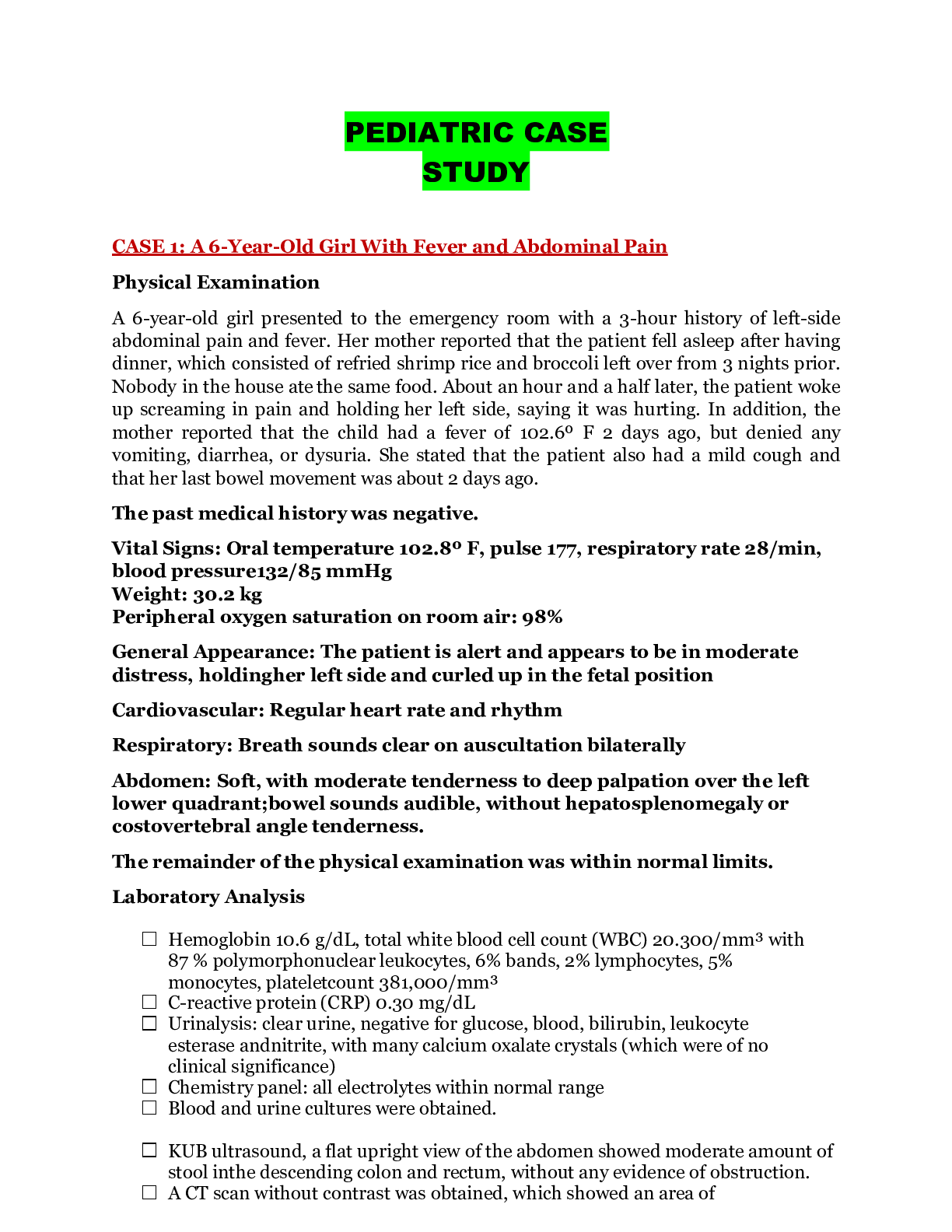
CASE 1: A 6-Year-Old Girl With Fever and Abdominal Pain Physical Examination A 6-year-old girl presented to the emergency room with a 3-hour history of left-side abdominal pain and fever. Her mother... reported that the patient fell asleep after having dinner, which consisted of refried shrimp rice and broccoli left over from 3 nights prior. Nobody in the house ate the same food. About an hour and a half later, the patient woke up screaming in pain and holding her left side, saying it was hurting. In addition, the mother reported that the child had a fever of 102.6º F 2 days ago, but denied any vomiting, diarrhea, or dysuria. She stated that the patient also had a mild cough and that her last bowel movement was about 2 days ago. The past medical history was negative. Vital Signs: Oral temperature 102.8º F, pulse 177, respiratory rate 28/min, blood pressure 132/85 mmHg Weight: 30.2 kg Peripheral oxygen saturation on room air: 98% General Appearance: The patient is alert and appears to be in moderate distress, holding her left side and curled up in the fetal position Cardiovascular: Regular heart rate and rhythm Respiratory: Breath sounds clear on auscultation bilaterally Abdomen: Soft, with moderate tenderness to deep palpation over the left lower quadrant; bowel sounds audible, without hepatosplenomegaly or costovertebral angle tenderness. The remainder of the physical examination was within normal limits. Laboratory Analysis • Hemoglobin 10.6 g/dL, total white blood cell count (WBC) 20.300/mm³ with 87 % polymorphonuclear leukocytes, 6% bands, 2% lymphocytes, 5% monocytes, platelet count 381,000/mm³ • C-reactive protein (CRP) 0.30 mg/dL • Urinalysis: clear urine, negative for glucose, blood, bilirubin, leukocyte esterase and nitrite, with many calcium oxalate crystals (which were of no clinical significance) • Chemistry panel: all electrolytes within normal range • Blood and urine cultures were obtained. • KUB ultrasound, a flat upright view of the abdomen showed moderate amount of stool in the descending colon and rectum, without any evidence of obstruction. • A CT scan without contrast was obtained, which showed an area of consolidation present in the left lung lower lobe medially and increased fecal material in the bowel. Remainder of the soft tissue and structures were within normal limits. [Show More]
Last updated: 1 year ago
Preview 1 out of 19 pages
Reviews( 0 )
Document information
Connected school, study & course
About the document
Uploaded On
Feb 20, 2023
Number of pages
19
Written in

Additional information
This document has been written for:
Uploaded
Feb 20, 2023
Downloads
0
Views
44
.png)