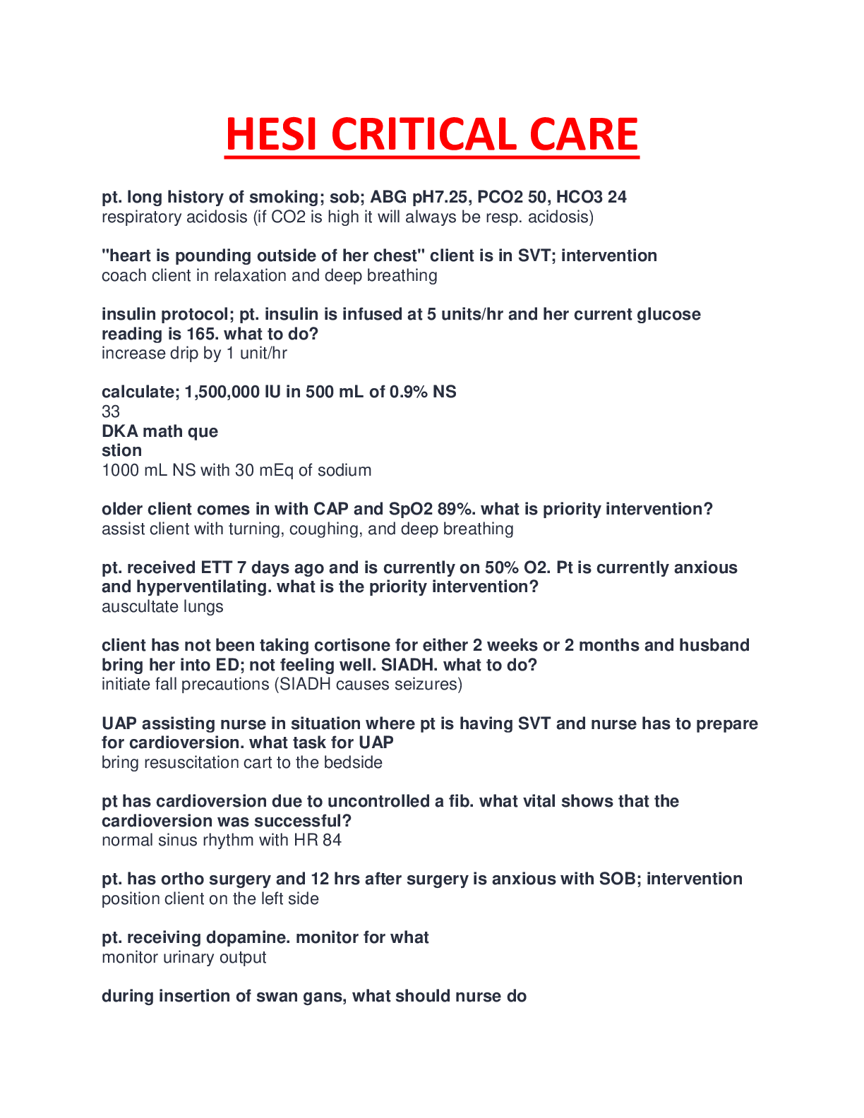
*NURSING > STUDY GUIDE > NURS 415 /313 cardio Exam_2 Study Guide,100% CORRECT (All)
NURS 415 /313 cardio Exam_2 Study Guide,100% CORRECT
Document Content and Description Below
First Degree Heart Block - Caused by a conduction delay through the AV node but all electrical signals reach the ventricles. - Every impulse is conducted to the ventricles but the duration of AV co... nduction is prolonged - The PR interval is prolonged to more than 0.20 second Second Degree Type II Heart Block - A P wave is nonconducted without progressive antecedent PR lengthening - A certain number of impulses from the SA node are not conducted to the ventricles - The QRS complex is almost always more than 0.12 second because of bundle branch block Third Degree AV Block - 3rd degree block or complete heart block occurs when atrial contractions are 'normal' but no electrical conduction is conveyed to the ventricles. - The ventricles then generate their own signal through an 'escape mechanism' from a focus somewhere within the ventricle. The ventricular escape beats are usually 'slow' Ch. 37 – Inflammatory and Structural Heart Disorders - Valvular Heart Disease functional alteration of one or more valves of the heart due to stenosis and regurgitation o Because of the 4 valves there can be 8 different diagnoses - Stenosis constriction or narrowing o Can be due to hardening o So narrow, it doesn’t close completely, allowing leakage o In a stenotic valve, the valve orifice is smaller, impeding the forward flow of blood and creating a press ure gradient difference across and open valve o The degree of the stenosis is seen in the pressure gradient differences the higher the gradient, the greater the stenosis - Regurgitation insufficiency that permits leakage or backward flow through the incompetent valve; blood goes back and forth o Incomplete closure of the valve leaflets results in the backward flow of blood o In a normal heart, blood coming out of the atrium causes the valve to open as the ventricle fills, the valve closes - Cardiac output decreases, LV has to work harder trying to compensate for decreased CO CHF evolves - Valvular disorders occur in children and adolescents primarily from congenital conditions such as tricuspid atresia, pulmonary stenosis, and aortic stenosis. Mitral Valve Stenosis shortening, thickening of the (mitral) valve - Most cases result from rheumatic heart disease o Due to mostly infection or complication of streptococcal infection o Tonsils are the first structures that filter microorganisms in your system if your immune system is already weak, the microorganism multiples and creates a colony o It begins to circulate and goes to the valves of the heart o It eventually heals but creates a scar resulting in stenosis - Patho: o Rheumatic carditis causes scarring of the valve leaflets and the chordae tendineae contractures and adhesions develop between the commissures (the junctional areas) stenosic mitral valve takes on a “fish mouth” shape because of the thickening and shortening of the valve structures o These structural deformities cause obstruction of blood flow and create a pressure difference between the LA and LV during diastole o LA pressure and volume elevation cause increased pulmonary vasculature pressure and subsequent hypertrophy of the pulmonary vessels o Chronic mitral stenosis pressure overload occurs in the LA, the pulmonary bed, and the RV - Less common causes are congenital mitral stenosis, rheumatoid arthritis, and systemic lupus erythematosus - Manifestations: o Exertional dyspnea due to reduced lung compliance o Fatigue and palpitations from atrial fibrillation due to cardiac hypertrophy (ventricle enlarges because of constant compensation; atria enlarges because of constant backflow of blood) o Heart sounds include a loud first heart sound and a low-pitched, rumbling diastolic murmur (best heard at the apex with a stethoscope) murmur caused by turbulence of blood passing through a narrow passageway o Hoarseness from atrial enlargement pressing on the laryngeal nerve o Hemoptysis from pulmonary HTN o Chest pain from decreased CO o Seizures or stroke from emboli that can arise from blood stasis in the LA Mitral Valve Regurgitation backward flow of blood from LV to LA - Mitral valve function depends on intact mitral leaflets, mitral annulus, chordae Tendineae, papillary muscles, LA, and LV any defect in any of these structures can result in regurgitation - Allows blood to flow backward from the LV to LA due to incomplete valve closure during systole - LV and LA both work harder to preserve an adequate CO - Causes: o MI o Chronic rheumatic heart disease o Mitral valve prolapse o Ischemic papillary muscle dysfunction o Inflammatory due to the damage from the valve lifting and due to infection o Degenerative o Infective o Structural/congenital it just doesn’t close or fit together - Manifestations: o Chronic MR: Atrial enlargement, ventricular dilatation, and eventual ventricular hypertrophy resulting from additional volume load Weakness, fatigue, palpitations, dyspnea, orthopnea, paroxysmal nocturnal dyspnea, peripheral edema secondary to LV failure Systolic murmur due to accentuated LV filling o Acute MR LA and ventricle do not abruptly dilate Pulmonary edema and shock due to sudden increase in pressure and volume that is transmitted to the pulmonary bed Thready peripheral pulses, cool clammy extremities secondary to decreased CO Mitral Valve Prolapse abnormality of the mitral valve leaflets and the papillary muscles or chordae that allows the leaflets to prolapse, or buckle, back into the LA during systole - Etiology unknown - Most patients asymptomatic for their entire lives - Characteristic murmur from regurgitation that gets more intense through systole Aortic Valve Stenosis causes obstruction of flow from the LV to the aorta during systole - Results in LV hypertrophy and increased myocardial oxygen consumption because of the increased myocardial mass - As the disease progresses and compensatory mechanisms fail, reduced CO leads to pulmonary HTN and HF - Mitral valve disease often accompanies this when occurs secondary to rheumatic fever - Causes: o Congenital o Older patients usually a result of rheumatic fever or senile fibrocalcific degeneration - Manifestations: o Symptoms develop when the valve orifice becomes about one third its normal size o Symptoms include classic triad angina, syncope, and exertional dyspnea o Systolic crescendo-decrescendo murmur Aortic Valve Regurgitation retrograde blood flow from the ascending aorta into the LV during diastole, resulting in volume overload - LV initially compensates for chronic AR by dilation and hypertrophy - Myocardial contractility eventually declines and blood volumes increase in the LA and pulmonary bed pulmonary HTN and RV failure - Causes: o Result of primary disease of the aortic valve leaflets, the aortic root, or both o Acute AR trauma, IE, or aortic dissection Life-threatening emergency o Chronic AR result of rheumatic heart disease, a congenital bicuspid aortic valve, syphilis, or chronic rheumatic conditions - Manifestations: o Acute AR sudden signs of cardiovascular collapse (severe dyspnea, chest pain, and hypotension) o Chronic AR water hammer pulse (a strong, quick beat that collapses immediately) Generally remains asymptomatic or years and is seen with exertional dyspnea, orthopnea, and paroxysmal nocturnal dyspnea only after considerable myocardial dysfunction has occurred TABLE 37-13 Clinical Manifestations of Valvular Heart Diseases Clinical Manifestations Mitral valve stenosis Dyspnea on exertion, hemoptysis; fatigue; palpitations; loud, accentuated S1; low-pitched, rumbling diastolic murmur; atrial fibrillation on ECG Mitral valve regurgitation Acute—generally poorly tolerated, with fulminating pulmonary edema and shock developing rapidly; new systolic murmur Chronic—weakness, fatigue, exertional dyspnea, palpitations; an S3 gallop, holosystolic or pansystolic murmur Mitral valve prolapse Palpitations, dyspnea, chest pain, activity intolerance, syncope; midsystolic click, late or holosystolic murmur Aortic valve stenosis Angina, syncope, dyspnea on exertion, heart failure; normal or soft S1, diminished or absent S2, systolic crescendo-decrescendo murmur, prominent S4 Aortic valve regurgitation Acute—abrupt onset of profound dyspnea, chest pain, left ventricular failure and shock Chronic—fatigue, exertional dyspnea, orthopnea, PND; water-hammer pulse; heaving precordial impulse; diminished or absent S1, S3, or S4; soft decrescendo high-pitched diastolic murmur, Austin-Flint murmur, systolic ejection click Tricuspid and pulmonic stenosis Tricuspid—peripheral edema, ascites, hepatomegaly; diastolic low-pitched, decrescendo murmur with increased intensity during inspiration Pulmonic—fatigue, loud midsystolic murmur Management of Valvular Heart Disease - Prevent recurrent rheumatic fever or infective endocarditis prophylactic antibiotic therapy - Focused on preventing exacerbations of HF, acute pulmonary edema, thromboembolism, and recurrent endocarditis - If manifestations of HF present vasodilators, positive inotropes, beta blockers, diuretics, and low-sodium diet - Anticoagulant therapy is used to prevent and treat systemic or pulmonary embolization and is also used prophylactically in patients with A. fib - Dysrhythmias treated with digoxin, antidysrhythmic drugs, or electrical cardioversion - Beta blockers may be used to slow the ventricular rate in pts with A. fib Nursing Therapeutics - Prevent acquired rheumatic valvular disease - Prevent recurrent infection through antibiotics prophylactic treatment should continue for life in individuals who develop rheumatic heart disease o Additional prophylaxis is necessary if a patient with known rheumatic heart disease has dental or surgical procedures involving the upper respiratory, GI, or GU tract - Hospitalization due to CHF and arrhythmia - Exercise plan to increase cardiac tolerance strenuous physical activity should be avoided because damaged valves may not be able to handle the demand for an increase in CO o Restrict activities that regurlarly produce fatigue and dyspnea - Smoking cessation may increase incidences of respiratory and upper respiratory infections - Assist in planning ADLs emphasis on conserving energy, setting priorities, and taking planned rest periods Ch 38 – Vascular Disorders Aortic Aneurysm - Out pouchings or dilations of the arterial wall - May involve the aortic arch, thoracic aorta, and/or abdominal aorta o Most aneurysms found in abdominal aorta below the level of the renal arteries - The larger the aneurysm, the greater the risk of rupture - Patho: o The dilated aortic wall becomes lined with thrombi that can embolize, leading to acute ischemic symptoms in distal branches o Primary causes: Atherosclerosis plaques deposit beneath the intima causing degenerative changes in the media leading to loss of elasticity, weakening, and eventual aortic dilation • When the high pressure gets to be too much, an out pouching forms Genetic predisposition Penetrating or blunt trauma Infection - Classifications: o True Aneurysm one in which the wall of the artery forms the aneurysm, with at least one vessel layer still intact Fulsiform aneurysm circumferential and relatively uniform in shape Saccular aneurysm pouch like with a narrow neck connecting the bulge to one side of the arterial wall o False Aneurysm not an aneurysm, but a disruption of all layers of the arterial wall resulting in bleeding that is contained by surrounding structures May result from trauma or infection, or occur after peripheral artery bypass graft surgery at the site of the graft to artery anastomosis May also result from arterial leakage after removal of cannulae such as upper or lower extremity catheters and intraaortic balloon pump devices - Manifestations: o Often asymptomatic because the blood still flows through Becomes symptomatic when the delivery at the distal part no longer receives oxygen o Deep, diffuse chest pain that may extend to the interscapular area o Hoarseness a result of pressure on the recurrent laryngeal nerve o Dysphagia pressure on esophagus o Distended neck veins and edema of the head and arms caused by decreased venous return if aneurysm presses on superior vena cava o Pulsatile mass in the periumbilical area never touch pulsation (can result in another out pouching or rupture) o Audible bruit from the turbulence of blood in the structure o Pain, abdominal or back r/t compression of nearby anatomic structures o Discomfort with or without alteration of bowel elimination from bowel compression - Complications: o Rupture/Bleeding/Death o Grey Turner’s Sign severe back pain secondary to rupture occurring into the retroperitoneal space o If rupture occurs anteriorly into the abdominal cavity, most pts font survive long enough to get to the hospital die from massive hemorrhage - Collaborative Care o Goal prevent rupture of aneurysm If aneurysm is < 4 cm conservative treatment (i.e., no lifting etc…anything that can increase abdominal pressure, basic lifestyle modifications) Greater that 5-6 cm surgical repair - Nursing Therapeutics: o Decrease risk factors associated with atherosclerosis o Pre-op support and teaching o ICU care post surgery o Maintain BP an adequate BP is important to maintain graft patency Prolonged hypotension may result in graft thrombosis Severe HTN may cause undue stress on the arterial anastomoses, resulting in leakage of blood or rupture at the suture lines o O2 Supply if any reason the oxygen level decreases, the heart will want to work harder, therefore increasing pressure and increasing the tension on the suture line resulting in an eruption o Prevention of infection/ ABT the development of a prosthetic vascular graft infection is a relatively rare but potentially life-threatening complication o Prevention of paralytic ileus through early ambulation – start with isometric movements (sitting in bed and letting their legs dangle before letting them walk) o Monitor peripheral perfusion status a decreased or absent pulse in conjunction with a cool, pale, mottled, or painful extremity may indicate embolization of aneurismal thrombus or plaque, or graft occlusion o Monitor renal perfusion urine output – one cause of decreased renal perfusion is embolization of a fragment of thrombus or plaque from the aorta that subsequently lodges in one or both of the renal arteries o Avoid heavy lifting 4-6 weeks post op o Monitor for S/S of infection Chapter 26 – Respiratory System Structures and Functions • Primary purpose gas exchange, which involves the transfer of oxygen and carbon dioxide between the atmosphere and the blood Two Parts of the Pulmonary System - Upper respiratory tract nose, pharynx, adenoids, tonsils, epiglottis, larynx, and trachea - Lower respiratory tract bronchi, bronchioles, alveolar ducts, and alveoli Physiology of Respiration - Ventilation involves inspiration and expiration o Air moves in and out of the lungs because intrathoracic pressure changes in relation to pressure at the airway opening - Perfusion how the oxygen crosses the capillaries o Oxygen is inhaled alveoli capillary circulation o How oxygen gets into the capillaries is perfusion - Diffusion oxygen and carbon dioxide move back and forth across the alveolar capillary membrane o Oxygen delivery to the tissue o O2 from alveolar gas into arterial blood o CO2 from arterial blood into alveolar gas Assessment of the Respiratory System - Inspection RR, depth, rhythm, and skin color - Palpation tracheal position, symmetry of chest expansion - Percussion assess density of aeration of the lungs - Auscultation Fine crackles Series of short-duration, discontinuous, high-pitched sounds heard just before the end of inspiration; result of rapid equalization of gas pressure when collapsed alveoli or terminal bronchioles suddenly snap open; similar sound to that made by rolling hair between fingers just behind ear Idiopathic pulmonary fibrosis, interstitial edema (early pulmonary edema), alveolar filling (pneumonia), loss of lung volume (atelectasis), early phase of heart failure Coarse crackles Series of long-duration, discontinuous, low-pitched sounds caused by air passing through airway intermittently occluded by mucus, unstable bronchial wall, or fold of mucosa; evident on inspiration and, at times, expiration; similar sound to blowing through straw under water; increase in bubbling quality with more fluid Heart failure, pulmonary edema, pneumonia with severe congestion, COPD Rhonchi Continuous rumbling, snoring, or rattling sounds from obstruction of large airways with secretions; most prominent on expiration; change often evident after coughing or suctioning COPD, cystic fibrosis, pneumonia, bronchiectasis Wheezes Continuous high-pitched squeaking or musical sound caused by rapid vibration of bronchial walls; first evident on expiration but possibly evident on inspiration as obstruction of airway increases; possibly audible without stethoscope Bronchospasm (caused by asthma), airway obstruction (caused by foreign body, tumor), COPD Stridor Continuous musical or crowing sound of constant pitch; result of partial obstruction of larynx or trachea Croup, epiglottitis, vocal cord edema after extubation, foreign body Absent breath sounds No sound evident over entire lung or area of lung Pleural effusion, mainstem bronchi obstruction, large atelectasis, pneumonectomy, lobectomy Pleural friction rub Creaking or grating sound from roughened, inflamed surfaces of the pleura rubbing together; evident during inspiration, expiration, or both and no change with coughing; usually uncomfortable, especially on deep inspiration Pleurisy, pneumonia, pulmonary infarct Bronchophony, whispered pectoriloquy Spoken or whispered syllable more distinct than normal on auscultation Pneumonia Egophony Spoken “e” similar to “a” on auscultation because of altered transmission of voice sounds Pne Pneumonia, pleural effusion Chapter 28 – Lower Respiratory Problems Pneumonia - An acute inflammation of the lung parenchyma (tissue) caused by a microbial organism - Consolidation of the lung tissue/lobe(s) - Pneumonitis inflammation of the lung tissue Acquisition of Organisms - Aspiration from nasopharynx or oropharynx - Inhalation of microbes present in the air - Hematogenous Spread from a primary infection elsewhere in the body Classification of Pneumonia - Community-Acquired Pneumonia onset in the community or during the first 2 days of hospitalization o Smoking is an important risk factor o If patient is hospitalized, IV antibiotics are initiated and may include 2 or 3 drugs o Antibiotic prescribed needs to be started “door to dose,” within 4 hours of arrival at the hospital early treatment reduces mortality - Hospital-Acquired occurs 48 hours or longer after hospital admission and not incubating at the time of hospitalization o Could be secondary to intubation as well suppresses cough and causes aspiration - Pneumonia in the Immunocompromised Host o Those at risk include those who have severe protein-calorie malnutrition; those who have immune deficiencies; those who have received transplants and been treated with immunosuppressive drugs; and patients who are being treated with radiation therapy, chemotherapy, and corticosteroids. - Aspiration Pneumonia refers to the sequelae occurring from abnormal entry of secretions or substances into the lower airway o Usually follows aspiration of material from the mouth or stomach into the trachea and subsequently the lungs o Patient typically has a hx of loss of consciousness (gag and cough reflexes depressed), and also tube feedings o Inert substance (i.e. barium) initial manifestation is usually caused by mechanical obstruction of airways o Toxic fluids (i.e. gastric juices) chemical injury to the lung with infection as a secondary event, usually 48-72 hours later o Bacterial infection usually from the normal oropharyngeal flora Risk Factors - Conditions that increase mucous or bronchial secretions smoking, COPD, etc. - Immunosuppressed patients - Smoking - Prolonged immobility - Depressed cough reflex patients on ventilators - NPO with placement of NG tube, ET tube - Supine position decreased lung expansion - Antibiotic therapy - Alcohol intoxication decreased cough reflex - Anesthetic agent - Advancing age - Nosocomial procedures done in the hospital including, NG tube placement, G-tube, and not washing hands Pathophysiology of Pneumonia - Risk factors (predisposing/precipitating) decrease immunologic defenses o Predisposing smoking o Precipitating intubation - Infectious agent enters the sterile lung field comes from the blood and makes its way to the lung - System microorganism from blood trapped in the alveoli-capillary bed - Inflammation of the alveoli increased circulation to that area creating increased amount of exudates - Exudate formation fluid, dead WBC’s, and dead bacteria create a solid medium/obstruction resulting in a decrease in oxygen perfusion and diffusion causing consolidation in the lobe - Interfere with diffusion of oxygen and carbon dioxide - Edema of lung tissue causing obstruction - Inflammation of exudate and edema - Hardening of lung tissue/lobe(s) o Lobar pneumonia the whole lobe is hardened due to exudate formation secondary to the infection Clinical Manifestations - Sudden onset of fever or chills - Chest pain (pleuritic) chest pain during respiratory activity - Tachypnea - Headache - Mucoid mucopurulent sputum yellow sputum - Central cyanosis - Poor appetite - Increased tactile fremitus, dullness to percussion - Bronchial breath sounds and crackles Complications of Pneumonia - Shock and respiratory failure - Pleural effusion - Atelectasis usually clear with effective coughing and deep breathing - Super infection Nursing Interventions - Improving airway patency o Encourage hydration loosens hardened or dense phlegm (2-3L/day unless contraindicated) o Humidification decreases thickness of mucous o Coughing/ deep breathing/ incentive spirometer helps increase pressure to expel mucous o Chest physiotherapy creates pressure to help mobilize secretions - Promote rest and conserve energy engage patient in moderate activity; do not over exert patient - Promote fluid intake replace insensible water loss secondary to increased RR - Maintain nutrition small, frequent meals don’t compromise breathing (health shake is a good option – less oxygen demand to consume) - Promoting patient knowledge costs, management, S/S, risk factors, and complications - Monitor/ management of potential complications observe responses to antibiotic therapy (change in fever etc.) o Shock/Respiratory Failure be prepared to intubate patient o Atelectasis/Pleural Effusion pt may need to undergo thoracentesis o Superinfection monitor for all s/s of infection; if fever doesn’t go down 24-48 hrs after initiation of antibiotics, then inform doctor o Confusion secondary to hypoxia; poor prognosis; sign of increased fever, dehydration Pulmonary Tuberculosis - Leading cause of death from infection in third world countries poor health practices - Infection/Infectious disease primarily affecting the lung parenchyma - Caused by Mycobacterium tuberculosis usually involves the lungs, but it also occurs in the larynx, kidneys, bones, adrenal glands, lymph nodes, and meninges and can be disseminated throughout the body - Transmitted via airbone person to person Conditions Associated with TB - Malnutrition - Overcrowding, poverty prisons, nursing homes - Substandard housing - Inadequate health care Risk Factor/Transmission - Contact with person having active TB - Immunocompromised status HIV, cancer, those taking immunosuppressive drugs - Substance abuse injection drug users, malnutrition, alcoholics - No adequate health care - Preexisting medical condition diabetics, organ transplant - Immigration illegal immigrants - Institution long-term psychiatric, prisons - Overcrowded/ substandard housing - High risk jobs medical field Pathophysiology - Person inhales M. tuberculosis bacilli commonly spread by repeated close contact with the infected person - Infection of the tracheobronchial tree (most common) - Multiply in the alveoli favorable environment (warm, moist, oxygenated, dark) - Transport to other body parts via lymph/blood - Inflammatory process increased circulation - Neutrophils/ macrophages engulf bacteria - Accumulation of exudate in the lung/lobe - Granulomas form formed from alveolar macrophages after the cellular immune system is activated o Dead bacteria, WBCs, neutrophils, and macrophages - Transformation to a fibrous mass (Ghon tuberculi) evident in CXR - Formation of cheesy mass and cavitation of lobe decreased oxygenation creating necrosis underneath the surface - Tissue necrosis - Calcification and formation of collagen scar uneven surface, very evident in CXR - Bacteria becomes dormant / no progression of the disease Clinical Manifestations (insidious or slow to develop) - Low grade fever - Cough usually starts as non-productive cough, then becomes frequent and produces white, frothy sputum o Hemoptysis can occur seen in more advanced cases; occurs because of cavitations (exposes an open capillary causing bright red blood to be spit up) - Night sweats - Fatigue - Weight loss TB Infection occurs when the bacteria are inhaled but there is an effective immune response and the bacteria become inactive - Positive PPD indicates the person has been exposed to TB and has developed antibodies active - Artificial positive PPD secondary to immunization - If positive, will do 2 step PPD to determine a consistent reaction if positive again, you have antibody showing you have been exposed to TB - CXR is next step to determine if its TB disease checking for cavitation TB Disease defined as active bacteria that multiply and cause clinically active disease Diagnostic Findings - History and Physicals - TB Skin Test Mantoux Test using purified protein derivative (PPD) o > 10 cm induration (regular population) not redness o > 5 cm induration for immunocompromised - QFT – G Test (Quantiferon TB – Gold) very specific, reacts in 24 hours; expensive so not widely used; reacts to TB bacilli only (not immunization, not infected people, only active TB disease) - Chest Xray suggestive of TB include upper lobe infiltrates, cavitary infiltrates, and lymph node involvement - AFB Smear confirms tubercle bacilli; can take up to 8 weeks - Sputum/Culture Sensitivity Classification of TB - Class 0 No TB exposure o No TB exposure, not infected (no history of exposure, negative tuberculin skin test) - Class 1 TB exposure, no infection o TB exposure, no evidence of infection (history of exposure, negative tuberculin skin test) - Class 2 Latent TB infection, no disease o TB infection without disease (significant reaction to tuberculin skin test, negative bacteriologic studies, no x-ray findings compatible with TB, no clinical evidence of TB) - Class 3 TB clinically active o TB infection with clinically active disease (positive bacteriologic studies or both a significant reaction to tuberculin skin test and clinical or x-ray evidence of current disease) - Class 4 TB, but not clinically active o No current disease (history of previous episode of TB or abnormal, stable x-ray findings in a person with a significant reaction to tuberculin skin test; negative bacteriologic studies if done; no clinical or x-ray evidence of current disease) - Class 5 TB suspect o TB suspect (diagnosis pending); person should not be in this classification for more than 3 months Management of TB - Treated primarily with chemo therapeutic agent for 6-12 months (short-term chemo therapy) - Use of multiple drug therapy TB has a chance to develop resistance over time Types of Drug Resistance - Primary drug resistance one drug; first line drug - Secondary drug resistance one or more TB agents - Multiple drug resistance resistance to 2 or more agents; Rifampin, INH First Line TB Medications Drug Mechanism of Action Side Effects Comments Isoniazid (INH) Bacteriocidal against rapidly dividing cells Asymptomatic elevation of aminotransferases, clinical hepatitis, fulminant hepatitis, peripheral neurotoxicity, hypersensitivity (skin rash, arthralgia, fever) Metabolism primarily by liver and excretion by kidneys, pyridoxine (vitamin B6) administration during high-dose therapy as prophylactic measure; routine monthly monitoring of liver tests not necessary unless preexisting liver disease or abnormal liver tests; safe in pregnancy. Increases liver enzymes – given at 300 mg/day max Rifampin (Rifadin) Bacteriocidal against rapidly dividing cells and against semidormant bacteria Cutaneous reactions, GI disturbance (nausea, anorexia, abdominal pain), flulike syndrome, hepatotoxicity, immunologic reactions, orange discoloration of bodily fluids (sputum, urine, sweat, tears); drug interactions Most common use with isoniazid; safe in pregnancy; low incidence of side effects; suppression of effect of oral contraceptives; possible orange urine. 600 mg/day max May develop hepatitis Ethambutol (Myambutol) Bacteriostatic for the tubercle bacillus Retrobulbar neuritis (decreased red-green color discrimination), peripheral neuritis (rare), skin rash Side effects uncommon and reversible with discontinuation of drug; most common use as substitute drug when toxicity occurs with isoniazid or rifampin; safe in pregnancy; baseline Snellen test and color discrimination and monthly if dose >15-25 mg/kg. Rare, but can cause blindness Pyrazinamide (PZA) Bacteriocidal effect against dormant or semidormant organisms Hepatotoxicity, GI symptoms (nausea, vomiting), polyarthralgias, skin rash, hyperuricemia, dermatitis No data on safety of PZA in pregnancy; World Health Organization recommends it for use in pregnancy. Nursing Interventions for TB - Promote airway clearance - Advocate adherence to treatment regimen noncompliance can create a multi-drug resistance form o Teach side effects to watch for o Stress that complying with meds is most sufficient form of treatment and most effective way to prevent transmission o Cover mouth, hand washing, properly disposing used tissues - Promote activity and adequate nutrition o Small, frequent meals - Monitor and mange for potential complications o Malnutrition high caloric intake along with supplements o Side effects of medication therapy take on an empty stomach or 1 hour before meals INH extreme headache, hypotension, diaphoresis • If mixed with foods containing tyramine (cheese, tuna, red wine, aged cheeses, soy sauce, foods with yeast) Rifampin increases metabolism of other medications (decreased effectiveness of those meds); adjust doses of other medications o Multiple drug resistance watch VS and spikes in temp (TB may be resisting current treatment) o Spread of TB infection monitor other organ involvement (urine output, bones, muscle, cognitive function) Pleural Effusion - The pleural space lies between the lung and chest wall and normally contains a very thin layer of fluid - Collection of fluid in the pleural space - Type of pleural effusion can be determined by thoracentesis o Exudates have high protein content fluid generally dark yellow or amber o Transudates have a low protein content or contain no protein fluid is clear or pale yellow - Empyema pleural effusion that contains pus Causes of Pleural Effusion - Imbalance in hydrostatic pressure pressure from outside is so great that it fits into the less pressured pleural space so fluid is able to move into that space o Transudate - Exudate and extravasation of fluids - Transudate occurs primarily in non-inflammatory conditions and is an accumulation of protein-poor, cell-poor fluid o Caused by increased hydrostatic pressure found in heart failure (HF) most common cause of pleural effusion o Or by decreased oncotic pressure (from hypoalbuminemia) found in chronic liver or renal disease fluid movement is facilitated out of the capillaries and into the pleural space - Exudative effusion accumulation of fluid and cells in an area of inflammation o Results from increased capillary permeability characteristic of the inflammatory reaction o Occurs secondary to conditions such as pulmonary malignancies, pulmonary infections, pulmonary embolization, and GI disease Clinical Manifestations - Symptoms of underlying disease process CHF (increased hydrostatic pressure), dyspnea o Adequate treatment of HF with diuretics and sodium restriction will result in decreased pleural effusion - Fever, chills, chest pain (pleuritic) - Orthopnea difficulty breathing when laying down - Absent breath sounds secondary to fluid accumulation - Dull, flat on percussion fluid in pleural space - Tracheal deviation in severe cases; d/t increased accumulation and creates pressure to that pressure and causing the trachea to deviate from the midline; goes to the unaffected side; may push the organs to the unaffected side as well (lungs, heart) - X-ray showed fluid accumulation Medical Management - Goal prevent accumulation of fluid to relive discomfort - Thoracentesis removal of fluid from pleural space with large needle o Diagnostic test needed to obtain pleural fluid for analysis o Can relieve symptoms (dyspnea) improves oxygenation o Nursing Responsibilities: Assist patient, relive anxiety, positioning, prepping Drape and prep site Have patient sit upright and lean forward on bedside table to help facilitate downward movement of fluid 16-18 gauge needle X-ray guided through 8th intercostal space Draw fluid slowly rapid removal can result in hypotension, hypoxemia, or pulmonary edema - Chest tube insertion - Pleurodesis o Induction of chemical agent to produce adhesion to close the space prevents fluid accumulation; irritates visceral and parietal layers (irritation causes layers to stick together) Nursing Management - Assist in thoracentesis positioning, documentation, obtain specimen - Monitoring/care of chest tube - Pain management analgesics Acute Respiratory Failure - A sudden and life threatening deterioration of gas exchange function in the lungs insufficient O2 is transferred to the blood or inadequate CO2 is removed from the lungs - A condition that occurs as a result of one or more diseases involving the lungs or other body systems Causes of Respiratory Failure - Decreased respiratory drive brain injury, lesions, overuse of sedative medications, hypothyroidism - Dysfunction of the chest wall musculoskeletal disorders that affect expansion, GBS (progressive paralysis), ALS (spinal cord disorder, spinal cord loses its control in the muscle, when nerve innervating diaphragm fails resulting in death) - Dysfunction of the lung parenchyma pleural effusion, pneumothorax, hemothorax, trauma to lung itself - Other causes analgesics, anesthetics, sedatives Predisposing Factors Mechanisms of Respiratory Failure Airways and Alveoli Acute respiratory distress syndrome •Direct lung injury: aspiration; severe, disseminated pulmonary infection; near-drowning; toxic gas inhalation; airway contusion •Indirect lung injury: sepsis/septic shock, severe nonthoracic trauma, cardiopulmonary bypass Fluid enters the interstitial space and subsequently the alveoli, markedly impairing gas exchange. The result is an initial ↓ in PaO2 and later an ↑ in PaCO2. A low-flow state to pulmonary capillaries can result in ischemic injury to lung tissues with loss of integrity of the alveolar-capillary membrane. Asthma Bronchospasm escalates in severity rather than responding to therapy. Bronchospasm, edema of the bronchial mucosa, and plugging of small airways with secretions greatly reduce airflow. Work of breathing increases, causing respiratory muscle fatigue. ↓ PaO2 and ↑ PaCO2. Chronic obstructive pulmonary disease (COPD) Alveoli are destroyed by protease-antiprotease imbalance or respiratory infection, or an exacerbation of COPD escalates in severity rather than responding to therapy. Secretions obstruct airflow. Work of breathing increases and causes respiratory muscle fatigue. ↓ PaO2 and ↑ PaCO2. Cystic fibrosis Abnormal Na+ and Cl− transport produces secretions that are viscous, poorly cleared, and therefore foci for infection. Over time the airways become clogged with copious, purulent, often greenish colored sputum. Secretions obstruct airflow. Repeated infections destroy alveoli. Work of breathing increases, causing respiratory muscle fatigue. ↓ PaO2 and ↑ PaCO2. Central Nervous System Opioid or other drug overdose with CNS depressant Respirations slowed by drug effect. Insufficient CO2 is excreted, resulting in ↑ PaCO2. Brainstem infarction, head injury Medulla cannot alter respiratory rate in response to changes in PaCO2. Total loss of respiratory drive secondary to severe brainstem injury. Chest Wall Severe soft tissue injury, flail chest, rib fracture, pain Prevent normal rib cage expansion resulting in inadequate gas exchange. Kyphoscoliosis Change in spinal configuration compresses the lungs and prevents normal expansion of the chest wall. Morbid obesity Weight of the chest and abdominal contents prevents normal rib cage movement. Neuromuscular Conditions Cervical cord injury, phrenic nerve injury Neural control is lost, preventing use of the diaphragm, the major muscle of respiration. As a consequence, the patient inspires a smaller tidal volume, which predisposes to an ↑ in PaCO2. Amyotrophic lateral sclerosis (ALS), Guillain-Barré, muscular dystrophy, multiple sclerosis, poliomyelitis, myasthenia gravis, myopathy, critical illness polyneuropathy, prolonged effects of neuromuscular blocking agents Respiratory muscle weakness or paralysis occurs, preventing normal CO2 excretion. Dysfunction may be slowly progressive (e.g., muscular dystrophy, multiple sclerosis), progressive with no potential of recovery (e.g., ALS), rapid with good expectation of recovery (e.g., Guillain-Barré), or stable for extended periods of time (e.g., poliomyelitis, myasthenia gravis). Clinical Manifestations - Restlessness - Fatigue - Headache - Dyspnea - Air hunger use of accessory muscles - Tachycardia compensation - Increased BP - Central cyanosis - Diaphoresis - Decreased breath sounds lungs are no longer expanding - Signs of SNS flight and fight because of decreased oxygenation and possibility of shock Medical Management - Goal correct the underlying cause, restore adequate gas exchange - Intubation - Mechanical ventilation o Both of which done in ICU Nursing Management - Assist in intubation positioning - Maintain mechanical ventilation ICU - Assess respiratory status - Monitor level of responsiveness, BG, pulse ox, VS - Turning schedule, mouth care prevent pneumonia - Skin care - ROM - Provide means of communication high tech communication, pen paper, picture boards, blinking, give PENCIL because ink by gravity won’t reach the paper, magnetic boards - Provide health teaching patient or family Lung Cancer - Proliferation of atypical cells in the lungs o Absence of predictability o One cell grows and spreads each cell has its own characteristic Pathophysiology - 80-90% caused by inhaled carcinogens o Cigarette smoking, asbestos, radon, nickel, iron and iron oxides, uranium, polycyclic aromatic hydrocarbons, chromates, arsenic, and air pollution - Damages the cell’s DNA - Result in cellular changes/abnormal growth of cell (uncontrolled) o Has ability to metastasize Risk Factors of Lung Cancer - Tobacco Smoking single most preventable cause o 10x more risk of developing lung cancer than a nonsmoker (identified by how many PPD/year history) o The younger the person starts smoking, the higher the risk - Second Hand Smoking passive smoking - Environmental and Occupational Exposure industrial carcinogens - Genetics family hx - Dietary Factors especially if low fruit and vegetable intake in addition to being a smoker Clinical Manifestations - Coughing dry, persistent, without sputum - Dyspnea - Hemoptysis coughing out blood o If dark, coffee ground color from GI o If bright red from lungs o Why? HCl coming from the stomach mixes with the enzymes and blood creating the coffee ground color - Chest and shoulder pain pleuritic (intensifies with respiratory effort) o Late sign with bone cancer indication of metastasis - Fever - Hoarseness - Dysphagia - Head and neck edema - Weakness, anorexia, weight loss general cancer manifestations Common Sites of Metastasis - Lymph nodes - Bone - Brain confusion, agitation, restlessness - Contra lateral lung opposite lung - Adrenal glands - Liver Medical Management - Surgical Management preferred, especially for localized tumor with no metastasis o Be sure to check cardiopulmonary status before surgery (needs clearance) pulmonary function tests, arterial blood gases, etc. o Lobectomy removal of lobe o Pneumonectomy removal of lung - Radiation Therapy useful for a neoplasm that cant be surgically removed o Can reduce size and reduce symptoms localized to area to shrink growth o Can damage normal or healthy cells and cause anemia making the patient prone for infection! o Monitor S/S for infection - Chemotherapy used to alter tumor growth o Indicated for distant metastasis o Given IV o More systemic can target metastasis o Can be used in combination with surgery and radiation - Palliative Therapy end of life/hospice care o Symptom control o Focus more on comfort o Chemotherapy and radiation can still be used to managed symptoms o Blood transfusion patient is anemic from radiation Transfusion will help reduce respiratory efforts by increasing H&H Nursing Management - Managing symptoms dyspnea, anorexia, etc. - Relieving breathing problem hydrate pt to loosen up secretions, proper positioning, suction if indicated, removal of excess secretions o Suction only in oral cavity the more you suction, the more secretions (suctioning irritates the lining causing more mucous production) - Reducing fatigue plan for activities - Providing psychological support counseling therapy or supporting religious preference Pneumothorax - Presence of air in the pleural space causing restriction of lung expansion and collapse - Usually complication of respiratory complication collapse of alveoli - Should be suspected after any blunt trauma to the chest wall Types of Pneumothorax - Simple or Closed Pneumothorax has no associated external wound o Spontaneous accumulation of air in the pleural space without an apparent antecedent event Typically caused by rupture of small blebs on the visceral pleural space o Complication of a respiratory problem and it causes the over distension o Other causes include: Injury to the lungs from mechanical ventilation Injury to the lungs from insertion of a subclavian catheter Perforation of the esophagus Injury to the lungs from broken ribs Ruptured blebs or bullae in a patient with COPD - Traumatic or Open Pneumothorax occurs when air enters the pleural space through an opening in the chest wall o Examples include stab or gunshot wounds and surgical thoracotomoy o Another example includes wrong placement of CPR (pushing the left side of the chest and pushing the rib cage, which will break and it’ll end up puncturing the lung) o Open pneumothorax should be covered with a vented dressing one that is secured on three sides with the fourth side left untaped This allows air to escape from the vent and decreases the likelihood of tension pneumothorax developing - Tension Pneumothorax pneumothorax with rapid accumulation of air in the pleural space causing severely high intrapleural pressures with resultant tension on the heart and great vessels o Can result from either an open or closed pneumothorax o Open chest wound a flap may act as a one-way valve; thus air can enter on inspiration but cannot escape The intrathoracic pressure increases, the lung collapses, and the mediastinum shifts towards the unaffected side which is subsequently compressed As the pressure increases, cardiac output is altered because of decreased venous return and compression of the vena cava and aorta o Can occur with mechanical ventilation and resuscitative efforts o Can also occur if chest tubes are clamped or become blocked in a patient with pneumothorax unclamping the tube or fixing the obstruction will fix this o To prevent this, you want to seal (emergency) 3 way -sided covering (before chest tube is done) Every time a pt inhales air from the outside gets sucked in and into the chest, putting this covering on will help prevent air from getting in, but let’s air get out through the one side so it doesn’t get trapped to prevent the mediastinal shift - Hemothorax an accumulation of blood in the intrapleural space o Frequently found in association with open pneumothorax hemopneumothorax (blood and air) o Causes include chest trauma, lung malignancy, complication of anticoagulant therapy, pulmonary embolus, and tearing of pleural adhesions - Mediastinal Flutter/Swing shift; pushes organs to other side Clinical Manifestations - Pain sudden and pleuritic - Tachypnea - Anxiety - Dyspnea - Use of accessory muscles - Central cyanosis - Diminished breath sounds on affected lung - Tracheal deviation/shift towards unaffected side - Agitation secondary to decreased oxygenation - Hypotension - Profuse diaphoresis Medical/Nursing Management - Goal to evacuate the air or blood from pleural space - Chest tube most definitive and common form of treatment - Autotransfusion patients own blood from chest tube drainage can be filtered and given back (especially when large amount of blood is lost) - Emergency care seal/cover the area or do a 3-side dressing - Thoracentesis large bore needle inserted at 2nd intercostal space, midclavicular ling (air is higher) to relieve air until chest tube can be inserted - Thoracotomy surgical opening into thoracic cavity (done if there is more than 1500 ml of blood) - Care for tension pneumothorax large bore needle into chest wall to release the trapped air (14 guage) Chest Tube/ Pleural Drainage - Purpose is to remove the air and fluid from the pleural space and restore normal intrapleural pressure to allow the lungs to reexpand - Done to decrease tension in pleural space - Can be inserted in ER (bedside), or OR o OR inserted via thoracotomy incision; done in open heart surgery (CABG) or any open chest surgery; out from OR with chest tube because they opened the chest and there could be bleeding and air that came in during surgery and need to evacuate it o ER patient is placed in a sitting position or is lying down with the affected side elevated - Air to be removed catheter is placed anteriorly through the second ICS to remove air (pneumothorax) - Fluid to be removed chest tube placed posteriorly through the 8th or 9th ICS to drain to and blood (hemothorax) 3 Basic Components of Pleural Drainage - Collection Chamber receives fluid and air from the chest cavity o Fluid drains through chest tube into chamber (can hold up to 2000 ml) o Fluid stays in chamber and air vents to the second compartment - Second Chamber/ Water Seal Chamber contains 2 cm of water, which acts as a one-way valve o The incoming air enters from the collection chamber and bubbles up through the water o The water acts as a one-way valve to prevent backflow of air into the patient from the system o Fluctuations or “tidaling” will be seen that reflect the pressures in the pleural space o No tidaling think obstruction o Bubbling (consistent) think air leak o Bubbling only occurs when there is still air in the space (this will be intermittent) - Third Chamber/ Suction Control Chamber applies controlled suction to the chest drainage system o Uses a column of water with the top end vented to the atmosphere to control the amount of suction from the wall regulator o Chamber typically filled with 20 cm of water when the negative pressure generated by the suction source exceeds the set 20 cm, air from the atmosphere enters the chamber through the vent on top and the air bubbles up through the water, causing a suction-beaker effect resulting in the relief of excess pressure o Amount of suction applied is regulated by amount of water in the chamber o Applies suction to the chest tube drainage o Bubbling occurs when negative suction pressure exceeds 20 cm it controls too much suctioning pressure o Monitor water level evaporation occurs secondary to bubbling o Amount of wall suction doesn’t matter amount of water controls amount of suction Nursing Therapeutics - Keep all tubing straight as much as possible below chest level When you get into the room, check it right away; check the level; follow-up with the CNA or transporters and keep them from putting the chest tube on the bed with the pt - the fluid will go up into the tube and into the lungs increasing the pressure - Keep all connection tight and sealed - Keep appropriate water level, use sterile water 2 cm of water (remember evaporation) - Mark the time of measurement and fluid level date and time - Observe air bubbling/tidaling in water seal chamber - Bubbling is intermittent in water seal, if continuous determine leakage by momentarily clamping tube distal from the patient until the bubbling stops must have 2 straight clamps at bedside (hemostat, Kelly clamps) one closest to the patient, and one clamped next to it - Monitor VS and chest movements - Never elevate drainage to the level of patient’s chest - Encourage deep breathing and ROM to affected side to improve circulation - Do not strop or milk chest tubes change the old drainage tube if obstructed - If drainage tube breaks place the distal end of the drainage tube in sterile water at 2 cm level without water seal, air can get in and create a pneumothorax again - Clamp with rubber stopper at bedside (2) if one would dislodge, you have another one clamped as a backup - Always have a Vaseline gauze at bedside to reinforce dressing if leakage is present will create a seal; do not remove to apply a new one (only used as reinforcement!) TABLE 28-21 Clinical Guidelines for Care of Patient with Chest Tubes and Water-Seal Drainage Drainage System 1. Keep all tubing as straight as possible and coiled loosely below chest level. Do not let the patient lie on it. 2. Keep all connections between chest tubes, drainage tubing, and the drainage collector tight and tape at connections. 3. Observe for air bubbling in the water-seal chamber and fluctuations (tidaling). •If no tidaling is observed (rising with inspiration and falling with expiration in the spontaneously breathing patient), the drainage system is blocked, the lungs are reexpanded, or the system is attached to suction. •If bubbling increases, there may be an air leak in the drainage system or a leak from the patient (bronchopleural leak). 4. If the chest tube is connected to suction, disconnect from wall suction to check for tidaling. 5. Bubbling in the water seal may occur intermittently. •When bubbling is continuous and constant, the source of the air leak may be determined by momentarily clamping the tubing at successively distal points, starting at the patient's chest site and ending at the drainage set, until the bubbling ceases. •When bubbling ceases, the leak is above the clamp. •Retaping tubing connections, replacing the drainage apparatus, or securing the chest tube with air-occlusive dressing may be necessary to correct the air leak. 6. Keep the water-seal chamber at the appropriate water level by adding sterile water as needed due to evaporation of water. 7. High fluid levels in the water seal indicate residual negative pressure. •The chest system may need to be vented by using the high-negativity release valve available on the drainage system to release residual pressure from the system. •Do not lower water-seal column when wall suction is not operating or when patient is on gravity drainage. Patient's Clinical Status 1. Monitor the patient's clinical status. •Take and document vital signs, auscultate lungs, observe chest wall. Document pain level. 2. Assess every shift for manifestations of reaccumulation of air and fluid in the chest (↓ or absent breath sounds), significant bleeding (>100 ml/hr), chest drainage site infection (drainage, erythema, fever, ↑ WBC), or poor wound healing. Notify physician for management plan. Evaluate for subcutaneous emphysema at chest tube site. 3. Encourage the patient to breathe deeply periodically to facilitate lung expansion and encourage range-of-motion exercises to the shoulder on the affected side. Incentive spirometry every hour while awake may be necessary to prevent atelectasis or pneumonia. 4. Chest tubes are not routinely clamped. They are not clamped for transport. A physician may require clamping for 24 hr to evaluate for reaccumulation of fluid or air prior to discontinuing the chest tube. A physician order is required. Chest Drainage 1. Never elevate the drainage system to the level of the patient's chest because this will cause fluid to drain back into the lungs. Secure the unit to the drainage stand. If the drainage chambers are full, change the system. Do not try to empty it. 2. Mark the time of measurement and the fluid level on the drainage bottle according to the prescribed orders. Marking intervals may range from once per hour to q8hr. Any change in the quantity or characteristics of drainage (e.g., clear yellow to bloody) should be reported to the physician and recorded. Notify physician if >100 ml/hr drainage. 3. Monitor the fluid drainage and evacuate no more than 1000 to 1200 ml of pleural fluid from the pleural space at one time to prevent rebound hypotension or reexpansion pulmonary edema. 4. Check the position of the chest drainage container. If the drainage system is overturned and the water seal is disrupted, return it to an upright position and encourage the patient to take a few deep breaths, followed by forced exhalations and cough maneuvers. 5. If the drainage system breaks, place the distal end of the chest tubing connection in a sterile water container at a 2-cm level as an emergency water seal. 6. Do not strip or milk chest tubes routinely since this dangerously increases pleural pressures. Stripping: pinch tubing close to chest with one hand and, using a thumb and forefinger, compress and slide fingers down toward receptacle; release pressure on tube and repeat stripping action down tube. Milking: alternately folding or squeezing and then releasing drainage tubing. Milk only if drainage and evidence of clots/obstruction. Take 15-cm strips of the chest tube and squeeze and release starting close to the chest and repeating down the tube distally. Suction Control Chamber in Wet Suction System 1. Keep the suction control chamber at the appropriate water level by adding sterile water as needed due to evaporation 2. Keep the muffler in place covering the suction control chamber opening to prevent more rapid evaporation of water and to decrease the noise of the bubbling. 3. After filling the suction control chamber to the ordered suction amount (generally 20 cm water suction), connect the suction tubing to the wall suction. 4. Dial the wall suction regulator higher than the ordered suction amount until bubbling is seen in the suction control chamber (generally 80-120 mm Hg). Vigorous bubbling is not necessary and will cause quicker evaporation of the water in the chamber. Use gentle bubbling. 5. This suction control chamber should have constant bubbling. This indicates the suction is functioning. 6. If there is no bubbling seen in the suction control chamber, this indicates (1) no suction/suction loss, (2) suction not high enough, or (3) pleural air leak so large that suction not high enough to evacuate it. Chest Tube Dressings 1. Change dressing when wet; change routinely Monday, Wednesday, and Friday unless ordered more frequently by physician. 2. Remove old dressing carefully to avoid removing unsecured chest tube. Evaluate the site and culture site if necessary. 3. Clean site with sterile normal saline. Apply sterile 4 × 4 gauze and tape to secure the dressing. Vaseline gauze may be used around the tube to prevent air leak. Date the dressing and document dressing change. Tracheostomy - A surgical incision into the trachea for purpose of establishing an airway o Double lumen outside part and holds it in place; and has an inner tube that fits inside; it can be changed because that’s where secretions are lodged; remove and replace with sterile technique o Single lumen has a balloon o Obturator only used for purpose of insertion otherwise it’s an obstruction Indications for a Tracheostomy - Bypass an upper airway obstruction - Facilitate removal of secretions - Permit long-term mechanical ventilation - Permit oral intake and speech in the patient who requires long-term mechanical ventilation Nursing Therapeutics Providing Tracheostomy Care - Educate patient/family prior to procedure won’t be able to speak while balloon is inflated (need doctors order to deflate balloon) - Explain to patient the type of tracheostomy being used Outer cannula, inner cannula (remove for cleaning), obturator (guide; has to be removed) o Face plate where you put the ties o Remove the tie by removing very carefully o Remove one, put the new one where the old one is so you have something to hold it in place o Need to have the same size or one size smaller at the bedside - Suction to remove secretions don’t apply suction while going in; circular motion going out 10-15 seconds; consider oxygenation hyperventilate the patient - Cleaning around the stoma - Change tracheostomy tie remember when changing the string to be sure to only untie one side first, replace, then do the other (don’t want trach to dislodge) - Check cuff inflation o Know the danger of an over inflated cuff risk for ischemia d/t occlusion; possibility of damaging the nerve of the trachea o Use MLT (minimal leak technique) inflate prescribed amount, then deflate 0.1 ml of air (reduces risk for ischemia) o Some cases, cuff is deflated to remove secretion, let patient cough out secretions and then suction to prevent aspirations Be ready to suction as soon as they cough it out when cuff is deflated (there is no seal when the cuff is deflated) If cuff is deflated and there’s secretions on the sides, the secretions can go down and create aspiration = pneumonia o Deflate cuff during exhalation, re-inflate cuff during inspiration o Monitor cuff pressure daily - Tape free ends of retention suture to patient’s skin black tie, not absorbable suture; tape down to prevent edges getting caught and suture pulled out - Do not dislodge trach tube from stoma during the first few days when stoma is not mature (healed) - Replace tube with equal or smaller size kept at bedside (easily accessible for emergency reinsertion) - Do not change tracheostomy tapes for at least 24 hours post insertion procedure - First tube change is done by doctor 7 days post trach - If tube is accidentally dislodged, RN should attempt to reinsert use hemostat to spread the opening to facilitate the insertion of the tube; use obturator to replace, lubricate with saline (lubrication makes it less traumatic) - Mild dyspnea position patient in semi-fowler’s - Respiratory arrest cover trach site with sterile dressing and use bag-mask ventilation until help arrives - The spontaneously breathing patient may be able to talk by deflating the cuff can be enhanced by occluding the tube deflate cuff first (if you don’t and you cover the tube, there’s no breathing facilitated or air exchange) TABLE 27-6 Procedure for Suctioning a Tracheostomy Tube 1. Assess the need for suctioning q2hr. Indications include coarse crackles or rhonchi over large airways, moist cough, increase in peak inspiratory pressure on mechanical ventilator, and restlessness or agitation if accompanied by decrease in SpO2 or PaO2. Do not suction routinely or if patient is able to clear secretions with cough. 2. If suctioning is indicated, explain procedure to patient. 3. Collect necessary sterile equipment: suction catheter (no larger than half the lumen of the tracheostomy tube), gloves, water, cup, and drape. If a closed tracheal suction system is used, the catheter is enclosed in a plastic sleeve and reused. No additional equipment is needed. 4. Check suction source and regulator. Adjust suction pressure until the dial reads 2120 to 2150 mm Hg pressure with tubing occluded. 5. Assess SpO2, heart rate and rhythm to provide baseline for detecting change during suctioning. 6. Wash hands. Put on goggles and gloves. 7. Use sterile technique to open package, fill cup with water, put on gloves, and connect catheter to suction. Designate one hand as contaminated for disconnecting, bagging, and operating the suction control. Suction water through the catheter to test the system. 8. Provide preoxygenation by (1) adjusting ventilator to deliver 100% O2; (2) using a reservoir-equipped manual resuscitation bag (MRB) connected to 100% oxygen; or (3) asking the patient to take 3–4 deep breaths while administering oxygen. The method chosen will depend on the patient's underlying disease and acuity of illness. The patient who has had a tracheostomy for an extended period of time and is not acutely ill may be able to tolerate suctioning without use of an MRB or the ventilator. 9. Gently insert catheter without suction to minimize the amount of oxygen removed from the lungs. Insert the catheter the length of the artificial airway. Stop if an obstruction is met. 10. Withdraw the catheter ½-¾ inch (1–2 cm) and apply suction intermittently, while withdrawing catheter in a rotating manner. If secretion volume is large, apply suction continuously. 11. Limit suction time to 10 seconds. Discontinue suctioning if heart rate decreases from baseline by 20 beats/minute, increases from baseline by 40 beats/minute, an arrhythmia occurs, or SpO2 decreases to less than 90%. 12. After each suction pass, oxygenate with 3–4 breaths by ventilator, MRB, or deep breaths with oxygen. 13. Rinse catheter with sterile water (if in suction kit). 14. Repeat procedure until airway is clear. Limit insertions of suction catheter to as few as needed. 15. Return oxygen concentration to prior setting. 16. Rinse catheter and suction the oropharynx or use mouth suction. 17. Dispose of catheter by wrapping it around fingers of gloved hand and pulling glove over catheter. Discard equipment in proper waste container. 18. Auscultate to assess changes in lung sounds. Record time, amount, and character of secretions and response to suctioning. TABLE 27-7 Tracheostomy Care 1. Explain procedure to patient. 2. Use tracheostomy care kit or collect necessary sterile equipment (e.g., suction catheter, gloves, water, basin, drape, tracheostomy ties, tube brush or pipe cleaners, 4 × 4 gauze pads, hydrogen peroxide [3%], sterile water, and tracheostomy dressing [optional]). Note: Clean rather than sterile technique is used at home. 3. Position patient in semi-Fowler's position. 4. Assemble needed materials on bedside table next to patient. 5. Wash hands. Put on goggles and clean gloves. 6. Auscultate chest sounds. If rhonchi or coarse crackles are present, suction the patient if unable to cough up secretions (see Table 27-6). Remove soiled dressing and clean gloves. 7. Open sterile equipment, pour sterile H2O and hydrogen peroxide in basins, and put on sterile gloves. 8. Unlock and remove inner cannula, if present. Many tracheostomy tubes do not have inner cannulas. Care for these tubes includes all steps except for inner cannula care. 9. If disposable inner cannula is used, replace with new cannula. If a nondisposable cannula is used: a.Immerse inner cannula in 3% hydrogen peroxide and clean inside and outside of cannula using tube brush or pipe cleaners. b.Drain hydrogen peroxide from cannula. Immerse cannula in sterile water. Remove from sterile water and shake to dry. c.Insert inner cannula into outer cannula with the curved part downward and lock in place. 10. Remove dried secretions from stoma using 4 × 4 gauze pad soaked in hydrogen peroxide. Rinse with another 4 × 4 soaked in sterile water. Gently pat area around the stoma dry. Be sure to clean under the tracheostomy faceplate, using cotton swabs to reach this area. 11. Maintain position of tracheal retention sutures, if present, by taping above and below the stoma. 12. Change tracheostomy ties. Use two-person change technique or secure new ties to flanges before removing the old ones. Tie tracheostomy ties securely with room for one finger between ties and skin (see Fig. 27-8). To prevent accidental tube removal, secure the tracheostomy tube by gently applying pressure to the flange of the tube during the tie changes. Do not change tracheostomy ties for 24 hr after the tracheotomy procedure. 13. As an alternative, some patients prefer tracheostomy ties made of Velcro, which are easier to adjust. 14. If drainage is excessive, place dressing around tube (see Fig. 27-8). A tracheostomy dressing or unlined gauze should be used. Do not cut the gauze because threads may be inhaled or wrap around the tracheostomy tube. Change the dressing frequently. Wet dressings promote infection and stoma irritation. 15. Repeat care 3 times a day and as needed. Chapter 29 – Obstructive Pulmonary Diseases Chapter 42 – Upper Gastrointestinal Problems Hiatal Hernia - Protrusion of a portion of the stomach into the esophagus through an opening, or hiatus in the diaphragm - Also referred to as diaphragmatic hernia and esophageal hernia - Most common abnormality found on x-ray examination of the upper GI tract 2 Types of Hiatal Hernias - Sliding the stomach “slides” into the thoracic cavity when the patient is supine and usually goes back into the abdominal cavity when the patient is standing upright o Increase in abdominal pressure causes it to roll into the thoracic cavity - Paraesophageal or Rolling the esophagogastric junction remains in the normal position, but the fundus and the greater curvature of the stomach roll up through the diaphragm, forming a pocket alongside the esophagus o May slide back and forth as well Clinical Manifestations - Heartburn especially after a meal or after laying supine - Dysphagia - Severe burning pain when bending over change of position and increase of intraabdominal pressure may cause this; usually relieved by sitting or standing Collaborative Care/Nursing Therapeutics - Reduce intraabdominal pressure eliminate constricting garments, avoid lifting or straining, eliminating alcohol and smoking, elevating the head of the bed (life-style modifications) - Use of antacid or antisecretory agent decreases burning sensation (PPIs, H2 receptor blockers) - Weight loss management heavy weight weakens the muscle wall and increases intraabdominal pressure Peptic Ulcer Disease - A condition characterized by erosion of the GI mucosa resulting from the digestive action of HCl acid and pepsin (responsible for digestion of food) - Any portion of the GI tract that comes into contact with gastric secretions is susceptible to ulcer development o GI is lined with mucous membranes when there is erosion in that mucous lining, you’re exposing that muscle layer to HCl; that muscle is not immune; starts to create the ulceration o Action of your mucous membrane is to prevent erosion/ulceration autodigestion – it’s your gastric acid that erodes its own muscle layer o Digestion ends in the ileum (cecum is next to it and is part of your large intestine) Your cecum is a passageway for the waste product and reabsorption of water Consistency of the waste product is watery; digestion is still occurring and it still has the acid and enzymes in it Potential problem for a pt with an ileostomy is irritation, impaired skin integrity, dehydration, nutrition deficit, comfort The farther it is in the colon, the more formed the stool is Types of Peptic Ulcer Disease - Acute ulcer associated with superficial erosion and minimal inflammation; it is of short duration and resolves quickly when the cause is identified and removed o Taking antacids or Mylanta would neutralize and help solve the problem - Chronic ulcer one of long duration, eroding through the muscular wall with the formation of fibrous tissue; it is present continuously for many months or intermittently throughout the person’s lifetime Etiology and Pathophysiology - Peptic ulcer develop only in the precence of an acid environment - The stomach is normally protected from autodigestion by the gastric mucosal barrier - The GI has a high cell turnover rate, and the surface mucosa of the stomach is renewed about every 3 days - As a result of this high turnover rate, the mucosa can continually repair itself except in extreme instances when the cell breakdown surpasses the cell renewal rate - The mucosal barrier prevents the back-diffusion of acid and pepsin from the gastric lumen through the mucosal layers to the underlying tissue - However, with certain conditions the mucosal barrier can be disrupted and back-diffusion of acid and pepsin can occur resulting in cellular destruction and inflammation - Histamine is released from the damaged mucosa, resulting in vasodilation and increased capillary permeability - The released histamine stimulates further secretion of acid and pepsin - H. pylori produces the enzyme, urease, which buffers the area around it through the production of ammonia, thus protecting itself from destruction (urease also mediates inflammation, which makes the gastric mucosa more vulnerable to other noxious substances) - Aspirin and NSAIDs inhibit synthesis of prostaglandins and cause abnormal permeability - Corticosteroids have the ability to decrease the rate of mucosal cell renewal and thereby decrease its protective effects - Lipid-soluble cytotoxic drugs can pass through the barrier and destroy it - Increased vagus nerve stimulation from a variety of causes (i.e. emotions) results in hypersecretion of HCl acid and can alter the mucosal barrier Types of Ulcerations - Gastric Ulcers can occur in any portion of the stomach; characterized by a normal to low secretion of gastric acid o H. pylori is present in 60-80% injury to the gastric mucosa by noxious agents such as drugs or smoking may be increased by the presence of H. pylori Can be enhanced by H. pylori (bacteria that is normal flora) • Defenses go down (stress, hydrostatic pressure decreases) – mucous membrane can’t protect • Normal flora proliferates and becomes an agent o Causative factors chronic alcohol abuse, chronic gastritis, bile reflux from an incompetent pyloric sphincter, cigarette smoking (nicotine seems to enhance reflux of duodenal contents into the atrium of the stomach), hot, rough, or spicy foods - Duodenal Ulcers associated with a high HCl acid secretion (80%) o Also associated with alcohol and smoking - Stress-Related Mucosal Disease (Physiologic Stress Ulcers) acute ulcers that develop following a major physiologic insult such as trauma or surgery o R/T development of ischemia of mucous membrane Predisposes a person to ischemia is hypotension secondary to blood loss, severe injury, extensive burn (develops stress, fluid shift, dehydration, decrease in hydrostatic pressure; ECF ICF), complicated surgery Burn: fluid in vascular; blister formation is an example of fluid going from ECF to ICF; more than 50% of body has burns and comes from ECF, the volume goes down; hydrostatic pressure decreases; oxygen decreases; mucous membranes gets ischemic; can’t protect and the HCl corrodes the GI lining Clinical Manifestations - May have no pain gastric and duodenal mucosas are not rich in sensory pain fibers - Burning or gaseous pain located high in the epigastrium and occurs about 1-2 hours after meals; associated with gastric ulcers o The pain can occur when the stomach is empty or when food has been ingested o Food tends to aggravate rather than alleviate the pain if the ulcer has eroded through the gastric mucosa - Burning or cramp-like pain most often located in the midepigastric region beneath the xiphoid process; most often associated with duodenal ulcers - Back pain secondary to ulcers located on the posterior aspect of the duodenum o Usually occurs 2-4 hours after meals o Relieved by antacids alone or in combination with an H2 receptor blocker and sometimes by foods that neutralize and dilute the HCl acid o Comes and goes some patients claim their symptoms worsen in the spring and fall R/T allergens in the spring because of the increased histamines; emotions and food in the fall because of the holidays Complications - Hemorrhage most common complication of PUD o Develops from erosion of the granulation tissue found at the base of the ulcer during healing or from erosion of the ulcer through a major blood vessel - Perforation most lethal complication o Commonly seen in large penetrating duodenal ulcers that have not healed and are located on the posterior mucosal wall o Perforation occurs when the ulcer penetrates the serosal surface, with spillage of either gastric or duodenal contents into the peritoneal cavity o Sepsis from the stomach content that goes into abdominal cavity – peritonitis and generalized sepsis occurs - Gastric Outlet Obstruction predisposed by ulcers located in the antrum and the pyloric areas of the stomach and the duodenum o Obstruction due to edema, inflammation, pylorospasm, as well as fibrous scar tissue formation narrowing of pylorus o Over time, increased contractile force needed to empty the stomach results in hypertrophy of the stomach wall o Long standing obstruction stomach dilates and becomes atonic Collaborative Care - Adequate rest (quiet, calm environment) eliminates or reduces stressors therefore helping decrease HCl acid secretion - Bland diet - Cessation of smoking smoking has an irritating effect on the mucosa, increases gastric motility, and delays mucosal healing - Discontinue use of aspirin and NSAIDs possibility of bleeding; give plavix instead - Drugs o H2R Blockers block the action of histamine on the H2 receptors and thus reduces HCl acid secretion Cimetidine (Tagamet), ranitidine (Zantac), famotidine (Pepcid), nizatidine (Axid) o PPIs block the ATPase enzyme that is important for the secretion of HCl acid More effective than H2R blockers in reducing gastric acid secretion and promoting ulcer healing Omeprazole (Prilosec), lansoprazole (Prevacid), pantoprazole (Protonix), rabeprazole (Aciphex), esomeprazole (Nexium) o ABT eradicates H. pylori; 7-10 day treatment o Antacids increase gastric pH by neutralizing the HCl acid Adjunct therapy Take on an empty stomach lasts 20-30 minutes o Anticholinergic decrease cholinergic (vagal) stimulation of HCl acid Not used when gastric outlet obstruction is a concern decrease gastric motility Side effects dry mouth and skin, flushing, thirst tachycardia, dilated pupils, blurred vision, and urine retention o Cytoprotective drug coats stomach to prevent erosion Sucralfate (Carafate) provides cytoprotection for the esophagus, stomach, and duodenum • Accelerates ulcer healing • Should be given at least 30 minutes before or after an antacid Misoprostol (Cytotec) synthetic prostaglandin anolog • Has protective and some antisecretory effects on gastric mucosa - Nutritional Therapy o Avoid foods and beverages irritating to the patient o Bland diet consisting of 6 small meals a day o Alcohol and caffeine containing products should be eliminated TABLE 42-19 COLLABORATIVE CARE: Peptic Ulcer Disease Diagnostic History and physical examination Upper GI endoscopy with biopsy H. pylori testing of breath, urine, blood, tissue Upper GI barium contrast study Complete blood count Urinalysis Liver enzymes Serum electrolytes Collaborative Therapy Conservative Therapy Adequate rest Bland diet (six small meals a day) Cessation of smoking Drug therapy •H2-receptor blockers (see Table 42-20) •Proton pump inhibitors (see Table 42-20) •Antibiotics for H. pylori (see Table 42-17) •Antacids (see Table 42-21) •Anticholinergics •Cytoprotective drugs (see Table 42-20) Stress management Acute Exacerbation without Complications NPO NG suction Adequate rest Cessation of smoking IV fluid replacement Drug therapy •H2-receptor blockers •Proton pump inhibitors •Antacids •Anticholinergics •Sedatives Acute Exacerbation with Complications (Hemorrhage, Perforation, Obstruction) NPO NG suction Bed rest IV fluid replacement (lactated Ringer's solution) Blood transfusions Stomach lavage (possible) Surgical Therapy Perforation—simple closure with omentum graft Gastric outlet obstruction—pyloroplasty and vagotomy Ulcer removal/reduction Chapter 43 – Lower Gastrointestinal Problems Inflammatory Bowel Disease Ulcerative Colitis - Characterized by inflammation and ulceration of the colon and rectum - Colon may become hyperemic and edematous - May develop abscess then ulceration - Usually starts in the rectum and moves in a continual fashion towards the cecum - The inflammation and ulcerations occur in the mucosal layer, the innermost layer of the bowel wall - Water and electrolytes are absorbed when the mucosal epithelium is healthy, but cannot be absorbed through inflamed mucosa diarrhea with large fluid and electrolyte losses is a characteristic feature Clinical Manifestations - Bloody diarrhea o Also mucoid because of the ulceration and edema formation o Diarrhea comes from the increase of peristaltic movement; movement comes from the irritation of the inflammatory process = decreases absorption = diarrhea = more peristaltic movement and bleeding - Abdominal pain cramping associated with diarrhea - 4-5 stools, BM/day (moderate ulcerative colitis) - Fever, malaise, anorexia (moderate ulcerative colitis) - Weight loss - Anemia, tachycardia o Compensation secondary to blood loss and decreased O2 - Dehydration diarrhea, FVD Collaborative Care/ Nursing Therapeutics - Goals rest the bowel, control inflammation, combat infection, alleviate stress, symptomatic relief - Drug Therapy o Antimicrobial flagyl, cipro o Corticosteroid decrease inflammation from the bacteria o Sedative promotes rest, decreases stress Diazepam, valium, xanax Pt anticipates pain and they fear the pain which creates more anxiety which creates more peristaltic movement which creates more pain o Antidiarrheal decreases GI motility to decrease discomfort Lomotil If you don’t replace your fluids and electrolytes you’ll run into dehydration which will decrease your mucous lining The ulceration creates the peristaltic movement secondary to the infection; want to decrease the discomfort associated with that - Surgical Intervention depends on where the ulcer was identified o Proctocolectomy with ileostomy removal of anus, rectum, and colon, with closure of the anus; ileostomy acts as a bypass Ileostomy (dehydration, skin irritation) vs. colostomy (formed stool) • Double lumen/barrel colostomy o Proximal and distal o Proximal: end attached to GI (stool and secretions, waste products) o Distal: attached to rectum - rests, no activity, no stool After the distal has rested they can put them all together again - Nutritional Therapy healthy diet with sufficient calories, protein, and nutrients Chron’s Disease - Chronic non-specific inflammatory disease of the bowel - Cause is unknown - Characterized by inflammation of segments of the GI tract - Most often seen in the terminal ileum, jejunum, and colon - Areas involved are usually discontinuous with segments of normal bowel skip lesions - Inflammation involves all layers of the bowel wall - Ulcerations are deep and longitudinal and penetrate between islands of inflamed edematous mucosa, causing the classic cobblestone appearance - Microscopic leaks can allow bowel contents to enter the peritoneal cavity and form abscesses or peritonitis Clinical Manifestations - Diarrhea - Fatigue - Abdominal pain - Weight loss, malnutrition, dehydration - Fever due to dehydration and diarrhea; secondary to inflammatory process Collaborative Care - Goals control of inflammatory process, relief of symptoms, correct nutritional and metabolic problems - Drug Therapy o Sulfasalazine MOA not known; effective if it involves large intestine o Corticosteroid inflammation o Flagyl wash or topical Wash in perineal area d/t frequency of diarrhea because the diarrhea irritates - Nutritional Therapy o Encourage low residue diet, low in fat, and roughage o Increase in calories and protein Diverticulosis and Diverticulitis - Diverticulum saccular dilatation or out pouching of the mucosa through the circular smooth muscle of the intestinal wall o Common in sigmoid colon - Diverticulosis non-inflamed diverticula o Diagnosis of the presence of diverticulum that can lead to the development of diverticulitis - Diverticulitis infection of the diverticular sacs that is thought to be caused by an obstruction with fecal matter - Cause is unknown, but deficiency in dietary fiber has been associated with its development o Know patients diet pre-disposing factor Pathophysiology - When diverticula form, the smooth muscle of the colon wall becomes thickened - Lack of dietary fiber slows transit time, and more water is absorbed from the stool making it more difficult to pass through the lumen - Decreased stool size raises intraluminal pressure promoting diverticula formation - Diverticulitis results from retention of stool and bacteria in the diverticulum Clinical Manifestations - Can be asymptomatic diverticulosis - Crampy, abdominal pain in the LLQ that is relieved by flatus or BM decreases intraluminal pressure - Alternating constipation and diarrhea know patients history and change in bowel habits - Fever, chills, N/V, anorexia - Increase in WBC secondary to ongoing inflammatory process Collaborative Care/ Nursing Therapeutics - Uncomplicated diverticula increase fiber in diet - Bulk laxative Metamucil; encourage increase fluid intake - Increase fluid intake < 8 glasses/day causes constipation (especially when taking Metamucil) - Weight reduction more prone to diverticulitis - Prevent intraabdominal pressure straining of stool, lifting, bending, avoid tight restrictive clothing - NPO and bed rest acute abdominal pain attacks o Given parenteral fluids for hydration - No seeds in the diet strawberries, sesame seeds, salad with baby tomatoes o Can get trapped in the diverticulum and cause inflammation - Know enema procedure o Position pt on the left side because of descending colon is on that side (left sims lateral position) Follows the track of your colon (rectum, sigmoid, descending, transverse, ascending) By gravity if you put the pt on the right side it’ll distend/extend (?) and when you insert the tube there will be a risk for perforation; might not be able to infuse all the fluid inside to create the cleansing Insert tube towards the navel 700-1000 cc of soap suds o Less than 700 cc you’re infusing to maximize the fluid to create cleansing Lower the bucket (12-18 inches) If pt has the urge to go, slow down the rate Breath through the mouth Clamp it Bring the buttocks together and let the pt breath through the mouth until the urge is gone and then you can start over again o Soap – irritates mucosa layer creating peristaltic movement o Fleet (less than 200 mL of fluid) = hypertonic solution Attracts fluid creating distention and creates the lubrication and defecation Chapter 44 – Liver, Pancreas, and Biliary Tract Problems Hepatitis - Liver is the largest internal organ in the body - The functional unit is the liver lobules o Liver synthesizes the bile stored in the gallbladder goes into the bile duct into the common pancreatic duct pancreatic duct is being controlled by the hepatopancreatic sphincter (contraction of sphincter is being controlled by your hormones – cholecystokinin or CCK) and into your duodenum CCK is secreted when chyme arrives in the duodenum; CCK gets triggered by the presence of fat in the diet CCK allows the contraction of the gallbladder to increase peristaltic movement of the bile duct and relaxation of the sphincter • Results in the release of bile in order to aid in fat digestion • Release of bile also comes the yellow/brown pigment = give normal color of stool o Obstruction occurs or inflammation of the bile duct As fat travels there will be relaxation, but the bile can’t be excreted because of the obstruction d/t inflammation Pain is felt in RUQ Bile builds up and gets circulated back into the body = jaundice occurs Stool is clay, whitish or ashen color o Pt comes in with RUQ pain; intensifies after a big meal (burger); change of stool color; sclera is yellowish - Hepatitis inflammation of the liver - Most common cause viral hepatitis o Types A, B, C, D, E, and G o May also be caused by drugs (including alcohol), chemicals, and autoimmune liver disease o Other viruses known to produce liver inflammation and damage include cytomegalovirus, Epstein-Barr virus, herpes virus, coxsackievirus, and rubella virus Hepatitis A - RNA virus is transmitted via oral fecal route - Greatest risk of transmission occurs before clinical symptoms are apparent - Frequently occurs in small outbreaks caused by fecal contamination of food or water Factors Related to Hepatitis - Poor hygiene - Crowded situation - Poor sanitary condition - Take patients history into consideration Prevention - Hepatitis A and immune globulin are used for prevention - IG gives passive immunity for 6-8 weeks if given 1-2 weeks after exposure o Passive immunity your body isn’t producing antibodies, it’s coming already prepared to you - Booster 6-12 months after the primary dose (antigen) - Immunization given on a 0-1-6 months schedule (antigen) o Antigen = active immunity, artificial = stimulates body to produce your own antibodies - Side effects of vaccine are usually limited to soreness and redness to injection site Hepatitis B - Primarily transmitted percutaneously IV drug use, needle stick punctures - Mucosal exposure to infectious blood, blood products or other body fluid o Can live on a dry surface for 7 days - Sexually transmitted disease - High risk of developing hepatocellular carcinoma Prevntion - Vaccination is the most effective prevention schedule in newborn and adolescent (CDC) - Recombivax HB/ Engerix – B given IM at 0-1-6 months - HBIG can be given for post exposure given 24 hours post exposure then vaccine series can be started Hepatitis C - Primarily transmitted percutaneously sharing of contaminated needles and paraphernalia among IV drug users o High-risk sexual behavior o Occupational exposure, hemodialysis, and perinatal transmission - No vaccine currently available Major Risk Factors - Direct percutaneous exposure - Transfusion of infected blood products - Hemodialysis - High risk sexual behavior - Organ transplant - Exposure to blood products by health care workers Hepatitis D - Requires the helper function of HBV to replicate cannot survive on its own - HDV can be acquired at the same time as HBV - Transmission via sexual activity is much less Hepatitis E - Transmitted via fecal-oral route - Most common mode of transmission is drinking contaminated water - No current serologic test to diagnose HEV Hepatitis G Virus - Can be transmitted via transfusion - Poorly characterized parenterally and sexually transmitted virus Pathophysiology - Liver cell damage results in hepatic cell necrosis due to the infection - Enlargement of kupffer cells - Inflammation may interrupt bile flow o May impede storage of bile Inflammation process cause the cells to swell Swelling of cells can obstruct the ducts causing bile flow obstruction • Bile will get reabsorbed into the body to cause jaundice Clinical Manifestations Classified in 3 Phases - Preicteric Phase o Lasts 1-21 days o Before jaundice occurs o Period of maximal infectivity for hepatitis o Generalized symptoms malaise, anorexia, fatigue, nausea, occasional vomiting, and abdominal (RUQ) discomfort - Icteric Phase o Lasts 2-4 weeks o Characterized by jaundice Results when bile diffuses into tissues will notice first in sclera Assess palate first in dark pigmented individuals Pruritus can occur secondary to bile salts in the tissue Fatigue Hepatomegaly, tenderness, weight loss o Urine may darken because of excess bilirubin being excreted by the kidneys o Light or clay colored stools if there’s obstruction or inflammation of the bile ducts - Posticteric Phase o Jaundice is disappearing o Lasts weeks to months (2-4 months) o Malaise o Easy fatigability o Hepatomegaly - Fulminant Hepatitis o Clinical syndrome that results in severe impairment or necrosis of liver cells and potential liver failure o A complication of HBV accompanied by infection of HDV Diagnostic Finding - Looking for Elevated liver enzymes; damage of the liver releases these enzymes to cause them to increase - Transaminases increased - Alkaline Phosphatase increased - Serum Protein decreased o Protein is exemplified by albumin which is synthesized in the liver; your liver can’t synthesize the albumin so the serum protein goes down - Serum Bilirubin increased o Goes into the circulation rather than being stored in the gallbladder - Urinary Bilirubin increased (excreted by the kidneys) - Prothrombin Time increased o Prolonged bleeding time o Less clotting factors liver can’t absorb the vitamin K from the GI, there’s a decrease in vitamin K, which decreases clotting of the blood Collaborative Care/ Nursing Therapeutics - No specific treatment or therapy for acute viral hepatitis - Emphasis is on measure to rest the body and assist the liver in regenerating o Rest is essential and is an important factor in promoting hepatocyte regeneration - Acute and Chronic o High caloric, high protein, high carbohydrate, and low fat diet High calories help regenerate the liver High protein helps replace the protein that’s been lost o Vitamin supplement o Rest o Avoid alcohol intake and drugs irritate liver; can’t detoxify o Antiviral agent Interferon • Decreases progression of hepatic damage • Reduces serum hepatitis virus • Decreases incidence of cirrhosis (as well as complications) and hepatocellular cancer • Antiproliferative action against tumor cell • Side effects N/V, depression Adefovir dipivoxil (Hepsera) • Slows down the progression of chronic HBV by interfering with viral replication Vaccination - Jaundice o Monitor occurrence sclera, palate o Small frequent feedings distaste of food can cause this to be a challenge; make meal attractive to patient o Avoid hot/cold foods these are irritants o Adequate fluid intake 2500-3000 ml/day (unless contraindicated) Cirrhosis of the Liver - A chronic progressive disease of the liver characterized by extensive degeneration and destruction of the liver parenchymal cells - The liver cells attempt to regenerate, but the regenerative process is disorganized, resulting in abnormal blood vessel and bile duct architecture - The over growth of new and fibrous connective tissue distorts the liver’s normal lobular structure, resulting in lobules of irregular size and shape with impeded blood flow - Eventually, irregular, disorganized regeneration; poor cellular nutrition; and hypoxia caused by inadequate blood flow and scar tissue result in decreased functioning of the liver - The liver is highly vascularized and the scar tissue disrupts circulation which will disrupt bile excretion = jaundice Four Types of Cirrhosis - Alcoholic (Laennec’s) Cirrhosis usually associated with alcohol abuse o First change in the liver is an accumulation of fat in the liver cells o Can be potentially reversible if the person stops drinking alcohol o If abuse continues, widespread scar formation occurs throughout the liver o Food you eat passes through your duodenum and digestion still continues d/t villi (responsible for the absorption); as it absorbs it has a lot of capillaries – goes through your veins and empties into your portal vein o Food gets absorbed goes to liver to be detoxified the toxins go to kidney for excretion • E.g. ammonia is a byproduct of protein metabolism; breaks down so it becomes urea and then it’s excreted out in the urine o A major source of ammonia is the bacterial and enzymatic deamination of amino acids in the intestine o The ammonia that results from this deamination process normally goes to the liver via the portal circulation and is converted to urea which is excreted by the kidneys In liver damage, the blood is shunted past the liver via the collateral anastomoses or the liver is unable to convert ammonia to urea Large quantities of ammonia remain in the systemic circulation Crosses the BBB and produces neurologic toxic manifestations • Build up of ammonia in the system is a CNS disruption (causes a comatose state) • Liver in normal in conditions breaks it down easier for your kidneys to excrete it out Alcohol gets absorbed and liver has to work; if it’s constant it irritates and damages the liver; starts the process of inflammation and fibrosis making scar tissue Creates hardened patches d/t liver regeneration - Postnecrotic Cirrhosis complication of viral, toxic, or idiopathic hepatitis o Broad bands of scar tissue form within the liver - Biliary Cirrhosis associated with chronic biliary obstruction and infection o Diffuse fibrosis of the liver with jaundice as the main feature - Cardiac Cirrhosis results from long-standing, severe right-sided heart failure in patients with cor pulmonale, constrictive pericarditis, and tricuspid insufficiency o Blood backs up to IVC = congestion = blood backs up to liver and congestion of the cell occurs = impedes its own O2 to the liver cell = damage of the cell because of hypoxia The congestion impedes arterial circulation to the liver cell itself Creates injury and ischemia Clinical Manifestations - Early Symptoms o GI disturbance occur as a result of altered metabolism of carbohydrates, fats, and proteins o Abdominal pain described as dull, heavy feeling in RUQ or epigastrium Pain due to swelling and stretching of the liver capsule, spasm of the biliary ducts, and intermittent vascular spasm o Fever o Weight loss o Palpable liver - Later Manifestations o Jaundice results from function derangement of liver cells and compression of bile ducts by connective tissue overgrowth o Peripheral edema circulation returning to the heart slowed down secondary to enlarged liver causing circulation to become congested Congestion in the lower extremities and with the low albumin the congestion seeps out creating the edema Less protein and less oncotic pressure and fluid seeps out o Ascites fluid accumulation in the abdominal cavity Blood is being absorbed in the capillaries in the direction towards the portal vein; the portal vein brings the blood to the liver for the liver to detoxify and later returns it to the IVC The liver has a lot of scar formation and it can’t receive the circulation coming from the portal vein blood going to the liver congests and backs up As it congests, the hydrostatic pressure increases and there’s continuous backing up creating portal HTN Portal vein enlarges and the pores open up and allows the fluid seeps out Oncotic pressure is low d/t the low protein in the blood that the blood and fluid can’t be held in goes to the abdominal cavity ascites!!! o Skin lesions spider angiomas, palmar erythema Attributed to an increase in circulating estrogen as a result of the damaged liver’s inability to metabolize steroid hormones Especially in the abdomen Secondary to venous congested due to portal HTN o Hematologic disorders thrombocytopenia, leucopenia, anemia, and coagulation disorders Thrombocytopenia, leucopenia, and anemia caused by splenomegaly • Back up of blood from the portal vein into the spleen over activity of the enlarged spleen results in increased removal of blood cells from circulation • Anemia is also due to inadequate RBC production and survival All blood components are decreased because of decreased synthesis in the liver o Pruritus secondary to an accumulation of bile salts underneath the skin o Coagulation problem result from the liver’s inability to produce prothrombin and other factors essential for blood clotting May experience nose bleeding, petichiae, BRUISING (d/t decreased clotting factors, platelets) Heavy menstruation Gingival bleeding – risk for aspiration –side lying position • Bleeds where the gum meets the teeth • Constant oral care • Portal vein gets congested, so do the venules throughout the GI and even into your esophagus veins o Leading to esophageal varices connecting to the capillaries in the gums With less clotting factors and increased congestion it will bleed continuously o Endocrine disorders liver is unable to properly metabolize hormones (mainly estrogen and testosterone) Men gynecomastia, loss of axillary and pubic hair, testicular atrophy, and impotence with loss of libido Women vaginal bleeding or amenorrhea Complications - Portal HTN characterized by increased venous pressure in the portal circulation, as well as splenomegaly, large collateral veins, ascites, systemic HTN, and esophageal varices o Structural changes in the liver cause compression and destruction of the portal and hepatic vein and sinusoids resulting in obstruction to the normal flow of blood through the portal system portal HTN - Esophageal varices a complex of tortuous veins at the lower end of the esophagus, enlarged and swollen as a result of portal HTN o Contain little elastic tissue and are quite fragile o Tolerate high pressure poorly, and the result is distended veins that bleed easily o Large varices are more likely to bleed Bleeding esophageal varices are the most life-threatening complication of cirrhosis They rupture and bleed in response to ulceration and irritation - Peripheral edema results from decreased colloidal oncotic pressure from impaired liver synthesis of albumin and increased portacaval pressure from portal HTN - Ascites accumulation of serous fluid in the peritoneal or abdominal cavity o When liver BP is elevated proteins move from the blood vessels via the larger pores of the sinusoids (capillaries) into the lymph space o When the lymphatic system is unable to carry off the excess proteins and water, they leak through the liver capsule into the peritoneal cavity o The osmotic pressure of the proteins pulls additional fluid into the peritoneal cavity o Second mechanism hypoalbuminemia resulting from the inability of the liver to synthesize albumin Results in decreased colloidal oncotic pressure o Third mechanism hyperaldosteronism, which occurs when aldosterone is not metabolized by damaged hepatocytes The increased level of aldosterone causes increased sodium reabsorption by the renal tubules Retention of sodium as well as an increase in antidiuretic hormone, causes additional water retention - Hepatic encephalopathy (coma) neuropsychiatric manifestation of liver damage o Liver damage causes ammonia to enter the systemic circulation without liver detoxification o Ammonia resulting from deamination processes normally goes to the liver via the portal circulation and is converted to urea, which is then excreted by the kidneys o When the blood is shunted past the liver via the collateral anastomoses or the liver is unable to convert ammonia to urea, large quantities of ammonia remain in the systemic circulation o Ammonia cross the BBB and produces neurologic toxic manifestations - Hepatorenal syndrome characterized by functional renal failure with advancing azotemia, oliguria, and intractable ascites o Portal HTN along with liver decompensation results in splanchnic and systemic vasodilation and decreased arterial blood volume renal vasoconstriction renal failure Diagnostic Studies - Elevated liver enzymes due to damage in liver (Kupffer) cells - Decreased total protein and albumin related to synthesis of albumin - Decreased cholesterol level secondary to metabolism of fat - Prothrombin time prolonged risk for bleeding Collaborative Care/ Nursing Therapeutics - Rest promote liver cell regeneration by decreasing metabolic demand to the liver o May be placed on complete bed rest - Ascites o Na restriction, diuretics 2g/day; fluid removal as needed o Restrict fluid in severe ascites o Paracentesis needle puncture of the abdominal cavity to remove fluid Reserved for the patient with impaired respiration or abdominal pain caused by severe ascites Risk for hypotension only remove a little bit of fluid at a time Can be done at bedside • RN’s role: preparing equipment and preparing the site • Once it’s inserted your responsibility is positioning the pt side to side so it facilitates drainage of ascetic fluid • Doctor gets the specimen; you label it right Done as palliative care • The bigger the protrusion of the abdomen, the more it restricts respiration of the lungs (decreases lung expansion) o Peritoneovenous shunt surgical procedure that provides continuous reinfusion of ascitic fluid into the venous system Laveen consists of a tube and a one-way valve • Tube runs from the abdominal cavity through the peritoneum, under the subcutaneous tissue, and into the jugular vein or superior vena cava Allows ascitic fluid to flow into the venous system Risk for infection or sepsis - Esophageal varices o Risk for rupturing – avoid bleeding and hemorrhage High pressure in the distended veins • If there’s prolonged bleeding and can’t stop the problem is shock o When you have low albumin you have an increased risk for shock d/t low oncotic pressure (hypotension and uncontrolled bleeding) o Avoid alcohol, aspirin, irritating foods May irritate a distended veins • Distension of veins of esophagus secondary to back up of blood coming from portal circulation o From GI it goes to liver, but the liver can’t accept too much blood and there’s portal HTN; later it backs up causing congestion throughout the venous system o Treat respiratory infections to prevent cough increased pressure can result in rupture o Beta blocker decreased bleeding/afterload = vasodilates which decreases pressure against the walls to prevent rupture o Bleeding therapy first step is to manage airway!! (risk for aspiration) IV therapy increased hydrostatic pressure and volume, prevent shock IV vasopressin prevents shock Ligation of veins tie up veins to stop bleeding Balloon tamponade (Sengstaken-Blakemore Tube) 2 balloons, 3 lumens (one for the gastric balloon, one for the esophageal balloon, and one for gastric aspiration) • One balloon presses up against the wall to stop the bleeding (esophageal) • The other one balloon anchors in the stomach (gastric balloon) • Aspiration of gastric contents o To monitor drainage o Most important purpose to prevent hepatic encephalopathy (sucking out blood) Pt can swallow the blood and is digesting protein from the blood which produces ammonia; CNS depressant = decreases LOC leading to coma state Liver isn’t able to convert ammonia to urea to excrete it out; ammonia will build up If ammonia level is still high even though you have pt on protein restriction it’s because they’re bleeding and swallowing (from gums, etc) You’ll be able to suck it out to prevent digestion of blood - Hepatic encephalopathy o Protein restriction ammonia is byproduct of protein breakdown o Lactulose in the colon is split into lactic acid and acetic acid, decreasing pH and discouraging bacterial growth The lactulose traps the ammonia in the gut, and the laxative effect of the drug expels the ammonia from the colon Usually given orally but may be given as a retention enema or via NG tube o ABT/ Neomycin Sulfate reduce bacterial flora of the colon Bacterial action on protein in the feces results in ammonia production Giving too many ABT kills normal flora; decreases nutrition; give lactobacillus to promote growth of bacteria • Lactobacillus would not be a good choice for a pt with a risk for hepatic encephalopathy o Prevent ingestion of blood attached to suction (balloon tamponade) - Nutritional Therapy o High calorie 3000 kcal/day from carbs (not protein) High carbs, moderate to low fat, very low protein o Vitamin supplement o Low protein Liver Cancer - Characterized by rapid tumor growth and metastasis Causes - Chronic liver disease - Hepatitis B and C - Cirrhosis of the liver alcoholism - Exposure to chemical and toxins aflatoxins – metabolite of fungus found as a toxic mold (usually on old nuts) Risk Factor - Cigarette smoking - Alcohol - Aflatoxin Clinical Manifestations - Pain early, dull continuous ache in RUQ (extends in epigastrium and back) - Weight loss - Loss of strength - Anorexia - Anemia decreased iron intake from food - Hepatomegaly - Jaundice when tumor occludes bile duct - Ascites later on (secondary to portal HTN and cirrhosis) Assessment and Diagnostic - Based on clinical s/s - History and physical identify exposure to risk factors - X-ray size and location of tumor - Lab findings bili level, increase of serum liver enzymes - Liver scan - CT scan - Ultrasound - PET scan can be used to identify undetermined metastasis - Biopsy position patient on affected side post biopsy so pressure from laying on that side (will help prevent bleeding) - Laparotomy they’ll do a “frozen section” (surgical team is frozen. Wait for the results to know extent of surgery… lab needs to prepare a complete team…STAT results determine what to remove/where to remove aspects of liver) Metastasis Sites - Lung - Regional lymph nodes - Adrenal - Bone generalized, deep bone pain - Kidneys - Heart - Pancreas - Stomach Medical Management - Resection if possible o Most pts that have liver cancer have underlying cirrhosis and that can increase surgical risk d/t more bleeding and complications don’t do resection is patient has underlying cirrhosis o Other palliation may prolong the survival and improve quality of life (controlling the symptoms like pain) - Radiation therapy stops growth of tumor, but may damage normal hepatocytes - Chemotherapy adjuvant therapy after surgical resection - Percutaneous biliary drainage transhepatic drainage o Bypasses obstructed bile duct o T-tube (trans pancreatic) relieves pressure and pain related to build up of bile facilitates drainage of bile, once inflammation decreases, the bile will drain into its normal cavity. NEVER ASPIRATE! Flushes = okay. o Decreases jaundice and pruitus = facilitates drainage of bile o Allows drainage to get reabsorbed into the system and relieves pressure to relieve discomfort o Complications of: Sepsis there’s a way of communication into the abdomen (T-tube) • Change dressing often Leakage of bile Hemorrhage Reobstruction - Other non-surgical management o Laser hyperthermia heat is directed to tumor to produce necrosis o Radiofrequency thermal ablation electrode inserted into liver tumor delivering heat to tumor and causing tumor death o Transcatheter arterial embolization interrupts arterial blood flow by placement of embolism in an artery that supplies blood to tumor causing necrosis - Surgical management o Surgical resection treatment of choice (think underlying cirrohsis) o Lobectomy removal of lobe of liver o Cryosurgery tumor is destroyed by liquid nitrogen (usually done with laparotomy; open up abdomen) o Liver transplantation lifelong immunosuppressant therapy; always at risk for infection Nursing Management - Glucose infusion 10% to prevent fall of blood sugar o Liver is responsible for glycogen storage o Liver is also responsible for glycolysis You’re removing your glycogen store when you’re resectioning Need to replace otherwise there will be decreased glucose (hypoglycemia) delivery to the brain = can lead to coma - Blood transfusion blood loss during surgery - Monitoring for hypothermia, hemorrhage, and bile leak - Self care o Teach pt to report complications d/t chemotherapy N/V o Stress follow-up visit o If there’s any tubing make sure to keep catheter clean and dry o Assess insertion site o Irrigate catheter with normal saline to allow it to drain Don’t aspirate because you can pull the GI content out and can ascend (fecal matter, digestive matter) which will create more infection Let it drain out by gravity Acute Pancreatitis - Acute inflammatory process of the pancreas - Inflammation varies from mild edema to severe hemorrhagic necrosis Causes - Biliary tract disease obstruction; most common cause in women - Alcoholism most common cause in men - Trauma post surgical, abdominal - Viral infection mumps, coxsackievirus B, HIV - Duodenal ulcer, abscess, Kaposi’s sarcoma - Drugs corticosteroids, thiazide diuretics, oral contraceptives, sulfonamides, NSAIDs - Auto digestion of the pancreas (bile back flow) most common; etiologic factors may cause injury to pancreatic cells or activation of the pancreatic enzymes in the pancreas rather than the intestine o Reflux of bile acids into the pancreatic ducts through an open or distended sphincter of Oddi may be due to blockage created by gallstones obstruction results in pancreatic ischemia o Bile descends to common pancreatic duct and irritates the pancreas causing inflammation Clinical Manifestations - Abdominal pain most predominant; usually located in LUQ, but may be in midepigastrium o Commonly radiates to the back because of the retroperitoneal location of the pancreas o Sudden onset and is described as severe, deep, piercing, and continuous or steady o Aggravated by eating and frequently has its onset when the patient is recumbent o Not relieved by vomiting o Pain is due to distention of the pancreas, peritoneal irritation, and obstruction of the biliary tract - N/V - Hypotension, tachycardia - Jaundice - Decreased bowel sounds - Abdominal distention caused by ileus - Low grade fever, leukocytosis - Grey Turner’s Sign bluish flank discoloration, especially with rupture or bleeding - Cullen’s Sign bluish periumbilical discoloration typically resulting from seepage of blood-stained exudate from the pancreas - Shock may occur due to hemorrhage into the pancreas, toxemia from the activated pancreas enzymes, or hypovolemia as a result of fluid shift into the retroperitoneal space (massive fluid shifts) Collaborative Care/ Nursing Therapeutics - Goals: o Relief of pain o Prevention or alleviation of shock o Reduction of pancreatic secretions (causes pain) o Control of fluid and electrolyte imbalances o Prevention and treatment of infection - Hydration - Pain management - Demerol decreases pain and spasm - IV MS longer effect - Shock give plasma expander (albumin, LR, dextrin) o LR is isotonic = doesn’t change concentration, increases volume = increases hydrostatic pressure = maintain volume and delivery of O2 into cells o Shock occurs d/t intensity of pain because pain will eventually lead to vasodilation, which will decrease BP and loss of diffusion of O2 - NPO suppresses pancreatic enzymes to decrease stimulation of the pancreas and allow it to rest - NGT decrease gastric distention, reduce vomiting, and prevent gastric acidic contents from entering the duodenum - Small frequent feedings bland diet with no stimulants because of risk for irritation - High carb diet least stimulating to already inflamed pancreas TABLE 44-20 COLLABORATIVE CARE: Acute Pancreatitis Diagnostic History and physical examination Serum amylase Serum lipase Two-hour urinary amylase and renal amylase clearance Blood glucose Serum calcium Triglycerides Flat plate of the abdomen Abdominal ultrasound Endoscopic ultrasound Contrast-enhanced CT (CECT) of the pancreas Magnetic resonance cholangiopancreatography (MRCP) Endoscopic retrograde cholangiopancreatography (ERCP) Chest x-ray Collaborative Therapy Pain medication (e.g., morphine) NPO with NG tube to suction Albumin (if shock present) IV calcium gluconate (10%) (if tetany present) Lactated Ringer's solution ranitidine (Zantac) or omeprazole (Prilosec) Antibiotics (if necrotizing pancreatitis) Cholelithiasis/ Cholecystitis - Cholelithiasis stones in the gallbladder o Most common disorder of the biliary tract o Can cause cholecystitis - Cholecystitis inflammation of the gallbladder (usually associated with cholelithiasis) Precipitating Factors - Cause unknown - Extensive burns - Recent surgery - Prolonged fasting or immobility - TPN - Infection (E. coli) bacteria reaches gallbladder via the vascular or lymphatic route - Stress and trauma Clinical Manifestations - Pain RUQ, referred to the right shoulder/scapula - Fever - Jaundice - Indigestion - RUQ pain/tenderness - N/V - Restlessness, diaphoresis - Increased WBC related to inflammatory process - Fat intolerance indicative of biliary pain - Change of stool color indicative of obstruction (no bile) Collaborative Care/ Nursing Therapeutics - Pain management (Demerol) - Gastric decompression used if N/V are severe to prevent further gallbladder stimulation - Biliary stone removal - Cholecystectomy removal of gallbladder o Post surgery increased drainage d/t inflammation from trauma of surgery As inflammation subsides, duct opens up = decrease drainage (progression of post operative period day) If drainage is similar or higher than the start of the post op day you have a problem inside; still an obstruction - T-placement connected to a bag; drains bile when there is an obstruction; prevents further obstruction - Questran treatment of pruritus o Binds with bile salts in intestine and increases their excretion in the feces so that it prevents pruritus - Low fat diet decreases stimulation of the gallbladder [Show More]
Last updated: 1 year ago
Preview 1 out of 61 pages
.png)
Reviews( 0 )
Document information
Connected school, study & course
About the document
Uploaded On
Jan 29, 2021
Number of pages
61
Written in

Additional information
This document has been written for:
Uploaded
Jan 29, 2021
Downloads
0
Views
42
.png)