NUR 101 (NUR101) NCLEX Study Notes Precautions (A.C.D.S.)
Document Content and Description Below
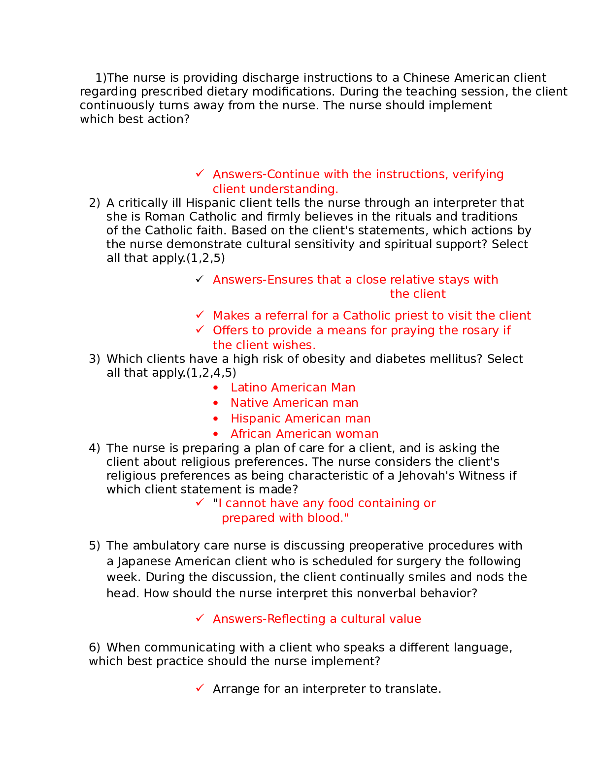
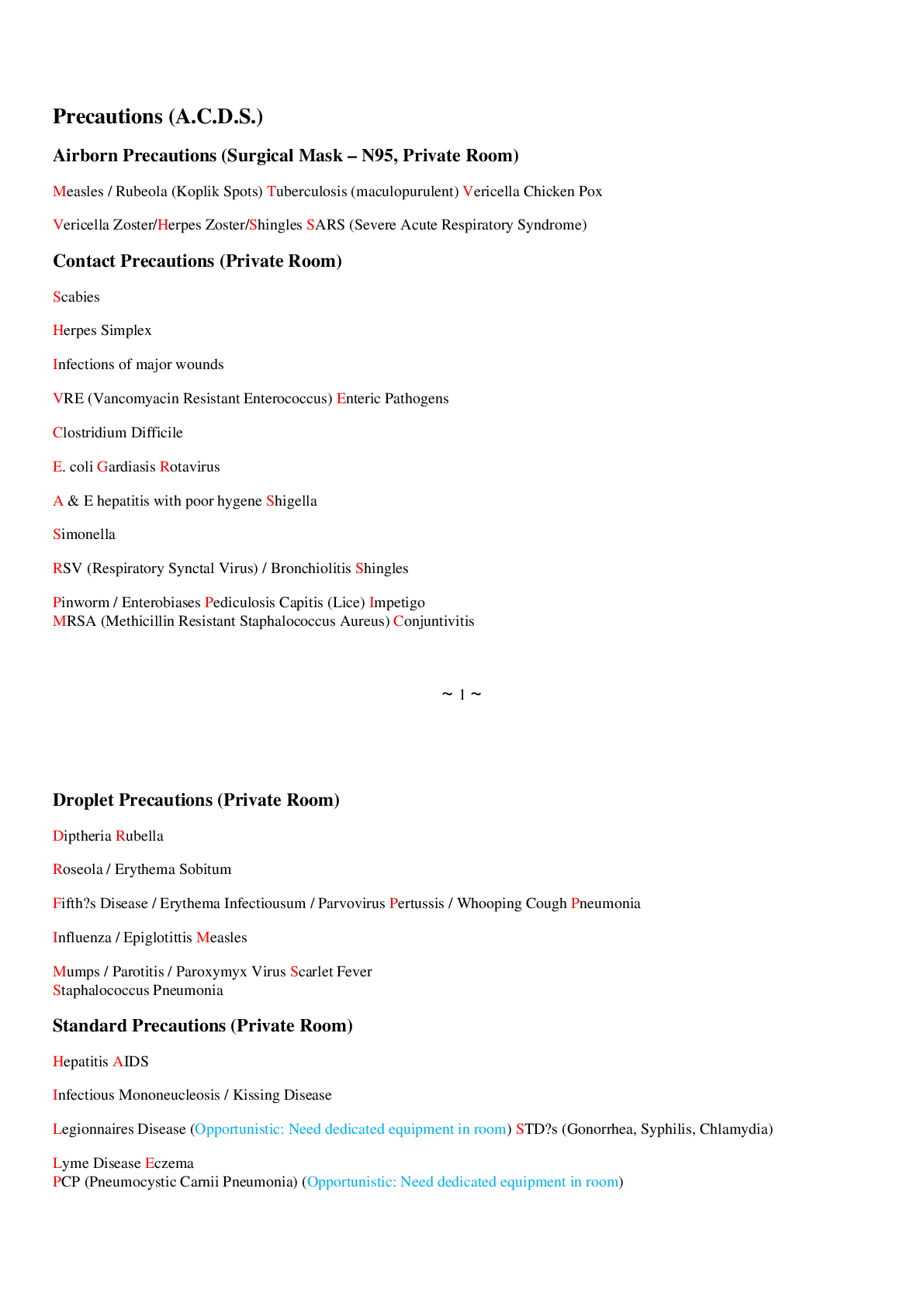
NUR 101 (NUR101) NCLEX Study Notes Precautions (A.C.D.S.) Precautions (A.C.D.S.) Airborn Precautions (Surgical Mask – N95, Private Room) Measles / Rubeola (Koplik Spots) Tuberculosis (maculopurulent... ) Vericella Chicken Pox Vericella Zoster/Herpes Zoster/Shingles SARS (Severe Acute Respiratory Syndrome) Contact Precautions (Private Room) Scabies Herpes Simplex Infections of major wounds VRE (Vancomyacin Resistant Enterococcus) Enteric Pathogens Clostridium Difficile E. coli Gardiasis Rotavirus A & E hepatitis with poor hygene Shigella Simonella RSV (Respiratory Synctal Virus) / Bronchiolitis Shingles Pinworm / Enterobiases Pediculosis Capitis (Lice) Impetigo MRSA (Methicillin Resistant Staphalococcus Aureus) Conjuntivitis Droplet Precautions (Private Room) Diptheria Rubella Roseola / Erythema Sobitum Fifth‟s Disease / Erythema Infectiousum / Parvovirus Pertussis / Whooping Cough Pneumonia Influenza / Epiglotittis Measles Mumps / Parotitis / Paroxymyx Virus Scarlet Fever Staphalococcus Pneumonia Standard Precautions (Private Room) Hepatitis AIDS Infectious Mononeucleosis / Kissing Disease Legionnaires Disease (Opportunistic: Need dedicated equipment in room) STD‟s (Gonorrhea, Syphilis, Chlamydia) Lyme Disease Eczema PCP (Pneumocystic Carnii Pneumonia) (Opportunistic: Need dedicated equipment in room) Psoriasis Tinea Capitis Karposi‟s Sarcoma (Opportunistic: Need dedicated equipment in room) Rocky Mountain Spotted Fever Airborn Precautions – Droplet organism very tiny capable of staying in air to infect others. 1. Private Room, negative pressure, vent outside of building, 6-12 air exchanges, UVLight, Door Closed 2. Wear N95 mask when entering room, particulate respirator mask, surgical mask. 3. When client leaves room client wears surgical mask. 4. Cohort only with same organism. 5. PPE when necessary. Contact Precautions – organism acquired by touching. 1. Private Room 2. Gloves & Gown when in contact with client 3. PPE when necessary. 4. Cohort only with same organism. Droplet Precautions – large droplet organism infects only within 3-6 feet. 1. Private Room, Door open, OK. 2. Wear mask when entering room. 3. Client wear mask when leaving room. 4. Cohort only with same organism. 5. PPE when necessary. Standard Precautions – promotes hand washing and use of PPE (eg mask, eye protection & gown) when appropriate for client. Apply to all blood and body fluids, non intact skin and mucus membranes. Use needless devices when appropriate, dispose of sharp instruments in puncture proof container. Don‟t recap dirty needles. Clean all blood spills with bleach. Antipsychotics / Neuroleptics End in “ine” A neuroleptic is used to treat schizophrenia Thorazine Haldol Inapsrine Risperdal Meldaril Olanzepine Stellazine, Seroquel, Serintil Sx: Anticholinergic – Dry symptoms, dry eyes, blurred vision, constipation, urinary retention. TE: Increase fluids, increase fiber, increase exercise. Can cause blood dyscrasia – sore throat, fever, malaise, bleeding. AE: Photosensitivity Orthostatic Hypotension Blood Dyscrasia Anticholinergic Galactorrhea EPS – Extra Pyramidal Symptoms Pseudo Parkinsonian Akathesia – inability to remain motionless (constantly moving) Dystonia – tortion or twisting of body parts. Tardive dyskinesia – tounge slapping, inability to perform voluntary muscle movements. EPS + Fever = NMS (Neuroleptic Malignant Syndrome) Causes are sudden decrease or change in THIRMOS drugs. Tx: Akineton Parlodel Artane (trihexyphenidyl HCl) Cogentin Kemadrin Anti Depressants Tricyclic Antidepressants Tofranil Anafranil Prozac – not a tricyclic Elivil Wellbutrin Zyban AE: Photosensitivity Orthostatic Hypotension Blood Dyscrasia Anticholinergic (most common) MAOI Parnate Nardil Marplan TE: Avoid foods rich in tyramine. Processed foods, cheese except cottage cheese, papayas, bananas, avocados, alcohol. Sx: Headache HTN Tachycardia N&V AE: Vomiting, Anorexia, Nausea, Diarrhea SSRI Paxil Prozac Serzone Zoloft Anti-alytic / Anti-anxiety Valium Ativan Librium Xanax / Alprazolam Anti-manic Lithium Given for Bipolar Disease AE: Vomiting Anorexia Nausea Diarrhea Tremors Ataxia Polyuria Tx: increase sodium, increase fluids, take oral contraceptives, do not use diuretics. TB Hepatotoxic Rifampin Inh – take with B6 to prevent peripheral neuritis. Pyrazinamide – PZA Elivil Streptomycin – (both nephro and ototoxic) Drugs that turn urine red/orange Dilantin Rifampin Macrodentin Pyridium PPD – Acid Fast Bacilli Test for TB has to be positive 3x. Check results 48-72 hours later. Wheal Induration Swelling Elevation Sx: TB Maculopurulent Sputum (bloody sputum) Anorexia Night Sweats Generalized Weakness / Fatigue Low grade fever Hepatotoxic Drugs Psychotics Anticoagulants* S anti-seizure TB Medications Acetamenophen / Tylenol L anti-Lipids Alcohol & Aventil Nifedipine Anticoagulants*: Fragman Aggrenox Ticlid Coumadin Heparin Integrilin Lovenox Dipyridamole Aspirin Plavix Signs of Liver Toxicity / Hepatotoxicity Jaundice Pururitis Pale colored stools Steatorrhea Dark colored urine Respiratory Drugs Anticholinergics – block parasympathetic nervous response. Atrovent / Inatropin Bromide SE: AE: Vomiting Anorexia Nausea Dizziness Tremors Tachycardia Restlessness Apprehension Irritablility Nervousness Beta Receptor Agonist “EROL” ending. Metaperenerol (Alupent) Albuterol (Proventil, Venteril) Levalbuterol (Xopenex) Terbutaline (Brethine) – given to pregnant women to delay labor. Broncho Dialators – give before ADL‟s Tx: activity induced asthma (xandine drugs) Aminophylline Theophylline (10-20 is therapeutic range), take with food. Glucocorticoids (inhalers) Beclamethasone Fluconasone TE: If no spacer then 1-2 inches from mouth. If spacer, then make sure they have a tight seal. Rinse mouth after each dose to prevent thrush. (Cushing Symptoms). Leukotriene inhibitors TE: Take daily dose at HS (Bedtime) Montelukast (Singulaire) Zafirlukast (Accolate) Mast Cell Stabilizer Cromylin Sodium (Intal) – Not effective during onset of asthma attack. Maintenance dose for COPD and Asthma. Patients with COPD need daily Peak Flow Rate. Green Zone 80-100% Yellow Zone 60-80% - pt needs to take meds within 2-3 hours then call M.D. Red Zone less than 60%, take meds then go to ER. HIV AIDS Viraimmune – take on time “Do Not Skip” AZT “Retrovir” take on empty stomach. Vivacept “use contraceptives” (causes birth deformities) Gancyclovier Acyclovir Zidovidine – “ZVD” to prevent neonate transmission. Given after 14 weeks gestation. IV during labor and in the form of syrup to neonate for 6 weeks after delivery. Patient can deliver natural childbirth but cannot breast feed. Patient cannot receive live vaccines (ex OPV, MMR) HIV Test to confirm infection: ELISA – Enzyme Linked ImmunoSorbent Assay – A single reactive result does not confirm alone. Need a second ELISA. Western Blot / IFA – Test for the presence of antibodies. CD4 (lymphocyst) count – Above 400 not concerned, Below 400 concerned. Viral load testing – measures the presences of HIV viral genetic material “RNA” TE: No fresh fruit No fresh flowers No raw meats Stay away from cat litter “toxoplasmosis” Stouvadine (D4T Zerit) is used for patients that don‟t respond / tolerate conventional therapy. AE: Peripheral Neuropathy, Monitor gait, Add paresthesia. Respiratory Ventilators – Causes of Ventilator Alarms Low Pressure Patient stops breathing spontaneously Disconnection or Leak Leak in the vent or patient airway cuff High Pressure Increased secretions or mucus plug Wheezing Endotracheal tube displacement H20 in the tube Kink in the tube Patient biting, coughing, or gagging on the tube. Anxiety or fighting vent. Modes of Ventilation: SIMV – Synchronized Intermittent Mandatory Ventillation – Allows patient to breathe on their own between ventilator breaths. (Ex. 8 breaths from patient, 8 breaths from vent). Used to wean patient off of ventilator. Assist Control – most commonly used mode. Ventilator is breathing for client if client does not initiate breath. PEEP Positive End Expiratory Pressure – to prevent closure of alveoli. Keep them open to prevent atelectasis. Controlled Ventilation (CV) – clients who are unable to initiate a breath . GB, TB, Polio, Total dependence on ventilator setting. Forms of O2 Masks: Non-rebreather mask – provides increased concentration of O2 90-100% on expiration. Bag does not deflate. Ventri Mask – delivers concentrated form of O2 40-60%. Used for short term emergencies. Renal System and Drugs Nephrotoxic Aminoglycosides “Nycin” Dye IV (angiogram) Antifungal Contraindicated in Renal Failure ACE Inhibitor – check creatinine Aldactone – check K+ MOM – check Mg+ Dialysate Solution Contents: 1. Albumen 2. Glucose 3. Insulin 4. Heparin 5. Electrolytes Renal Drugs 1. Colace (laxative) 2. Drugs to lower Phosphorus (↑ Calcium) Renagel (Sevelormer) Os-cal (Calcium Carbonate) – Take with meals. Phoslo (Calcium Acetate) – Take with meals. Aluminum Oxide (Amphogel) – Take with meals. Colace (Stool Softener 3. Drugs for Anemia a. Procrit b. Epoeiten (Epogen) c. Folic Acid d. Feosol (Iron) 4. To Prevent GI Bleeding a. H2 Blockers 5. Drugs for UTI / Cystitis a. Bactrim (Sulfa /TMT) b. Fluro-quinolone (Ofoxacin) i. Levofloxacin ii. Ciprofloxacin c. Macrodentin d. Pyridium 6. Drugs for ICP patients a. Mannitol b. Steroids with anti-ulcer c. Anti – Seizure meds (See 11 Neuro Drugs) 7. Drugs for Renal Transplant a. Steroids – for life b. Immunosuppressants i. Imuran (Cyclosporine) 8. Drugs for BPH (Benign Prosthetic Hyperplagia) a. Flomax (Tamsulosin) – Take with a full glass of water. b. San Palmetto / Saw Palmetto c. Alpha Receptor d. Proscar 9. Drugs contraindicated for BPH (Benign Prosthetic Hyperplagia) Patients a. Anticholinergics i. Atropine ii. Probantine / Ditropan b. Antihistamines (nasal decongestants) with pseudophedrine. Urinary Diversion Techniques Ileal Conduit No risk for fluid and electrolyte imbalance Continuous drainage Drain bag needed @ all times Stoma Care Koch Pouch Internal Ileal Conduit Self Catheter, bladder training Neobladder Nephrotomy Connected directly to kidney Continuous drainage In AM attach “saddle” bags, pouch attached to thigh. In PM drain into foley bag during HS. TPN Contents of TPN: Lipids Insulin Vitamins Electolytes Carbohydrates H20 Heparin Amino Acids Minerals Complications of TPN: 1. Air Emboli related to tubing / disconnection of tubing a. Tx: Clamp tubing, place on left side lying and call M.D. 2. Pneumothorax - Puncture from insertion of central line. 3. Infection a. Tx: To prevent use sterile dressing site change q 48h b. Solution IV tubing change q 24h 4. Hyperglycemia – Dry and Hot Give a Shot a. Causes i. Infusion of TPN too rapidly ii. Not enough insulin iii. Infection b. Tx: i. Slow infusion rate ii. Administer regular insulin 5. Hypoglycemia – Cold and Clammy Give Some Candy a. Causes i. Abrupt discontinuation or too much insulin b. Tx: i. ↓ flow of TPN ii. Run D10W iii. ↓ insulin 6. Hypervolemia – Fluid overload a. Tx: i. ↓ TPN Flow Rate ii. Administer diuretics Neurological Drugs Learn Neurological Disorders and their symptoms (Parkinson‟s, Guillian Barre, ALS, MS). 1. Mannitol 2. Steroids “Sone” 3. Antacids, PPI (Proton Pump Inhibitor), H2 Blocker, PGI (Prostiglandin Inhibitor) 4. Anti-seizure Meds – Cause blood dyscrasia (sore throat, fever, bleeding, malaise). a. Benzodiazepines i. Valium ii. Ativan iii. Librium iv. Xanax / Alprazolam v. Clonazepam b. Depakote (Valproic Acid) c. Carbamazepine (tegretol) d. Keppra e. Neurontin f. Dilantin (Phentoin) g. Lamictal 5. SCI (Spinal Cord Injury) drugs a. Stool Softener b. Muscle Relaxants (VALX) c. Steroids “Sone” 6. Anti-hypertensives – for autonomic dysreflexia a. Isosorbide Dinitrole b. Isosorbide Mononitrate c. Nitro (Paste) d. Nitro (Patch) 7. Antiviral (encephalitis) a. Acyclovir 8. Anticholinesterase (MG) a. Neostigmine b. Pyridostigmine c. Physostygmine d. Edrophonium Chloride (tensilon) – Test for MG. i. In MG, muscle strength will improve immediately after injection of tensilon. 9. Atropine Sulfate – cholinergic crisis 10. Anti Parkinson Drugs a. Levadopa b. Amantidine (Symmetrel) c. Carbidopa (Sinemet) d. Comtan e. Eldepryl 11. Anti EPS (Extra Pyrimidal Symptomes) a. Akinton b. Parlodel c. Artane (trihexyphenidyl HCl) d. Cogentin e. Kemadrin 12. Drugs for MS a. Steroids b. Muscle Relaxants i. Baclofen ii. Valium iii. Flexoril iv. Soma Neurological Disorders (GB, ALS, MG, MS, Parkinsons) Guillain-Barré Syndrome (GB) Acute infectious neuritis of the cranial and peripheral nerves. Recovery can take years Reversible NurDx: Impaired breathing pattern. Ascending paralysis (starts from the lower extremities and goes up) Sx: Paresthesia Weakness of the lower extremities Progressive weakness of the upper extremities and facial muscles Tx: Monitor respiratory status Monitor for autonomic dysreflexia Monitor for impaired mobility Monitor cardiac status Assess for gag reflex Avoid infection Plasmaphoresis, immunoglobulin Prepare to initiate respiratory support (02, ventilation, incentive spirometer). Amyotrophic Lateral Sclerosis (ALS) Lou Gehrig‟s Disease Progressive degeneration of the motor system that causes muscle weakness and atrophy. Irreversible NurDx: Impaired Respiratory Pattern Sx: Difficulty chewing Dysarthria Dysphagia Dysphonia Tongue Atrophy Weakness of the hands and feet Tx: Monitor respiratory status Monitor for autonomic dysreflexia Monitor for impaired mobility Monitor cardiac status Assess for gag reflex Avoid infection Plasmaphoresis, immunoglobulin Prepare to initiate respiratory support (02, ventilation, incentive spirometer). Myasthenia Gravis (MG) Not enough acetylcholine at the myoneural junction. Defect in the transmission of nerve impulses (Acetylcholine is the excitatory impulse). Dx: Impaired breathing r/t respiratory paralysis and failure. (Decending disease). Sx: Diplopia Dysphasia Difficulty Chewing Difficulty Breathing Diminished Breath Sounds Ptosis Weakness Weak Hoarse Voice Fatigue Tx: Monitor respiratory status Monitor for autonomic dysreflexia Monitor for impaired mobility Monitor cardiac status Assess for gag reflex Avoid infection Plasmaphoresis, immunoglobulin Prepare to initiate respiratory support (02, ventilation, incentive spirometer). Multiple Sclerosis(MS) Demyelenation of the neurons. Chronic progressive disease of the CNS. Sensory Motor loss. Dx: Potential For Injury Sx: Bladder, Bow, and Sexual Dysfunction Blurred Vision Decreased Sensory Perception (touch, pain, temp) Diplopia Dysphagia Emotional Changes (Depression, Euphoria, Apathy, Irritability) Fatigue Nystagmus Tremors, Ataxia Weakness Tx: Stationary exercise, swimming, cycling. Space exercise apart. ↑ Fluids before exercise. Bowel regimen. Parkinson‟s Disease Not enough dopamine at the receptor sites to inhibit the excitatory impulses. This results in a dysfunction of the Extrapyramidal System (EPS) and crippling disability. Dx: Potential for Injury Sx: Blank facial expression Bradykinesia Broad based gait Drooling Dysphagia Difficulty Swallowing Handwriting becomes smaller – micrographia Involuntary Tremors / Pill Rolling Tremors Monotonous Speech Muscle Rigidity Stooped shoulders / shuffling gait Walk with broad based gait Tx: Anti-parkinson drugs Levadopa Amantidine Carvedopa Eldepryl Comtan Ae: Confusion, Depression, Sleep Alteration Musculoskeletal Drugs 1. Herniated Intervertebrae a. Muscle Relaxants i. AE: Drowsiness /Sedation 1. Soma 2. Baclofen 3. Flexeril 4. VALX – (antialytics) b. Steroids “Sone” c. Pain Meds i. ASA (Aspirin) ii. NSAIDS iii. Narcotics 2. Osteoporosis a. Teaching ABCDEFGH i. Alcohol ii. Bone density iii. Calcium iv. D - Vitamin D v. Exercise vi. FACEC vii. Gain Weight b. Drugs i. Fosamax – take with full glass of H20. ii. Actonel iii. Calcitonin iv. Evista v. Calcium Carbonate vi. HRT ex Premarin c. If patient is at risk for Osteoporosis then take these medications i. Dilantin ii. Heparin iii. Lasix iv. Steroids v. Synthroid 3. Osteoarthritis Meds a. NSAIDS i. Feldene ii. Ibuprofen iii. Indomethacin iv. Naproxen b. Aspirin c. Steroids d. Muscle Relaxants 4. Gouty Arthritis a. Drugs i. Colchicine ii. Allopurinol b. AE: i. Vomiting ii. Anorexia c. Paget‟s Disease / Osteitis Deformus i. Fosamax ii. Actonel iii. Calcitonin 5. Fibromyalgia a. Antidepressants 6. SLE (Systemic Lupus Erythematosus) Meds a. Darvocet b. Tylenol 3 c. Oxycodone d. Fentanyl 7. Scleroderma a. Penicillamine b. Azathioprine c. Methotrexate 8. Polyarthritis Dodosa a. Steroids „Sone” 9. Rheumatoid Arthritis a. Sedimentation Rate is ↑ b. Tx i. Gold Salts ii. Monitor for Blood Dyscrasia – check CBC. Endocrine Drugs 1. Growth Hormone a. Somotropin 2. Drugs for Hyperthyroidism a. PTU (Propyl thiouracil) = blood dyscrasia b. Tapazole c. Beta Blockers (↓HR) i. Propanolol d. Sedatives - VALX e. KISS i. K – Potassium ii. Iodine iii. Saline iv. Solution - Lugol‟s Solution 3. Drugs for hypothyroidism a. Synthroid (Livothyroxin) b. Cytomel (Liothyronine T3) 4. Parathyroidectomy a. Calcium Gluconate at beside 5. Addison‟s Disease a. Prednisone b. Deltisone c. Dexamethasone d. Fluticasone e. Hydrocortisone f. Meythylprednisone Other Names a. Corticosteroids b. Glucocorticoids c. ACH Hormones d. Mineral Corticoids – contraindicated in patients with PUD (Peptic Ulcer Disease) /GI irritant. 6. Cushing‟s Disease – Diuretics a. Potassium sparring i. Spironolactone ii. Amiloride iii. Triamterene Gastrointestinal -GI 1. Upper and Lower GI Series a. Laxatives 2. EGD, ERCP a. Xylocaine Spray 3. Colonoscopy a. Laxative - Golytely (Mix 4L tap H20 with 1 glass every hour) 4. Cholecystography a. Iapanoic tabs (6 telepaque tabs. 1 tab q 5 min with full glass of water) 5. Liver Biopsy a. PASTALA 6. Drugs for peptic ulcer disease (PUD) a. Antacids – (Maalox /TUMS) b. H2 Blockers end in “tidine” i. Ramitidine ii. Cemetidine iii. Famitidine iv. Mizatidine c. Proton Pump Inhibitors (AE: VAND + Headache) end in “Prazole” PPI i. Pantaprazole ii. Omeprazole iii. Lansoprazole iv. Esomeprazole d. Proton Inhibitor PI i. Cytotec (Mysoprostol) – can cause abortion. e. Sucralfate (carafate) coats lining. f. Reglan (metoclopramide) – 30 min AC. 7. Drugs for Ulcerative Colitis a. Steroids b. Albumen c. Antidiarrheal (Lomotil, Immodium) 8. Drugs for Hemorrhoids a. Colace b. Metamucil – Drink with a full glass of water and follow up with another. c. Senokot 9. Cholecystitis a. Demerol (Avoid MS) 10. Liver Cirrhosis a. Vit K b. Portal Systemic Encephalpathy i. Neomycin Sulfate ii. Lactulose iii. Aldactone iv. Vitamin K v. Anti Pruritic (Benedryl) vi. Neomycin vii. Anti-emetics viii. Vitamin Supplements ix. Antacids Avoid PASTALAN and Sedatives / Narcotics 11. Pancreatitis – Do not give morphine. a. Antacids b. H2 Blockers c. PPI d. Prostiglandin Inhibitors PGI e. Demerol 12. Complications / Seizures a. Sedation / Anti-seizure i. Phenobarbitol – sedation / anti-seizure ii. Anti-anxiety iii. Mg MSO4 iv. B1 Thiamine b. Pancreatic Enzymes i. Viokase ii. Pancrease 13. Hepatitis Vaccination a. Immunoglobulin H2 Blockers ↓ HCL → Never take it together with Iron (Fe), antibiotics, antacids, give 2 hrs apart. 1. Zantac 2. Tagamet 3. Axid 4. Pepcid PPI ↓ HCL AE: VAND + Headache: coats lining of stomach (Sucralfate, Carafate). End with “Prazole”. 1. Pantoprazole 2. Omezprazole 3. Lansoprazole 4. Esomeprazole Contraindicated in PUD 1. NSAIDS – could cause ↑ bleeding a. Feldene b. Naprexyn c. Endomethacin d. Ibuprofen 2. Anti-coagulants 3. Steroids 4. Thermolytics Antacids Neutralize HCL, take 1-2 hours after meals 1. Maalox – never take together with Fe Antibiotics 2. Tums – H2 blocker, give 2 hours apart. Prostiglandins: Cytotec (Mysoprostol) can cause abortion. Vitamin B12 B12 (Cyanocobalamin) treats both Pernicious Anemia, Megaloblastic Anemia Patient with total gastrectomy are at risk for ↓ B12 and anemia. Give B12 injection IM q wk for 1 month then monthly for life. Dx: Schilling Test A Schilling test may be given in two parts. Part one measures the amount of vitamin B12 passed in urine after a known amount of the vitamin tagged with a radioactive substance is swallowed. If the intestines absorb vitamin B12 normally, a certain amount of the vitamin (up to 25% of the amount swallowed) will be passed in the urine. If the intestines cannot absorb the vitamin normally, very little or no vitamin B12 will be present in the urine. A Schilling test with abnormal results (no vitamin B12 in the urine) may be repeated after giving an oral dose of intrinsic factor and radioactive B12. This is called part two of the test, and it tells whether the vitamin deficiency is caused by a lack of intrinsic factor or from a problem with the intestines. Why It Is Done The Schilling test is done to: Determine the cause of a low level of vitamin B12. Check for vitamin B12 deficiency anemia in people at high risk for developing this anemia, such as those who have had stomach or intestinal surgery, small intestine problems, or people with a family history of this anemia. Help diagnose pernicious anemia, a serious blood disease caused by a lack of intrinsic factor. Risk Factors for Colorectal Cancer 1. ↑ in age over 40. 2. Family Hx of polyps 3. Previous colon cancer diagnosis 4. Hx of IBS (Irritable Bowel Syndrome) 5. Increase fat, protein, and ETOH intake. No Beef. PUD / Gastric Ulcer Disease – is aggravated by food. Sx: weight loss, patient eats small frequent meals DUO / Duodenal Ulcer – food relieves pain. Sx: Gain weight, pain at night. Gerd and Hiatal Hernia – minimize liquid intake, no eating or drinking 2 hours before bedtime, decrease fat intake, increase fiber, avoid tobacco, caffeine, carbonated beverages. No tight close, elevate HOB 6-8 inches (don‟t lie down after eating.) Colon Ascending Colon: Ileostomy – liquid stools, no irrigation needed. Transverse Colon: Semi-formed stool Descending Colon: Colonoscopy / sigmoid colon Formed Stool Give Yogurt Dumping syndrome – physiologic response to rapid emptying of gastric contents into the jejunum. Occurs in patients who have had partial gastrectomy and gastrojejunostomy. Sx: Nausea Weakness Sweating Palpitations Tachycardia Syncope Diarrhea Preventing Dumping Syndrome in Tube Feeding 1. Slow the formula rate to provide time for the carbohydrates and electrolytes to be diluted. 2. Administer feedings at room temperature. 3. Continuous drip if tolerated instead of bolus feeding. 4. Semi-Fowlers position for 1 hour after feeding. 5. Flush with minimal amount of water possible before and after the feeding. Diverticulitis Diverticulosis – multiple outpouching of the lining of the bowel that extends through a defect in the muscle layer and are without inflammation or symptoms. Diverticulitis – when food and bacteria are retained in the outpouchings and cause inflammation or infection and impede drainage and lead to perforation or abscess formation. Skip Lesions – if present then it is Crohn‟s Disease and not Diverticulitis. Asterixis – abnormal muscle tremor consisting of involuntary muscle movements in the hands and sometimes the feet and tongue. Usually found in patients with liver disease. Portal HTN – increased pressure in the portal vein caused by blockage of blood flow through the liver. Portal HTN is found in diseases such as cirrhosis, which is causes ascities, splenomegaly, and verices. Hepatic Encephalopathy – liver cannot process protein and causes ammonia levels to rise. Suction Chamber PLEUR-EVAC has 3 chambers ↓TO WALL↓ Suction Chamber Water Seal Chamber Drainage Chamber ↑TO PATIENT↑ H20 Should bubble constantly. If it is not bubbling then the lung has re-expanded (good). 10 – 20 cm H20 that fluctuates with respirations. If it is not fluctuating or tidaling then the lung has re- expanded (good). Not expected: constant bubbling (air leak). Mark at beginning of shift. Should be filling up (more at end of shift than at beginning). 100mL/hr. Serosangenous drainage. Do not empty, when it is full you replace the whole unit. Need to have a vaso-occlusive dressing at the bedside. It is usually a vaseline gauze dressing. If tube is dislodged from patient, cover it with vaseline gauze and call Dr. If tube is dislodged from wall. Put it in sterile H20 to clean it before reconnecting it. If water is not fluctuating and bubbling then the lung has re-expanded. Keep patient in semi-fowlers position to permit air and H20 in the pleural space. EENT Drugs 1. Eye Disorders a. Cataract i. Mydriatics (dilators) 1. Atropine sulfate ii. Colace b. Glaucoma i. Miotics (Constricts Pupil) 1. Pilocarpine (outflow of aqueous humor) ii. Carbonic Anhydrase Inhibitor 1. Diamox (↓ Production of AH) iii. Beta Blocker 1. Timola (↓ IOP ↓ Production of AH) iv. Xalatan (latanoprost ↓ IOP) – take once a day ↑ outflow of AH. Avoid: Anticholinergics, Nasal Decongestants, Anti-histamines. 2. Ear Disorders: a. Meniere‟s Disease i. Antivert (meclizine) Avoid: Ototoxic drugs like aminoglycosides “Nycin”, Lasix, Aspirin. 3. Nose Disorders a. Allergic Rhinitis i. Loratidine ii. Allegra iii. Steroids b. Epistaxis – Epistaxis Derma 4. Burns a. Silver Nitrate b. Marfedine (sulfamylon) 5. Decubitus Ulcers a. Dressing i. Hydrocolloid ii. Hydrophyllic (duodenal) 6. Cancer a. Chemotherapy i. Antiemetic 1. Zofran (odansetron) ii. Multiple Myeloma 1. Allopurinol EYES Cataract – Opacity of lens. Assessment: Blurred vision Decreased Color Persistent vision is better in dim light. Tx: Mydriatics / Dilate Eye Cataract Surgery on one eye at a time. Glaucoma – it is a medical emergency. Cannot be repaired can only be slowed. Tonometer 10-20 normal. ↑ IOP because of decreased drainage of aqueous humor. Increased production of aqueous humor. Assessment: Tunnel vision Loss of peripheral vision (Can see center objects, cannot see outside of periphery). Halo‟s in bright light Vision is worse in the afternoon. Tx: Miotics - Constrict Retinal Detachment – Layer of retina separates Assessment: See floaters, flashes of light, curtains being drawn. Loss of portion of visual field. Tx: Patch both eyes, Bed rest, Decrease IOP, Do not sneeze cough or strain to upset the eye. Penetrating objects of the eye – Cover eye and go to E.R. Chemical burn of eye – flush with water for 15-20 min. Cardiac Medications 1. Stress Test – Physical / Chemical a. Persantine b. Adenosine c. Dobutamine 2. Cardiac Arrythmias / Dysrithmias a. If symptomatic give the following and check K+. i. Atropine ii. Digoxin iii. Anticoagulants (FATCHILDAP) iv. Beta-Adrenergic Agonist Atropine, Digoxin, Anticoagulants, and Beta-Adrenergic Agonist are contraindicated in BPH and Glaucoma b. Amiodarone (Cordarone) – v tach, PVC‟s, V-fib. 3. CAD (Coronary Artery Disease) a. Anti-lipidemics “Statin” ↓ LDL, give at night. i. Rosovastatin ii. Atrovastatin iii. Lovastatin iv. Pravastatin v. Fluvastatin vi. Simvastatin b. Vasodilators – Nitrates ↓ BP i. Nitroglycerin – Patch ii. Nitroglycerin – Paste iii. Isosorbide – Mononitrate iv. Isosorbide – Dinitrate c. Anti-hypertensives – (See Neuro Drugs) d. Calcium channel blocker “dipine” i. Amlodipine ii. Dilitazem (cardizem) iii. Felodipine iv. Nifedipine v. Nicardipine e. Anti-coagulants (FATCHILDAP) i. Contraindicated in Peptic Ulcer Disease (PUD) or hx of bleeding. 4. Angina a. Nitroglycerine – vasodilate b. Beta-blocker “olol” c. Anti-coagulants 5. MI a. Tx: 1. Morphine 2. Oxygen 3. Nitrates 4. Aspirin 5. Thrombolytics 6. Heparin 7. Beta-blockers b. Anti-anxiety 6. Heart Failure a. Diuretics b. Digoxin c. Morphine Sulfate d. Inotropics - ↑ BP ↑ Contraction i. Dobutamine ii. Dopamine 7. Cardiogenic Shock – Cardiac Tamponade a. MSO4 b. Digoxin c. Inotropics (Dobutamine, Dopamine) d. Diuretics e. Vasodilators 8. Pericarditis a. Antibiotics b. Anti-inflammatory – Steroids 9. Endocarditis a. Antibiotics – (PCN) Penicillin 10. Valvular Disease a. Beta-blockers b. Digoxin 11. Cardiomyopathy – hospice a. Digoxin b. Lasix c. Diuretic 12. Raynauld‟s Disease, PAD a. Anti-platelet i. Trental (Pentoxifylline) ii. Pletal (Cilostazol) 13. Buerger‟s Disease / Thromboangitis Obliterans a. Peripheral Vasodilators i. Trental ii. Pletal 14. AAA a. Anti-hypertensives b. Anti-coagulants 15. Hypertensive Crisis ↓ BP ↑ Urinary Output, change positions slowly a. Nipride – wrap in dark foil b. Nitroglycerin IV Anti-hypertensives – Cardiac Continued – Will cause ↓ BP, dizziness, lightheadedness, syncope. 1. Alpha Adrenergic “Zosin” a. Doxazosin b. Prazosin c. Terazosin 2. ACE Inhibitors “Pril” (angiotensin Converting enzyme inhibitors). Low K+, check creatinine, persistent cough. a. Catopril (Capoten) b. Benzepril (Lotensin) c. Enalapril (Vasotec) d. Fosinopril (Monopril) e. Lisinopril (Prinvil / Zesteril) f. Quinapril (Accupril) g. Ramipril (Altace) 3. ARB (Angiotensin Receptor Blocker) “sartan” a. Volsartan b. Olmesartan c. Candisartan d. Losartan 4. Beta Blocker “olol” ↓ Heart Rate a. Metoprolol b. Acebutolol c. Labetalol d. Atenolol e. Nadolol (Corgard) f. Timolol g. Sotalol h. Carvedilol (Coreg) i. Propranolol 5. Calcium Channel Blockers “Dipines” a. Amlodipine (Norvasc) b. Diltiazem (Cardizem) c. Felodipine (Plendil) d. Nifedipene (Procardia) e. Nicardipine (Cardene) f. Verapamil (SE: Constipation) 6. Diuretics a. Loop ↑ urinary output (eat bannana‟s oranges, cantaloupe) i. Bumetanide (Bumex) ii. Furosemide (Lasix) iii. Torsenanimide (Demadex) b. Osmotic (tx: cerebral edema; check BP and K+) i. Mannitol (Osmitrol) c. Carbonic Anydrase Inhibitor i. Acetazolemide (Diamox); Tx: IOP, Glaucoma d. Potassium Sparing i. Spironolactone (Aldactone) ii. Amiloride iii. Triamterene e. Thiazides – Assess for sulfa allergy i. Chlorothiazide (Diuril) ii. Hydrochlorothiazide (Hydrodiuril) f. Zaroxolyn (Metolazone) causes ↓ SOB 7. Sympatholytics a. Clonidine (Catapress) b. Methyldopa (Aldomet) c. Hydrolizene 8. Peripheral Vasodilators & Anti-platelet a. Trental (Pentoxyfilline) b. Pletal (Cilostazol) ↑ microcirculation and tissue perfusion; antiplatelet. Source of Vitamins – Vitamin Enriched Foods 1. Vitamin B1 = Thiamin = Energy a. Thiamin is given to : i. Diabetics ii. Alcoholics b. Foods Rich in Thiamin i. Tomatoes ii. Tuna iii. Eggplant iv. Asparagus v. Mushrooms vi. Sunflower vii. Spinach viii. Romaine Lettuce ix. Green Peas x. Brussel Sprouts xi. Pork xii. Nuts xiii. Whole Grain xiv. Legumes 2. Vitamin B6 (Pyridoxine) – given to patients with TB specifically taking Inh because of peripheral neuropathy or neuritis. a. Meat b. Poultry c. Fish d. Corn e. Yeast 3. Vitamin B8 (Biotin). Lower digestive tract utilization of proteins folic acid and B12. a. Whole Brown Rice b. Wheat Germ c. Legumes d. Egg Yolk e. Sprouted Seeds f. Cauliflower g. Fruits h. Nuts 4. Vitamin B9 = Folic Acid ; given to a. Prenatal Moms to prevent neural tube defects b. Sickle Cell Anemia patients c. Hemophiliacs 5. Vitamin B12 (Cobalamin) – Given to patients that undergo gastrectomy. IM for life. a. Liver b. Organ Meats c. Poultry d. Dried Beans e. Egg Yolk f. Brewers Yeast g. Milk h. Nuts i. Green Leafy Vegetables j. Citrus Fruits 6. Billroth I / Gastroduodenostomy – Anastomosis of the upper portion of the stomach to the duodenum. 7. Billroth II / Gastroduodenostomy – A connection usually constructed surgically between the stomach and the jejunum. Diabetic Medications 1. Alpha glucoside inhibitors – delays absorption of carbohydrates & digestion. a. Glycel (Milgital) – take with milk b. Acarboose Milgital – take with meals or first bite. 2. Biguanides ↓ Promote insulin a. Glucophage (Metformin) – take with meals BID. Hold 48 hours prior to angiogram and surgery (Risk for metabolic acidosis) i. Check Creatinine ii. AE: VAND 3. Sulfonylurent – take once a day; no alcohol (Disulfiram like reaction if ETOH) a. Glyburide b. Amaryl c. Glipizide 4. Thiazoline diones - ↓ insulin resistance in muscle a. Actose – hepatoxic b. Avandia (Golitazam) 5. Meglitinides a. Prandin – 1-30 mins before meals b. Starlix – with meals (rapid onset of medication). Can cause patient to be hypoglycemic – 15gm of carbs. Calcium & Magnesium 1. Hypocalcemia and Hypomagnesemia both have - 3T‟s a. Tetany b. Trousseau‟s Sign (BP cuff) c. Chvostek‟s Sign (twitches) 2. Hypercalcemia Symptoms (normal 8.5 – 10) a. Bone pain b. Back/ Joint/ Flank c. Constipation d. Renal Stones e. Fractures 3. Hypomagnesemia a. Tall T-waves b. Tachycardia c. Hypertension d. Decreased bowel sounds e. Anorexia f. Shallow respirations g. twitching 4. Hypermagnesemia (normal 1.6 – 2.6) a. Neurological depression b. Drowsiness – Lethargy c. Loss of Deep Tendon Reflex d. Respitory Insufficiency e. Bradycardia f. Hypotension 5. Magnesium Toxicity – (Greater than 2.6) a. Blood pressure < 90/60 b. Urine Output < 30 mL/hr c. Respiratory Rate < 12 min d. Reflex (O +/-) Deep Tendon Reflex e. Pulmonary Edema (Crackles with fever) Drugs and their Antidotes 1. Acetaminophen - Acetylcycteine 2. Benzodiazepine - Flumazenil 3. Coumadin - Vitamin K 4. Curare - Tensilon 5. Cyanide poisoning - Methylene Blue 6. Digitalis - Digibind 7. Ethylene poisoning - Antizol 8. Heparin - Protamine Sulfate 9. Iron - Desferal 10. Lead - Edetate Disodium (EDTA), Dimercaprol (BAL), Succimer (CHEMET) 11. Lovenox - Protamin Sulfate 12. Magnesium sulfate - Calcium Gluconate 13. Morphine sulfate - Naloxone Hydrochloride 14. Methotrexate - Leucovorine 15. Mestinon - Atropine Sulfate 16. Neostigmine - Pralidoxime Chloride (PAM) 17. Penicillin - Epinephrine Drugs which are best TAKEN ON AN EMPTY STOMACH 1. Ampicin (Ampicillin) 2. Chloromycetin 3. Erythrocin 4. Ferrous Sulfate 5. Inh 6. Isordil 7. Penicillin 8. Rifadin Drugs which are best TAKEN BEFORE MEALS 1. Atropine Sulfate 2. Bactrim 3. Dalmane 4. Insulin 5. Mestinon 6. Valium Drugs which are best TAKEN AFTER MEALS 1. Artane (trihexyphenidyl HCl) 2. Cogentin 3. Clozaril 4. Deltasone 5. Elavil 6. Haldol 7. Lithium 8. MAOI 9. Nardil 10. Pyridium 11. Ritalin 12. Streptomycin 13. Thorazine 14. Tofranil Normal Lab Values Lab Test Value Low Meaning High Meaning Acid Phosphatase 11 - .60 U/L Alkaline Phosphatase 30 – 85 ImU/mL Ammonia Level 15 -110 ug./dL Amylase 56 – 190 Anti-streptolysin O (ASO) Titer ≤ 160 Todd Units /mL Arterial oxygen pressure (PaO2) 80-100 mm Hg Bicarbonate (Plasma) 22 -26 mEq/L Bilirubin direct 0 – 0.3 mg /dL Bilirubin Indirect 0.1 – 1 mg /dL Bilirubin Total < 1.5 mg/dL Bleeding time 1-9 minutes Blood Urea Nitrogen (BUN) 10 – 20 mg /dL Calcium (Ca) 9 – 10.5 mg /dL Chloride (Cl) 90 – 110 mEq /L Cholesterol Level < 200 mg /L Cortisol level 8am 6 -28 ug/dL Creatinine 0.5 – 1.5 mg /dL Muscle Damage Renal Disease Creatinine phosphokinase (CPK) Male: 12–70 U/mL Female: 10–55 U/mL Erythrocyte Sedimentation Rate (ESR) Male: < 15 mm/hr Female: < 20 mm/hr Decreased RBC‟s, Sickle Cell Folate 5-20 ug/mL Liver Disease Pernicious Anemia Glucose level (GTT) 70 – 115 Hematocrit (Hct) Male: < 47% Female: < 42% Anemia Poycythemia Hepatitis B surface antigen (HBsAg) Negative High Density Lippoprotein > 40 mg/dL Human Growth Hormone (HGH) Male: < 5 ug/L Female: < 10 ug/L Immunoglobulin E (Ig E) < .55 mg /dL Immunoglobulin M (Ig M) 55-375 mg /dL Ketosteroids Male: 7-25 mg /24hr Female: 4-15 mg/24hr LDH 45 -90 U/L Lead < 20 ug/dL Low Density Lipoprotein (LDL) < 100 mg/dL Myoglobin 0 – 85 ng/mL Magnesium 1.6 – 2.6 Hypotension Hypertension, loss of deep tendon reflex Oxygen saturation of arterial blood (SaO2) 95-98% Partial Thromboplastin Time (Coumadin) 60-70 seconds PCO2 35-45 mm Hg Percent Hydrogen (ph, blood) 7.35-7.45 Phenylalanine level < 2 mg /dL Phosphorus 3 – 4.5 mg /dL Platelet Count 150,000 – 450,000 Potassium (K+) 3.5-5.0 mEq/L Protein (Urine) 6.8-8.3 g/dL Prothrombin Time (PT) Male: 4.7 – 6.1 M/cu Female: 4.2 – 5.4 M/cu Sodium (Blood) 135 – 145 mEq/L SGOT/AST 10–50 IU/L or 8-20 U/L SGPT/ALT 5-35 IU/L or 8-20 U/L T3 (Triiodo Thyronine) 75-220 ng/dL T4 (Thyroxine) 4-11 ug/dL Troponin < 0.6 ng/mL Urine Osmolality 50 -1400 mOsm/kg Urine Specific Gravity 1.005 – 1.025 Edema, overhydration Dehydration Uric Acid Level 250 – 750 ml/24hr White Blood Cells (WBC) 5,000 – 10,000 Immunosuppressed Infection Adrenal Disorders Addison’s Disease and Cushing’s Syndrome / Disease Addison‟s Disease Addison‟s disease is failure of the adrenal glands to produce sufficient amount of steroids. The body does not make enough glucocorticoids and mineralcorticoids. This is caused by tuberculosis, cytomegalovirus, and histoplasmosis. Addisonian Crisis is caused by (S.I.T.S.) Stress, Infection, Trauma, and Surgery. Assessment: headache, severe abdominal, leg and lower back pain, generalized weakness, irritability and confusion, severe hypotension, shock. TX: Corticosteroids like Prenisone DX: Cosyntropin Stimulation Test is used to confirm Addison‟s Disease. Cushing‟s Syndrome / Disease Cushings Syndrome is a result of excessive glucocorticoid exposure. Usually as a result of pharmacological treatment of RAD or arthritis. Cushing‟s Disease is caused by pituitary or adrenal adenomas or excessive production of ACTH (adrenal corticotropic hormone). SX: Moon Face, weight gain, DM symptoms polydipsia, polyphagia, and polyuria because glucocorticoids oppose the action of insulin. TX: Reduce the amount of steroid or administer every other day. Perform surgery to remove the responsible adenoma. Diet ↑ protein and potassium ↓ calories, carbohydrates, and sodium. Pituitary Disorders – Diabetes Insipidus and Syndrome of Inappropriate Anti-diuretic Hormone DI – Diabetes Insipidus DI is caused by failure of the posterior pituitary to secrete ADH. It is treated with synthetic vasopressin. It is usually caused by hypothalamic injury (brain trauma or neurosurgery) or by drugs (lithium or demeclocyclene). This results in polyuria that is caused by either inadequate amount of ADH (hypothalamic DI) or failure of the kidneys to respond to ADH (nephritic DI). Urine is dilute and between 5-10 liters per day. Urine specific gravity is below 1.005. P.U.S.H up ↑ (Plasma Osmolarity, Urine Output, Serum Sodium, Hematocrit) = Dehydration. SIADH – Syndrome of Inappropriate Anti-diuretic Hormone Syndrome of increased ADH activity despite of reduced plasma osmolarity. Usually indicated by hyponatremia and it is associated with disorders of the central nervous system, various tumors, anxiety, pain, pneumonia, and drugs. P.U.S.H. down ↓ (Plasma Osmolarity, Urine Output, Serum Sodium, Hematocrit) = Overhydration. Arterial Blood Gases ABG Chart Uncompensated NORMAL RANGE Compensated Uncompensated Acidosis (acid) Normal pH Alkalosis (base) pH Acidosis 7.35 7.40 7.45 Alkalosis PC02 (Respiratory) Acidosis 45 40 35 Alkalosis HC03 (Metabolic) Acidosis 21-24 - 25-28 Alkalosis The pH determines the first name of either (compensated or uncompensated) and the last name of either (acidosis, or alkalosis). The PC02 and HCO3 determine the middle name of either (respiratory or metabolic). If the PC02 name matches the pH last name then it is Respiratory. If the HC03 name matches the pH last name then it is Metabolic. If the pH is normal range then it is Complete Compensation. If pH is not normal and either the pC02 or HC03 are normal range then there is NO Compensation. Practice: pH of 7.18, PC02 of 68, and HC03 of 25 = Uncompensated Respiratory Acidosis. pH of 7.51, PC02 of 40, and HC03 of 30 = Uncompensated Metabolic Alkalosis pH of 7.18, PC02 of 85, and HC03 of 24 = Uncompensated Respiratory Acidosis pH of 7.36, PC02 of 49, and HC03 of 28 = Compensated Respiratory Acidosis pH of 7.19, PC02 of 82, and HC03 of 10 = Respiratory and Metabolic Acidosis (Mixed Acidosis because both PC02 and HC03 names match the pH last name). Common Causes: Respiratory Acidosis Due to respiratory depression (drugs, CNS trauma), pulmonary disease (pneumonia COPD), respiratory hyperventilation. Respiratory Alkalosis Due to hyperventilation (emotions, pain). Metabolic Acidosis Due to diabetes, shock, renal failure, intestinal fistula, Diarrhea (ASSidosis). Metabolic Alkalosis Due to sodium bicarbonate overdose (TUMS, antacids), prolonged vomiting, nasogastric drainage. When pt is intubated PaCO2 should be 50 or greater and PaO2 should be less than 50. Fetal Monitoring 1. Description a. The fetal monitor displays the fetal heart rate (FHR). b. The device monitors uterine activity. c. The monitor assesses frequency, duration, and intensity of contractions. d. The monitor assesses FHR in relation to maternal contractions. e. Baseline FHR is measured between contractions; the normal FHR at term is 120 to 160 beats per minute. 2. External Fetal Monitoring a. External fetal monitoring is noninvasive and is performed using a tocotransducer or Doppler ultrasonic transducer. b. Perform Leopold‟s maneuvers to determine on which side the fetal back is located, and place the ultrasound transducer over this area (fasten with a belt). c. Place the toctransducer over the fundus of the uterus where contractions feel the strongest (fasten with a belt). d. Allow the client to assume a comfortable position, avoiding vena cava compression. 3. Internal fetal monitoring a. Internal fetal monitoring is invasive and requires rupturing of the membranes and attaching and electrode to the presenting part of the fetus. b. Mother must be dilated 2 to 3 cm to perform internal monitoring. 4. Periodic patterns in the FHR a. Fetal bradycardia and tachycardia i. Bradycardia: The FHR is less than 120 beats per minutes for 10 minutes or more. ii. Tachycardia: The FHR is greater than 160 beats per minute for 10 minutes or more. iii. Change position of the mother and administer oxygen. iv. Notify the physician. b. Variability i. Absent variability: undetected variability. ii. Minimal variability: greater than undetected but not more than 5 beats per minute. iii. Moderate variability: fetal heart rate fluctuations from 6 to 25 beats per minute. iv. Marked variability: fetal heart rate fluctuations greater than 25 beats per minute. v. Fluctuations in the baseline FHR may include irregular fluctuations of 2 cycles per minute or greater. vi. Decreased variability can result from fetal hypoxemia, acidosis, or certain medications. vii. A temporary decrease in variability can occur when the fetus is in a sleep state (sleep states do not usually last longer than 30 minutes). c. Accelerations i. Accelerations are brief, temporary increases in the FHR of at least 15 beats greater than the baseline and lasting at least 15 seconds. ii. Accelerations usually are a reassuring sign, reflecting a responsive, nonacidotic fetus. iii. Acceleration usually occur with fetal movement. iv. Accelerations may be nonperiodic (having no relation to contractions) or periodic. v. Accelerations may occur with uterine contractions, vaginal examinations, or mild cord compression, or when the fetus is in a breech presentation. d. Early decelerations i. Early decelerations are decreases in FHR below baseline; the rate at thelowest point of the deceleration usually remains greater than 100 beats per minute. ii. Early decelerations occur during contractions as the fetal head is pressed against the woman‟s pelvis or soft tissues, such as the cervix, and return to the baseline FHR by the end of the contraction. iii. Tracing shows a uniform shape and mirror image of uterine contractions. iv. Early decelerations are not associated with fetal compromise and require no intervention. e. Late decelerations i. Late decelerations are nonreasuring patterns that reflect impaired placental exchange or uteroplacental insufficiency. ii. The patterns look similar to early deceleration but begin well after the contraction begins and return to baseline after the contraction ends. iii. The degree of fall in the heart rate from baseline is not related to the amount of uteroplacental insufficiency. iv. Interventions include improving placental blood flow and fetal oxygenation. f. Variable decelerations i. Variable decelerations are caused by conditions that restrict flow through the umbilical cord. ii. Variable decelerations do not have the uniform appearance of early and late decelerations. iii. Their shape, duration, and degree of fall below baseline heart rate are variable; they fall and rise abruptly with the onset and relief of cord compression. iv. Variable decelerations also may be nonperiodic, occurring at time unrelated to contractions. v. One considers baseline rate and variability when evaluating variable decelerations. vi. Variable decelerations are significant when the FHR repeatedly decreases to less than 70 beats per minute and persist at that level for at least 60 seconds before returning to the baseline. g. Hypertonic uterine activity i. Assessment of uterine activity includes frequency, duration, intensity of the contractions, and uterine resting tone. ii. The uterus should relax between contractions for 60 seconds or longer. iii. Uterine contraction intensity is about 50 to 75 mm Hg (with the intrauterine uterine catheter) during labor and may reach 110 mm Hg with pushing during the second stage. iv. The average resting tone is 5 to 15 mm Hg. v. In hypertonic uterine activity the uterine resting tone between contractions is high, reducing uterine blood flow and decreasing fetal oxygen supply. h. Interventions for nonreassuring patterns i. Identify the cause (assess for cord prolapsed). ii. Discontinue oxytocin (pitocin) if infusing as prescribed. iii. Change the mothers position (avoid the supine position for patterns associated with cord compression). iv. Administer oxygen by face mask a 8 to 10L per minute. v. Increase intravenous (IV) fluids as prescribed. vi. Notify the physician or nurse midwife as soon as possible. vii. Prepare to initiate continuous electronic fetal monitoring with internal devices if not contraindicated. viii. Prepare to obtain a fetal scalp pH monitor to determine a blood pH value. ix. Prepare for cesarean delivery if necessary. i. Nonreassuring Patterns i. Tachycardia ii. Bradycardia iii. Decreased or absent variability iv. Late decelerations v. Variable decelerations falling to less than 70 beats per minutes for longer than 60 seconds. vi. Prolonged decelerations vii. Hypertonic uterine activity Stages of Labor 1. Stage 1 – Stage 1 is cervical dilation from 0 – 10 cm in three phases: latent, active, and transition. a. Latent Phase i. Cervical dilation is 1 to 4 cm. ii. Uterine contractions occur every 15 to 30 minutes and are 15 to 30 seconds in duration and of mild intensity. iii. Offer fluids and ice chips iv. Encourage voiding every 1 to 2 hours. b. Active Phase i. Cervical dilation is 4 to 7 cm. ii. Uterine contractions occur every 3 to 5 minutes and are 30 to 60 seconds in duration and of moderate intensity. iii. Encourage maintenance of effective breathing patterns. iv. Promote comfort with backrubs, sacral pressure, pillow support, and position changes. v. Offer fluids and ice chips vi. Encourage voiding every 1 to 2 hours. c. Transition Phase i. Cervical Dilation is 8 to 10 cm. ii. Uterine contractions occur every 2 to 3 minutes and are 45 to 90 seconds in duration and strong intensity. iii. Offer fluids and ice chips iv. Encourage voiding every 1 to 2 hours. 2. Stage 2 – Stage 2 is from complete dilation (10cm) to birth of the baby. a. Cervial dilation is complete b. Monitor fetal and mother vital signs c. Assist with positioning d. Prepare for birth of the baby. 3. Stage 3 – Stage 3 is delivery of the placenta to 1 hour after delivery of the baby. a. Placenta is delivered between 5 and 30 minutes after the birth of the baby. b. Shultze mechanism: Center portion of placenta separates first, and its shiny fetal surface emerges from the vagina. c. Duncan mechanism: Margin of placenta separates, and the dull, red, rough maternal surface emerges from the vagina first. d. Assess uterine status and maternal vital signs. e. Following birth of the placenta, uterine fundus remains firm and is located 2 finger breadths below the umbilicus. f. Examine placenta for cotyledons and membranes to verify that it is intact. g. Assess mother for shivering and provide warmth. h. Promote parental-neonatal attachment. 4. Stage 4 – Period of time from 1 – 4 hours after delivery of the baby. a. Blood pressure returns to prelabor level. b. Fundus remains contracted, in the midline, 1 to 2 fingerbreadths below the umbilicus. c. Lochia is moderate scant and red. d. Perform maternal assessments every 15 minutes for 1 hour, every 30 minutes for 1 hour, and hourly for 2 hours. e. Provide warm blankets f. Apply ice packs to perineum. g. Massage the uterus if needed and teach the mother to massage the uterus. h. Provide breast-feeding support as needed. AUTONOMIC NERVOUS SYSTEM Consists of the Sympathetic and Parasympathetic Nerves. Both impulses control internal organs and both are regulated by impulses from the hypothalamus and other parts of the brain. Cardiac muscle, smooth muscle, and glandular epithelial tissue receive impulses only via the autonomic nervous system. Sympathetic (synonyms) Adrenergic Sympathomimetic Anticholinergic Vagalytic Parasympathetic (synonyms) Cholinergic Sympatholytic Antiadrenergic Vagametic Sympathetic Parasympathetic Stimulates Stimulates most systems; Inhibits GI and Urinary Inhibits most systems; Stimulates GI and Urinary Main Function Fight or Flight response; mobilizes reserves; prepares body to meet emergencies. Repair response; promote vegative function; SLUD: Salivate, Lacrimate, Urinate, Deficate. Secretion Adrenalin Acetylcholine Eyes Pupil Dilation: Mydriatic Pupil constriction, Miotic; Decreases intraocular pressure. Saliva Stops secretion Stimulates secretion Heart Heart rate increases: Increases SA node rate; Increases contractibility Vasodilation of coronary vessels. BP increases. Heart rate decreases: Decrease SA node rate; Decrease contractibility. Vasoconstriction of coronary vessels. BP decreases. Lung Bronchial tube dilation Bronchial tube constriction Stomach Decrease muscle activity; slows glandular secretion; delays gastric emptying. Increase muscle activity; Increases secretion: Speeds gastric emptying. Intestine Decreases paristalsis Increases paristalsis Pancreas Decreases enzyme secretion Increases enzyme secretion Gallbladder ------ Biliary ducts stimulated Blood sugar Increases blood sugar Decreases blood sugar Liver Hepatic glycogenolysis ------ Kidney Vasoconstriction; decrease urine output, urine retention Vasodilation; Increase urine output, urine excretion Skin Vasoconstriction Vasodilation ←↑→↓ ≤ ≥ Dry Erase Board Contents Addisons Graphic Cushings Graphic Diabetes Insipidus Graphic SIADH Graphic Arterial Blood Gas Graphic Suction Chamber Graphic Standard Precautions Conversions 1kg = 1000g 1000mL = 1qt 1g = 15gr 1g = 1000mg 1tsp = 5mL 0.3g = 5gr 1mg = 1000mcg 1tbs = 15mL 60mg = 1gr 1mcg = 1000ng 60m = 1dr = 4mL 30mg = 1/2gr 1L = 1000mL 1floz = 30mL 1cc = 1cc 30mL = 1oz 1mL = 16m 1cc = 1mL 240mL= 8oz = 1cup 4mL = 1dr 1kg = 2.2lbs 500mL = 1pint 1kg = 2.2lbs 100mL = 1dL 4g = 60gr D/H x S (Desired-Dr. / what you Have x Stock) = Dosage D/M x S (Dr's Order divided by minutes x SDF) = Gtt's per minute. [Show More]
Last updated: 1 year ago
Preview 1 out of 74 pages
Instant download
Buy this document to get the full access instantly
Instant Download Access after purchase
Add to cartInstant download
Reviews( 0 )
Document information
Connected school, study & course
About the document
Uploaded On
Mar 11, 2021
Number of pages
74
Written in

Additional information
This document has been written for:
Uploaded
Mar 11, 2021
Downloads
0
Views
32