*NURSING > ATI MEDICAL SURGICAL > NURS 325 Adults Test 2 SG Questions and Answers,100% CORRECT (All)
NURS 325 Adults Test 2 SG Questions and Answers,100% CORRECT
Document Content and Description Below
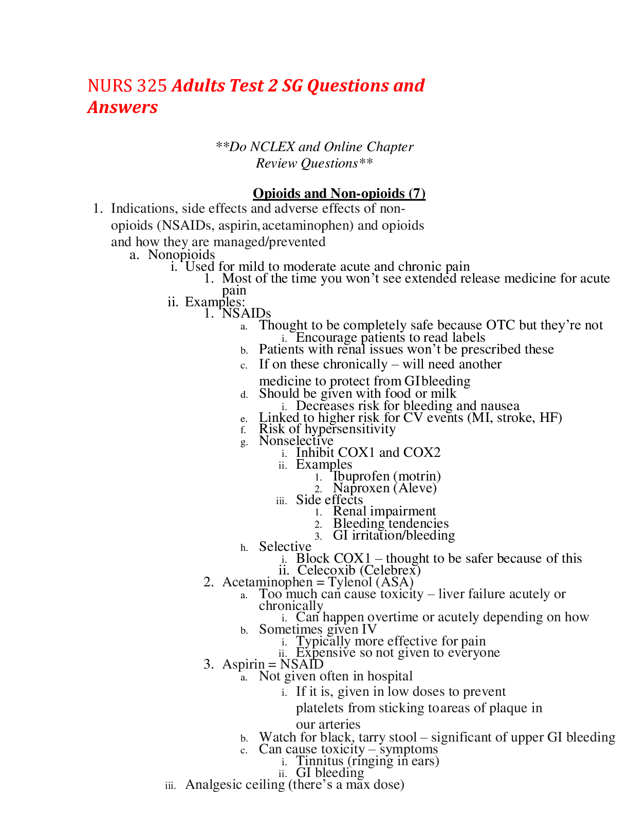
NURS 325 Adults Test 2 SG Questions and Answers **Do NCLEX and Online Chapter Review Questions** Opioids and Non-opioids (7) 1. Indications, side effects and adverse effects of non-opioids (NSA... IDs, aspirin, acetaminophen) and opioids and how they are managed/prevented a. Nonopioids i. Used for mild to moderate acute and chronic pain 1. Most of the time you won’t see extended release medicine for acute pain ii. Examples: 1. NSAIDs a. Thought to be completely safe because OTC but they’re not i. Encourage patients to read labels b. Patients with renal issues won’t be prescribed these c. If on these chronically – will need another medicine to protect from GI bleeding d. Should be given with food or milk i. Decreases risk for bleeding and nausea e. Linked to higher risk for CV events (MI, stroke, HF) f. Risk of hypersensitivity g. Nonselective i. Inhibit COX1 and COX2 ii. Examples 1. Ibuprofen (motrin) 2. Naproxen (Aleve) iii. Side effects 1. Renal impairment 2. Bleeding tendencies 3. GI irritation/bleeding h. Selective i. Block COX1 – thought to be safer because of this ii. Celecoxib (Celebrex) 2. Acetaminophen = Tylenol (ASA) a. Too much can cause toxicity – liver failure acutely or chronically i. Can happen overtime or acutely depending on how b. Sometimes given IV i. Typically more effective for pain ii. Expensive so not given to everyone 3. Aspirin = NSAID a. Not given often in hospital i. If it is, given in low doses to prevent platelets from sticking to areas of plaque in our arteries b. Watch for black, tarry stool – significant of upper GI bleeding c. Can cause toxicity – symptoms i. Tinnitus (ringing in ears) ii. GI bleeding iii. Analgesic ceiling (there’s a max dose) 1. Acetaminophen max dose = 4,000mg/day 2. Ibuprofen max dose = 2,400mg/day iv. No tolerance/physical dependence – no withdrawal v. Many OTC – encourage patients to read medications so they know what they’re taking vi. For mild to moderate pain vii. Commonly used in conjunction w/ opioids b. Opioids i. Bind to receptors in CNS resulting in 1. Inhibition of the transmission of nociceptive input from the periphery to the spinal cord 2. Altered limbic system activity a. Has a lot to do with emotional responses to pain 3. Activation of descending inhibitory pathways that modulate transmission in the spinal cord 4. Overall = Effects transmission of pain stimulus AND emotional response to pain ii. Used for moderate to severe acute pain (usually immediate release PO, IV, or PCA) iii. For moderate to severe chronic pain – extended-release or long-acting PO medications iv. Tolerance is expected over time 1. Tolerance a. Occurs with chronic exposure, is expected b. Need an increased dose to maintain the same degree of analgesia 2. Physical dependence a. Occurs with ongoing exposure, is expected b. Manifested by withdrawal syndrome that occurs when blood levels of the drug are abruptly decreased c. Need to taper when opioids are no longer needed v. Always higher dose if PO because of first pass effect of liver vi. Examples 1. Morphine a. Drug all other opioids are compared to for dosage 2. Hydromorphone (Dilaudid) – IV and PO 3. Methadone (Dolophine) – long acting a. Commonly seen in those with chronic pain 4. Levorphanol (Levo-Dromoran) 5. Fentanyl a. IV with transcutaneous patch for chronic pain b. Lollipops c. Can cause overdoses if people aren’t using it correctly d. Can be used for acute and chronic pain 6. Oxymorphone 7. Oxycodone (Percocet, Oxycontin) a. Percocet = oxycodone and Tylenol b. Sustained release and immediate release c. Can come as just oxycodone without Tylenol 8. Hydrocodone (Lortab, Vicodin) a. Hydrocodone and Tylenol 9. Codeine (Tylenol #3) vii. Reversal Agent = Naloxone (Narcan) 1. 0.4 – 2 mg IV/SC repeat q2-3min PRN 2. No more than 10mg 3. Also given down tracheal tube or as nasal spray if needed 4. Want to push it slowly a. Reverse analgesic effect and respiratory effect 5. May need to have another dose based on how much they’ve taken viii. Side effects (Most get better with time) 1. Constipation – dose not diminish with continued use (doesn’t get better) a. Talk with patient regarding their BM – color, frequency, consistency 2. Nausea and Vomiting 3. Sedation 4. Respiratory depression a. Monitor closely especially if they go up a dose 5. Pruritus (itching) a. Need to monitor to make sure it’s not an allergic reaction b. Difficult to treat b/c can make patient sleepy 6. Less common side effects a. Urinary retention – Monitor I/O b. Myoclonus c. Dizziness d. Confusion e. Hallucinations ix. Opioid to avoid – Meperidine (Demerol) 1. Associated with neurotoxicity (seizures) 2. May be used acutely but don’t use ongoing 2. Mechanism of Action a. Non-opioids b. Opioids Care of the Older Adult (6) 3. Common due to aging a. Pressure ulcers b. Incontinence c. Falls d. Delirium e. Others i. Functional decline ii. Malnutrition iii. Eating and feeding problems iv. Sleeping problems v. Dizziness and syncope vi. Self-neglect 4. Know how to assess for, prevent, and manage falls, pressure ulcers, urinary incontinence, and delirium a. Falls i. Risk factors 1. Intrinsic (patient related) a. Age (esp. > 75) b. History of recent fall c. Dementia, hip function, type 2 DM, parkinson’s, arthritis, depression, osteoporosis d. Use of assistive device e. Altered LOC or cognitive impairment f. Urge urinary incontinence g. Physical restraints h. Bare feet or inappropriate footwear i. Anticoagulant use 2. Extrinsic (environment related) a. Floor surface issues (spills, wet areas, unevenness) b. Poor or nonfunctional lighting c. Furniture, adaptive aids, or IV poles in poor working order d. Long gowns or clothing that may cause tripping ii. Assessment: Modified Egress Test 1. Provide standby assistance for Safety 2. Steps a. Have patient independently roll onto side and sit on edge of the bed b. Have the patient independently stand at the side of the bed c. Have the patient independently take 2 steps forward and 2 steps back 3. Patient must complete all 3 tasks to pass test a. If they need more than standby assistance they don’t pass b. All or nothing test 4. Failure a. Doesn’t mean can’t ambulate b. Needs to ambulate with assistant iii. Alternative assessment = get up and go (more subjective) iv. Fall precautions 1. Low beds – PAY ATTENTION TO THIS 2. Floor mats if high risk of injury a. Shouldn’t be down when patient is out and walking around (then it’s a trip hazard) 3. Easy access to call bell 4. Avoid physical restraints if possible 5. Use of personal or pressure sensor alarms 6. Increased observation/surveillance 7. Use of rubber-soled heeled shoes or nonslip slippers 8. Regular toileting at set intervals and/or continence programs 9. Urinals and bedpans within easy reach 10. Use corrective glasses when ambulating 11. Reduce clutter; esp. in high traffic areas 12. Early mobility programs 13. ALWAYS ask patient if there’s anything you can do before leaving room v. If fall occurs 1. Perform a physical assessment at time of fall (VS, neurologic, evaluation of head, neck, spine, and extremities) 2. Call for help 3. Observe patient for 48 hours after a fall 4. Once injury has been ruled out a. Obtain history of fall from patient and witnesses b. Note circumstances of fall, location, activity, time of day, and any significant symptoms c. Review of underlying illness and problems d. Review medications e. Assess functional, sensory, and psychological status f. Evaluate environmental conditions g. Review risk factors for falling b. Pressure ulcers i. Use Braden Scale 1. Assesses risk in six areas a. Sensory perception b. Activity c. Mobility d. Nutrition e. Friction/shear 2. Nursing Interventions based on score a. At risk (15-18) i. Frequent repositioning with written schedule ii. Maximize mobility iii. Protect heels iv. Use pressure reducing support surface (if bed or chairbound) b. Moderate risk (13-14) i. Everything from “at risk” PLUS ii. Use foam wedges for 30 degree lateral positioning c. High risk (10-12) i. Everything from “at risk” and “moderate” PLUS ii. Increase turning frequency (every 2 hours = bare minimum) iii. Do small shifts of position d. Very high risk (9 or less) i. All “ at risk,” “moderate,” and “high risk” PLUS ii. Manage moisture, nutrition, and friction and shear 3. Braden Scale Interpretation a. Scoring i. Assigns an item score ranging from 1 (highly impaired) to 3 or 4 (no impairment) ii. 15-18 = at risk iii. 13-14 = moderate risk iv. 10-12 = high risk v. 9 and below = very high risk **Most nurses score patients higher than they really are. Pay close attention to criteria under each category when scoring** c. Urinary Incontinence Assessment i. Notes: 1. Determine if it’s a new problem or ongoing 2. Don’t assume it to be chronic or normal ii. Part I = Transient Urinary Incontinence 1. Focuses on assessing for contributing causes of transient UI iii. Part II = Established/chronic urinary incontinence iv. Assessment/attempt to determine etiology with interprofessional team v. Avoid medications that contribute to UI vi. Avoid urinary catheters when possible (UTI risk) vii. Limit bladder irritants (there are a lot) viii. If BMI > 27, consider weight-loss plan ix. Provide patient with usual undergarments in expectation of incontinence 1. Try to avoid “diaper” term 2. Don’t want to use them in hospital due to skin breakdown 3. If patient uses at home, encourage patient to change them ASAP 4. Provide privacy and sensitivity a. Some are incontinent because they don’t want to bother you x. Provide skin care immediately after each UI episode xi. Prevention 1. Take patient to bathroom on schedule a. Especially if it’s not a functional/physical problem d. Delirium i. Disturbance of consciousness with impaired attention and disorganized thinking that develops rapidly and with evidence of an underlying physiologic or medical condition ii. Different from dementia 1. Delirium is rapidly progressing and has some sort of condition contributing to it iii. Characterized by 1. Reduced ability to focus, sustain, or shift attention 2. Memory impairment 3. Disorientation a. Able to get all 4 questions but as get more specific may not be able to answer those 4. Visual or other hallucinations 5. Misperceptions of stimuli 6. Clinical manifestations fluctuate over course of the day a. May have periods where they’re more clear and some when they’re less clear iv. Assessment: CAM Method 1. Clinician assesses for the prescence or absence of delirium by assessing for the following 4 features: a. Mental status altered from baseline (acute onset or fluctuating) b. Inattention c. Disorganized thinking d. Altered LOC 2. Results a. Patient has delirium if there is evidence of: i. 1 and 2, and 3 or 4 ii. Need ¾ of the features above v. Prevention: Eliminate or minimize risk factors 1. Avoid or minimize high-risk medications 2. Treat infections, dehydration, and electrolyte imbalances 3. Provide adequate pain control 4. Prevent/treat hypoxemia 5. Use sensory aids 6. Promote regular bowel and bladder function 7. Provide adequate nutrition vi. Treatment: Provide a therapeutic environment 1. Foster orientation 2. Provide appropriate sensory stimulation 3. Facilitate sleep 4. Foster familiarity 5. Maximize mobility 6. Avoid physical restraints 7. Communicate clearly 8. Minimize invasive interventions 9. Use psychotropic medication as a last resort for agitation 5. Know the difference between IADLs and ADLs a. Assess using Katz ADL and Lawton IADL index b. IADLs i. Have physical AND cognitive component to perform skill ii. Assesses: 1. Use a telephone 2. Shopping 3. Food prep 4. Housekeeping 5. Laundry 6. Transportation 7. Responsibility for medications 8. Finances c. ADLs i. Only a physical component ii. Assesses: 1. Bathing 2. Dressing 3. Transferring 4. Toileting 5. Continence 6. Feeding 6. Mini-cog a. Steps i. Ask patient to listen to and remember 3 unrelated words and then repeated ii. Instruct patient to draw the face of a clock on a blank sheet or sheet with clock circle iii. Ask patient to repeat the 3 words previously b. Scoring i. 1 point for each recalled word after the clock (possible 0-3) ii. 2 points for normal clock, 0 for abnormal c. Results i. 0-2 points = positive screen for dementia ii. 3-5 points = negative screen for dementia d. Not negatively impacted by education, language, or age Nutritional Assessment and Laboratory Values (2) 7. Nursing interventions and assessment when repleting potassium, magnesium, calcium, and phosphorus a. Potassium (3.6-5.0) i. Hyperkalemia 1. EKG changes 2. Cardiac arrest 3. Neuromuscular 4. Treatment a. Hold K-contained meds b. Diuretics, dialysis c. IV insulin d. Albuterol e. IV calcium chloride or gluconate f. Continuous ECG monitoring g. Sodium Polystyrene sulfonate (Kaexylate) ii. Hypokalemia 1. EKG changes 2. Cardiac arrest 3. Neuromuscular 4. Treatment (Hypo) a. Oral repletion i. Liquid needs to be diluted ii. May cause GI upset b. IV repletion i. Irritating to veins ii. Rate no more than 10mEq/hr c. Need adequate urine output and creatinine clearance for repletion b. Magnesium (1.6 -2.5) i. Hypermagnesemia 1. Often associated with renal impairment 2. Neuromuscular impairment 3. Hypotension 4. Risk of coma, respiratory, and cardiac arrest 5. Treatment a. Avoid magnesium-containing medications b. IV/oral fluids OR dialysis c. IV calcium gluconate ii. Hypomagnesemia 1. Often associated with low intake or GI/renal issues 2. Similar manifestations as hypocalcemia 3. Risk of dysrhythmias and cardiac arrest 4. Treatment a. Oral supplementation b. IV supplementation (give over 30-60 mins, depending on dose) c. Calcium i. Hypercalcemia 1. Decreased Neuromuscular excitability 2. Cardiac dysrhythmias 3. Risk of seizures and coma 4. Treatment a. Fluids (3-4L/day) b. Severe: Isotonic saline, bisphosphonate, and calcitonin c. ECG monitoring d. Seizure precautions ii. Hypocalcemia 1. Increased neuromuscular excitablility 2. ECG changes 3. Risk of seizures 4. Treatment a. Calcium and vitamin D supplements b. IV calcium gluconate c. ECG monitoring d. Seizure precautions e. d. Phosphorus (2.4 – 4.4) i. Hyperphosphatemia 1. Common with renal impairment 2. Often asymptomatic unless patient also has hypocalcemia due to phosphate -calcium binding 3. Treatment a. Restrict dairy products b. Binding agens i. Sevelamer (Renagel) ii. Calcium carbonate iii. Dialysis ii. Hypophosphatemia 1. Often due to nutritional absorption deficiencies or shift from ECF to ICF 2. Mild – moderate = asymptomatic 3. CNS depression 4. Muscle weakness 5. Respiratory and heart failure 6. Treatment a. IV repletion i. Potassium phosphate ii. Sodium phosphate b. Oral repletion i. Mix powder or liquid with an adequate amount of fluid 8. Interpretation of Total CO2 content laboratory values a. Normal = 23-39mmol/L b. Total CO2 content includes serum bicarbonate and available forms of CO2 (dissolved, carbonic acid, etc.) c. Serum bicarbonate comprises about 95% of the total CO2 i. Can be used to evaluate acid balance 1. High levels = metabolic alkalosis 2. Low levels = metabolic acidosis 9. Interpretation of BUN and creatinine levels a. BUN i. Normal = 6-20mg/dL ii. Measures concentration of urea in the blood 1. Urea concentration regulated by renal excretion iii. Nonrenal factors may also be involved 1. BUN ↑ with dehydration 2. BUN ↓ with liver cirrhosis b. Creatinine i. Normal = 0.6-1.3mg/dL ii. End product of muscle and protein metabolism iii. Released at a constant rate iv. More reliable than BUN when assessing renal function v. Monitor trends 1. If increased a. Avoid nephrotoxins b. Monitor I/O c. Daily weights 2. Decreased levels are usually not significant and only indicative of low muscle mass 10. Anticoagulant monitoring and labs (PT/INR for warfarin, PTT for heparin) **look more into** a. Prothrombin time (PT) i. Normal = 11-16 seconds ii. Assessment of extrinsic coagulation by measurement of factors I, II, V, VII, and X iii. Used in assessment of Warfarin (Coumadin) b. International Normalized Ration (INR) i. PT used to calculate independent of reagents or methods used ii. Normal = 0.8 – 1.1 iii. Warfarin is considered therapeutic if 2-3 c. Activated partial thromboplastin time (aPTT) i. Normal = 25-30 seconds ii. Assessment of intrinsic coagulation by measurement of factors I, II, R, VIII, IX, X, XI, XII iii. If on heparin therapy, goal is 1/5-2x control value 11. Normal values for CBC results, interpretation a. White blood cells (WBC) i. Normal levels = 4,000-1,000uL ii. Total number of leukocytes (all types) b. Hemoglobin (Hgb) i. Normal levels 1. Female = 11.7 – 16 2. Male = 13.2 – 17.3 ii. Measurement of gas-carrying capacity of the RBCs c. Hematocrit (Hct) i. Normal levels 1. Female = 35-47% 2. Male = 39-50% 3. Measure of packed cell volume of RBCs expressed as percdentage of the total volume 4. Generally 3 times greater than hemoglobin d. Platelets (Plt) i. Normal = 150,000-400,000uL ii. Number of platelets able to maintain platelet clotting functions iii. Does not measure quality of platelet function 12. Indications for nutritional assessment a. Older adults (65 and older) – Mini-Nutritional Assessment Short Form i. Based on food intake due to 1. Appetite 2. Digestive problems 3. Chewing or swallowing 4. Weight loss 5. Mobility 6. Psychological stress or acute disease 7. Neuropsychological problems 8. BMI ii. Scores 1. 12-14 points = normal 2. 8-11 = at risk 3. 0-7 = malnourished iii. BMI 1. Below 18.5 = underweight 2. 18.5-24.9 = normal 3. 25-29.9 = overweight 4. 30 = obese iv. Patient shouldn’t lose 10-15 pounds in 3-6 months unintentionally v. Includes 1. Diet history 2. Health history 3. Diet history 4. Physical exam 5. Lab data b. TJC requires nutrition screening for all patients within 24 hours of admission 13. Lab results found in malnutrition states a. Lab results found in malnutrition states i. Increased liver enzymes (inflammation ii. Increased BUN (dehydration) iii. Decreased lymphocytes/WBCs iv. Decreased fat- and water-soluble vitamins v. Decreased hemoglobin vi. Albumin – protein levels not specific to nutrition 1. Good for changes in illness state vii. Pre-albumin 1. Half life = 2 days 2. Good for nutritional status viii. Transferrin 1. Protein that transports iron 2. Decreases when protein is low End of life care (3) 14. Brain death criteria a. Brain Death Is: i. Irreversible loss of all brain functions (including brain stem) ii. Clinical diagnosis that can be made in patients whose hearts continue to beat and who are maintained on mechanical ventilation in the ICU iii. Cerebral cortex stops functioning or is irreversibly damage 15. Advanced directives a. Living will b. Allows you to document your medical wishes at end of life care c. Legal document d. If no person appointed goes in this order i. Guardian ii. Spouse iii. Adult child iv. Parent v. Adult sibling 16. Do not resuscitate orders a. “no code” b. AND = allow natural death c. Only in event of respiratory or cardiac arrest d. “Comfort care” i. Individualized for each person ii. No law/standards for this 17. Hospice a. Patients with life expectancy of 6 months or less b. Medicare benefit intended to support those in last 6 months of life c. Eating and drinking at end of life doesn’t cause pain/suffering d. Subset of palliative care (symptom management for those with serious illness) i. Reducing severity of disease symptoms rather than trying to delay or reverse progression of the disease and provide a cure 18. Physiologic and psychological changes at end of life and associated nursing care – LOOK AT CHARTS a. Goals i. Provide support during dying process ii. Improve quality of patients remaining life iii. Help ensure dignified death iv. Provide emotional support to family v. Physical care means vi. Skillful communication b. Nursing Implementation: Psychosocial i. Some things cannot be changed or fixed ii. We cannot change the inevitability of death. iii. We cannot erase the anguish that is felt when someone is dying or has died. iv. We all must face the fact that we will die. v. No matter how hard we try, the perfect words or gestures to relieve a patient or family’s distress rarely exist. vi. It is most often enough to be compassionately with the person 19. Care of family, communication a. SPIKES = A Six-step Protocol for Delivering Bad News i. Setting up the interview 1. Privacy 2. Involve significant others 3. Sit down 4. Make connection with patient/family 5. Manage time constraints and interruptions ii. Assessing the patients perception 1. Use open-ended questions iii. Obtaining the patients Invitation 1. Determine whether the patient wants specific details or a more generalized overview iv. Giving knowledge and information to the patient 1. Provide warning 2. Avoid using medical terms 3. Don’t be overly blunt 4. Provide info in small chunks and assess understand periodically 5. Don’t say “there’s nothing else we can do…” v. Addressing the patient’s Emotions and Empathetic 1. These may include silence, disbelief, crying, denial, anger 2. 4 steps to empathetic response a. Observe the emotion b. Identify the emotion c. Identify the reason for the emotion d. Allow patient to express feelings, make a connected statement vi. Strategy and Summary 1. Ask if patient is ready to discuss treatment option 2. Allow patient to express fears and conerns Diabetes (15) 20. Primary, secondary, tertiary prevention (including metabolic syndrome) a. Primary = exercise a. Secondary = diabetes screening c. Tertiary = insulin treatment 21. Medications that can cause hyperglycemia and mask hypoglycemia a. Beta-adrenergic blockers (end in -olol) i. Mask symptoms of hypoglycemia ii. Prolong effects of insulin b. Corticosteroids i. Can increase blood glucose levels in people who already have and people who don’t have DM 22. Diagnostic tests: HbA1C, fasting plasma glucose a. Hemoglobin A1C i. Based on the attachment of glucose to hemoglobin 1. RBC average lifespan = 3 months 2. A1C = average blood glucose levels over past 3 months ii. Regular assessments required iii. Goal = 7 or less 1. Goal may be 6.5 or less or 8 or less in some populations iv. Normal values reduces risks of retinopathy, nephropathy, and neuropathy v. Scores 1. Normal = 5.7 or less 2. Prediabetes = 5.7-6.4 3. Diabetes = 6.5 or above b. Fasting Plasma Glucose i. Fast then measure blood glucose ii. Normal = 99 or below iii. Prediabetes = 100-125 iv. Diabetes = 126 or above c. Oral glucose tolerance test i. Need to be NPO ii. Drink glucose then measure sugar in intervals iii. Normal = 139 or below 23. Complications: Hypoglycemia, macrovascular, microvascular (how to prevent, screen for) a. Hypoglycemia = low blood sugar i. Occurs when: 1. Too much insulin in proportion to glucose in the blood 2. Blood glucose level less than 70 mg/dL ii. Manifestations 1. Confusion 2. Irritability 3. Diaphoresis 4. Tremors 5. Hunger 6. Weakness 7. Visual disturbances iii. Untreated can progress to loss of consciousness, seizures, coma, and death iv. Check blood glucose at the first sign/symptom 1. If < 70mg/dL, begin treatment 2. If > 70 mg/dL, investigate further for cause of signs/symptoms a. If monitoring equipment not available, treatment should be initiated i. 15-20g of simple carb ii. Avoid food with fat iii. Recheck in 15 minutes iv. Repeat until over 70mg/dL v. After 2-3 doses of simple carb call provider b. Treatment if unable to swallow i. 1mg of glucagon IM ii. 20-50mL of 50% dextrose IV push iii. After awake ingest complex carb b. Hyperglycemia i. Diabetic retinopathy 1. Microvascular damage to retina a. Result of chronic hyperglycemia 2. Earliest and most treatable stages often produces no changes in vision 3. Screening = dilated eye examinations annually ii. Diabetic nephropathy 1. Associated with damage to small blood vessels that supply the glomeruli of the kidney 2. Leaded cause of end-stage renal disease 3. Critical factors for prevention/delay a. Tight glucose control b. Blood pressure management 4. Yearly screening a. Microalbuminuria in urine b. Serum creatinine iii. Sensory neuropathy: Distal symmetric 1. Affects hands and/or feet bilaterally 2. Loss of sensations, abnormal sensations, pain and paresthesia 3. Usually worse at night 4. Foot injury and ulcerations can occur without the patient having pain 5. Treatment a. Tight blood glucose control b. Drug therapy iv. Autonomic neuropathy 1. Can affect nearly all body systems 2. Complications a. Gastroparesis – delayed gastric emptying b. Cardiovascular abnormalities c. Sexual function d. Neurogenic bladder v. Infection 1. Diabetic individuals = more susceptible 2. Defect in mobilization of inflammatory cells 3. Impairment of phagocytosis by neutrophils and monocytes 4. Loss of sensation may delay detection 5. Treatment must be prompt and vigorous 24. Insulin (rapid, short, intermediate, long)-onset, peak, duration, nursing considerations, administration a. Onset = important with timing of meals (especially with short and rapid) b. Peak = important because that is when hypoglycemic reactions usually occur i. Hypoglycemic risk = low with long-acting c. Mixing precautions i. NEVER MIX LONG-ACTING ii. Rapid and short acting – not given together EVER iii. Intermediate can be mixed with rapid or short acting 1. Cloudy to clear d. Administration i. Cannot be taken orally ii. SubQ injection for self-administration iii. IV administration – regular insulin; for complications iv. Fastest absorption = abdomen (preferred), then arm thigh and butt v. Rotate injections within one particular site (checkerboard) vi. Do not inject in site to be exercised e. Insulin syringes i. U-100 syringes (100untils/mL) 1. 30 units (1/2 and whole markings) 2. 50 unites 3. 100 units f. Insulin pump i. Continuous subcutaneous infusion ii. Battery operated device iii. Connected via plastic tubing to a catheter inserted into subcutaneous tissue in abdominal wall iv. Potential for tight control v. Not every is good fit 1. Need to be knowledgeable, have psychomotor skills to deliver doses; cost prohibited g. Types of Insulin i. Rapid-acting (bolus) 1. Lispro, aspart, glulisine 2. Inject (subQ) 0-15 mins before meal 3. Onset = 15 minutes 4. Peak = 60-90 minutes 5. Duration = 3-4 hours ii. Short acting (bolus) 1. Regular 2. Injected (subQ) 30-45 mins before meal 3. Onset = 30 – 60 minutes 4. Peak = 2-3 hours 5. Duration = 3-6 hours iii. Intermediate 1. Onset = 2-4 hours 2. Peak = 4-10 hours 3. Duration = 10-16 hours iv. Long-acting (basal) 1. Glargine and detemir 2. Injected (subQ) once a day at bedtime or in the morning 3. Released steadily and continuously 4. Onset = 1-2 hours 5. No peak action 6. Duration = 24+ hours 7. Cannot be mixed with any other insulin or solution a. 2 injections; can be administered at same time 25. Oral medications: Mechanism of action, nursing considerations a. Not insulin b. Work to improve mechanisms by which insulin and glucose and produced and used by the body c. Work on 3 defects of type 2 diabetes i. Insulin resistance ii. Decreased insulin production iii. Increased hepatic glucose production 26. Nutritional Therapy a. Diet teaching i. Dietician initially provides instruction ii. Options 1. Carb counting (15g of carbs) 2. Plate method (help patient visualize the amounts of vegetable, starch, and meat that should fill 9 inch plate 27. Exercise a. Essential part of diabetes management i. Increases insulin receptor sites ii. Lowers blood glucose levels iii. Contributes to weight loss b. Several small crbohydrate snacks can be taken every 30 minutes during exercise to prevent hypoglycemia c. Best done after meals d. Exercise plans should be started i. After medical clearance ii. Slowly with gradual progression e. Monitor blood glucose levels before, during and after exercise 28. Foot care a. Wash feet daily b. Pat feet dry, especially between toes c. Examine feet daily for cuts, blisters, swelling, and red, tender areas d. Use lanolin on feet for prevent skin from drying and cracking e. Use mild foot powder on sweaty feet f. Don’t use commercial remedies to remove calluses g. Cleanse cuts with warm water and mild soap; cover with a clean dressing h. Report skin infection and nonhealing sores to HCP i. Cut toenails evenly with rounded contour of toes j. Separate overlapping toes with cotton k. Avoid open-toe, open-hell, and high-heel shoes l. Wear clean, absorbent socks or stockings m. Don’t wear clothing that leaves impressions, hindering circulation n. Guard against frostbit o. Don’t use heating pads or hot water bottles to warm feet p. Exercise feet daily by walking or flexing and extending feet in suspended position q. Avoid prolonged sitting, standing, and crossing of legs 29. Nursing diagnoses/evaluation a. Alteration in nutrition b. Less/more than body requirements c. Ineffective health maintenance d. Implementation i. Screen for type 2 DM should begin at age 45 ii. Especially if overweight/obese or with risk factors iii. Sick day rules – plan of care implemented when they are sick iv. Medical identification and travel card v. Hospitalization 1. Patients may not be able to take oral meds 2. Use sliding scale 3. Patients with procedures using contrast should hold metformin Stroke 30. Nursing care in acute vs. rehabilitation phase a. Acute Care i. Goals during acute phase 1. Preserving life 2. Preventing further brain damage 3. Reducing disability ii. Treatment differs according to type of stroke and as patient changes iii. Spot a stroke: FAST 1. Face drooping 2. Arm weakness (have patient close eyes and hold arms up – arm will drift down) 3. Speech difficulty 4. Time to call 911 iv. Begins with managing the ABCs v. Assessment findings 1. Altered level of consciousness 2. Weakness, numbness, or paralysis 3. Speech or visual disturbances 4. Severe headache 5. Unequal pupils 6. Facial drooping on affected side 7. Difficulty swallowing (dysphagia) 8. Seizures 9. Increase or decrease HR 10. Respiratory distress 11. Hypertension 12. Depends on type of stroke 13. This will determine where you want your BP to sit 14. Bladder or bowel incontinence 15. Nausea and vomiting 16. Vertigo 17. Important due to fall risk vi. Initial interventions 1. Ensure patent airway 2. Call stroke code or stroke team 3. Perform pulse oximetry 4. Maintain adequate oxygenation 5. Perform baseline laboratory tests. a. Check blood sugar b/c low blood sugar can present with similar symptoms 6. Obtain IV access with normal saline a. If emergent and no patent IV = bad 7. Maintain BP according to guidelines 8. Obtain CT scan immediately. 9. Anticipate thrombolytic therapy for ischemic stroke. 10. Position head midline. 11. Elevate head of bed 30 degrees if no symptoms of shock or injury occur. 12. Institute seizure precautions. a. May put them on medicine to prevent seizures 13. Remove dentures - May need to put something in their mouth 14. Remove clothing - It gets in the way vii. Hypertension = common immediately after stroke 1. Drugs to lower BP are used only if BP is markedly increased 2. Different in ischemic vs hemorrhagic viii. Fluid and electrolyte balance must be controlled carefully 1. Adequate hydration promotes perfusion and decreases further brain injury ix. Ongoing interventions – if patient has one stroke they’re at risk for another 1. Monitor vital signs and neurologic status a. Level of consciousness b. Monitor and sensory function c. Pupil size and reactivity d. O2 saturation e. Cardiac rhythm x. Aspirin 1. Used within 24-48 hours of stroke 2. Platelet inhibitors and anticoagulants may be used in thrombus and embolus stroke patients after stabilization a. Contradicted for patients with hemorrhagic stroke xi. Surgical interventions for stroke 1. Ischemic stroke – MERCI (Merci embolus retriever in cerebral ischemic stroke) a. Wire latches onto clot and pulls clot to prevent it from breaking off 2. Hemorrhagic stroke a. Immediate evacuation of aneurysm – induced hematomas b. Cerebral hematomas > 3cm b. Rehabilitation – After stroke has stabilized i. Shifts from preserving life to lessening disability and attaining optimal functioning ii. Patient may be transferred to a rehabilitation unit, outpatient therapy, or home care-based rehabilitation 31. Risk factors, clinical manifestations (thrombotic vs. hemorrhagic), prevention, diagnostic testing a. Risk Factors i. Nonmodifiable 1. Age 2. Gender 3. Race 4. Heredity/family history ii. Modifiable 1. Hypertension 2. Metabolic syndrome 3. Heart disease 4. Heavy alcohol consumption 5. Poor diet 6. Drug abuse 7. Sleep apnea 8. Obesity 9. Physical inactivity 10. Smoking b. Clinical Manifestations i. Right brain damage 1. Paralyzed left side (hemiplegia) 2. Left-side neglect 3. Spatial-perceptual deficits 4. Tends to deny or minimize problems 5. Rapid performance, short attention span 6. Impulsive, safety problems a. More at risk for falls 7. Impaired judgment 8. Impaired time concepts ii. Left-brain damage 1. Paralyzed on right side (hemiplegia) 2. Impaired speech/language aphasis 3. Impaired right/left discrimination 4. Slow performance, cautious 5. Aware of deficits: depression, anxiety 6. Impaired comprehension related to language, math iii. Motor function 1. Most obvious effect of stroke 2. Impairments a. Mobility b. Respiratory function c. Swallowing (dysphagia) and speech (dysphasia) d. Gag reflex e. Self-care abilities 3. Characteristic motor deficits a. Loss of skilled voluntary movement b. Impairment of integration of movements c. Alterations in muscle tone d. Alterations in reflexes 4. An initial period of flaccidity a. May last days-weeks b. Related to nerve damage 5. Spasticity of the muscles follows flaccidity a. Related to interruptions in upper motor neuron iv. Communication 1. Aphasia – total loss of comprehension and use of language a. When stroke damages the dominant hemisphere of brain 2. Dysphasia – difficulty related to the comprehension or use of language and is due to partial disruption or less a. Can be classified as non-fluent or fluent b. Expressive – can’t understand but can communicate to you c. Respective – don’t understand what we’re saying to them d. Global = combination of both 3. Dysarthria – disturbance in muscular control of speech (patient doesn’t have muscle control to safely eat and drink without aspirating) a. How a patient speaks determines how well they can control their muscles to drink or eat b. Experienced by many patients c. Impairments may involve i. Pronunciation ii. Articulation iii. Phonation v. Affect 1. May have difficulty controlling their emotions 2. Emotions may be exaggerated or unpredictable vi. Intellectual function 1. Both memory and judgement may be impaired 2. Left-brain stroke – more likely to result in memory problems related to language vii. Spatial-Perceptual Alterations (depth perceptions; perceptions of self) 1. Stroke on right side more likely to cause these problems a. But can occur on left-side strokes 2. Really hard to orient these patients a. They think they can do everything they used to be able to do 3. Can be divided into 4 categories a. Incorrect perception of self and illness b. Erroneous perception of self in space c. Inability to recognize an object by sight, touch, or hearing i. Sight - Hold up common item and ask what it is ii. Touch – Qtip test with patietns eye’s closed d. Inability to carry out learned sequential movements on command viii. Elimination 1. Problems with urinary and bowel elimination – typically temporary 2. When a stroke only affect one hemisphere – the prognosis for bladder function is excellent 3. Skin breakdown may be an issue 4. Be proactive – take patient to bathroom at least every 2 hours ix. Overall stroke: 1. Death of brain cells 2. Known as a brain attack 3. Functions are lost or impaired 4. Severity of the loss of function varies according to the location and extent of the brain involved c. Artery and manifestations (Circle of Willis) i. Anterior cerebral – motor and/or sensory deficit ii. Middle cerebral – dominant side (aphasia, motor and sensory deficit); nondominant side (neglect, motor and sensory deficit) 1. Most common iii. Posterior cerebral – hemianopsia, visual hallucination, spontaneous pain, motor deficit iv. Vertebral – cranial nerve deficits, diplopia, dizziness, nausea, vomiting, dysarthria, dysphagia, and/or coma d. Types of Strokes i. Thrombotic – blood clot forms in vessels in brain 1. Process of clot formation resulting in the narrowing of the lumen a. Blocks the passage of the blood through the artery ii. Ischemic – loose blood flow to the brain 1. Transient episode of neurologic dysfunction caused by focal brain, spinal cord, or retinal ischemia, without acute infraction of the brain 2. Symptoms last < 1 hour iii. Hemorrhage – bleeding (typically more critical) 1. Burst blood vessel may allow blood to seep into and damage brain tissues until clotting shuts off the leak 2. Subarachnoid hemorrhage a. Intracranial bleeding into cerebrospinal fluid-filled space between the arachnoid and pia mater b. Commonly caused by rupture of a cerebral aneurysm i. Majority of aneurysms are in the circle of willis c. “Worst headache of one’s life” e. Prevention i. Goals 1. Health promotion for the well individual 2. Education and management of modifiable risk factors to prevent a stroke ii. Antiplatelet drugs usually chosen treatment to prevent stroke in patients who have had a TAI 1. Aspirin = drug of choice a. Don’t give to patient with hemorrhagic stroke – it can increase bleeding iii. Surgical interventions for the patient with TIAs from carotid disease 1. Carotid endarterectomy a. A tube is inserted above and below the blockage to reroute the blood flow b. Atherosclerotic plaque in the common carotid artery is removed c. Once artery is stitched closed, the tube can be removed 2. Transluminal angioplasty 3. Stenting a. Balloon catheter used to implant the stent into an artery of the brain b. Balloon catheter is moved to the blocked area of the artery and then is inflated (stent expands) c. Balloon is deflated and withdrawn, leaving the stent permanently holding the artery open and improving the flow of blood 4. Extracranial-intracranial bypass f. Diagnostic testing i. Done to 1. Confirm stroke 2. Identify cause of stroke ii. CT = primary diagnostic test iii. Others 1. CTA 2. MRI, MRA 3. Cerebral angiography 4. Digital subtraction angiography 5. Transcranial Doppler ultrasonography 6. Lumbar puncture 7. LICOX system iv. Cardiac Assessment 1. Electrocardiogram 2. Chest x-ray 3. Cardiac enzymes 4. Echocardiography 5. Holter monitor v. 32. Know the purpose, action, side effects of antiplatelet drugs, indications for tPA (thrombolytic therapy). a. Recombinant tissue plasminogen activator (tPA) i. Used to reestablish blood flow through a blocked artery to prevent cell death in patients with acute onset of ischemic stroke symptoms ii. Must be administered within 3 to 4.5 hours of onset of clinical signs of ischemic stroke iii. Since it breaks up blood clots it can cause issues with bleeding 1. Want to administer all IV, blood draws, foleys, etc. before administration iv. One-time medication 33. Deficits after stroke, treatment and nursing care a. Movements, sensation, emotions Seizures 34. Drugs to use for: a. Acute seizures that doesn't spontaneously resolve and status epilepticus (benzodiazepines) i. Diazepam (Valium) ii. Midazolam (Versed) iii. Aadvin iv. Status epilepticus 1. State of constant seizure or condition when seizures recur in rapid succession without return to consciousness between seizures a. Neurologic emergency b. Can involve any type of seizure 2. Causes brain to use more energy than is supplied a. Neurons become exhausted and cease to function i. More excitable during a seizure – medications suppress them so not as excited b. Permanent brain damage can result b. Drugs need to have blood levels monitored (these are starred in your PowerPoint slides) i. Carbamazepine (Tegretol) ii. Phenytoin (Dilantin) iii. Fosphenytoin (Cerebyx) iv. Valproic acid (Depakene) v. Ethosuximide (Zarontin) c. Common CNS and other system side effects (in the PowerPoint) i. CNS side effects 1. Diplopia 2. Drowsiness 3. Ataxia 4. Mental slowing ii. Others 1. Rashes 2. Hyperplasia of gingiva 3. Blood dyscrasias 4. Effects on liver and kidneys d. Nursing implications i. Prevention 1. Wear helmet if risk for head injury 2. General health habits (diet, exercise) 3. Assist to identify events or situations precipitating seizures 4. Instruct to avoid excessive alcohol, fatigue, and loss of sleep ii. Acute intervention 1. Observation and treatment of seizure a. Maintain patent airway, support head, turn to side, loosen constrictive clothing, ease to floor b. May require suctioning or oxygen after seizure c. Can put nasal cannula on if needed 2. Assessment of level of understanding a. Talk with patient afterwards about their seizure (symptoms, etc) 3. Ambulatory and Home care a. Instruct on importance of adherence to medication, not to adjust dose without physician b. Keep regular appointments. c. Teach family members emergency management. d. Emotional support and identification of coping mechanisms e. Medical alert bracelets f. Referrals to agencies and organizations g. 35. Nursing care during and after a seizure, patient teaching a. After seizure – evaluation i. Appropriate HR/rhythm, depth of respirations ii. No injury iii. Verbalization of knowledge of potential injury iv. Arrangement of environment to minimize injury v. Acceptance of disorder vi. Acknowledgment seizure has occurred vii. Therapeutic drug levels viii. Compliance with therapeutic regimen Parkinson’s 36. Know how the drugs work in general (i.e. Dopamine vs. anticholinergic), know side effects of carbidopa/levodopa and dopamine receptor agonists (see page 1389-1390 in the Lewis text). a. Dopamine vs. anticholinergic i. Dopamine 1. Enhance or release supply of dopamine (DA) 2. Antagonize or block the effects of overactive cholinergic neurons in the striatum ii. Anticholinergic 1. Decrease the activity of acetylcholine to provide balance between cholinergic and dopaminergic actions b. Side effects of carbidopa/levodopa and dopamine receptor agonists i. Levodopa with carbidopa (Sinemet) 1. First drug used 2. Precursor of DA and crosses the blood-brain barrier 3. Converted to DA in the basal ganglia 4. Carbidopa inhibits an enzyme that breaks down levodopa before it reaches the brain 5. Effectiveness could wear off after a few years of therapy c. Antihistamines with anticholinergic properties or beta-adrenergic blockers i. Can be used for tremors d. Antivirals promote DA release from neurons e. Monoamine oxidase type (MAO-B) inhibitor i. Degradative enzyme for DA – so inhibiting MAO-B increases DA levels f. Catechol O-Methyl Transferase (COMT) inhibitors i. Block the COMT enzyme that breaks down levodopa in the peripheral circulation g. Within 3 – 5 years of treatment i. Patients experience episodes of hypomobility 37. Parkinson’s assessment findings, nursing care to prevent complications, home care a. Clinical manifestations i. Onset = gradual and insidious ii. Classic Triad – most patients will have all of these at some point (not adverse effects) 1. Tremor 2. Rigidity 3. Bradykinesia iii. Beginning stages 1. Mild tremor 2. Slight limp 3. Decreased arm swing iv. Later stage 1. Shuffling 2. Propulsive gait with arms flexed 3. Loss of postural reflexes b. Nursing Care i. Surgical therapy 1. Procedures aimed at relieving symptoms 2. Used in patients who are usually unresponsive to drug therapy or have developed severe motor complications ii. Ablation surgery – recently been replaced with deep brain stimulation 1. Thalamotomy – part of thalamus destroyed 2. Subthalamotomy – part of subthalamus nucleus destroyed 3. Pallidotomy – involves insertion of a probe into a tiny hole in the skull to destroy a small part of the globus pallidus iii. Deep brain stimulation 1. Involves placing an electrode in the thalamus, globus pallidus, or subthalamic nucleus 2. Connected to a generator placed in the upper chest 3. Device is programmed to deliver specific current to targeted brain location iv. Nutritional therapy 1. Malnutrition and constipation common. 2. Patients with dysphagia and bradykinesia need food that is easily chewed and swallowed. 3. Several small meals should be taken to prevent fatigue. 4. Provide ample time to avoid frustration. 5. Levodopa can be impaired by protein and B6 ingestion c. Nursing Diagnoses i. Impaired nutrition: Less than body requirements ii. Risk for injury iii. Planning 1. Maximize neurologic function. 2. Maintain independence in activities of daily living (ADLs) for as long as possible. 3. Optimize psychosocial well-being iv. Nursing Implementation 1. Promote physical exercise and a well-balanced diet. a. Limit the consequences from decreased mobility b. Specific exercises to strengthen muscles involved with speaking and swallowing 2. Teach maintenance of good health, independence, and avoidance of complications. 3. Problems secondary to bradykinesia can be alleviated by a. Consciously thinking about stepping over a line on the floor b. Lifting toes when stepping c. One step back and . . . d. Two steps forward 4. Get out of a chair by using arms and placing the back legs on small blocks. 5. Remove rugs and excess furniture. 6. Simplify clothing from buttons and hooks. 7. Use elevated toilet seats. 8. Assist patients as they make adjustments to their lifestyle to accommodate symptoms. 9. Caregivers may also experience stress associated with disease progression (i.e., dementia). a. Patient may end up in long-term care facility Vision/Hearing Impairment 38. Be able to describe visual deficits for cataracts, glaucoma, macular degeneration and associated nursing care a. Cataracts i. An opacity of the lens ii. Distorts image projected into the retina iii. Slightly blurred vision can progress to blindness iv. Decreased color perception v. Glare vi. Lens cloudiness vii. Cataract surgery – Postoperative Care 1. Antibiotic and corticosteroid eye drops a. Prevent infection and decrease inflammation d/t surgery 2. Avoid activities that increase IOP a. Bending, stooping, coughing, lifting 3. F/U in 6-8 weeks b. Glaucoma i. Group of ocular diseases resulting from increased intraocular pressure (IOP_ ii. Etiology 1. Degenerative changes from hypertension, obesity, diabetes 2. Trauma iii. Asymptomatic to pain = clinical manifestations iv. Pathophysiology 1. Increased IOP due to: a. Decrease in the outflow of aqueous fluid through the anterior chamber OR b. Overproduction of aqueous fluid 2. Increased IOP inhibits supply to optic nerve and retina a. Tissue becomes ischemic and dies b. Can result in blindness v. Primary open-angle glaucoma (POAG) 1. Most common 2. Usually bilateral 3. Usually asymptomatic early** 4. Reduced outflow of aqueous fluid through chamber angle 5. Gradual increase in IOP 6. Laser Surgery a. Increases size of spaces between fibers b. Allowing outflow of aqueous fluid and decreased IOP vi. Collaborative Care 1. Reduce intraocular pressure 2. Promotes aqueous flow a. Macular degeneration i. Deterioration of the macula ii. Types 1. Non-exudative (dry) a. Most commons (90% of all cases) b. Want to maximize remaining vision 2. Exudative (wet) – more severe a. Medications injected directly into the vitreous cavity to inhibit endothelial growth factor and slow vision loss b. Laser therapy to seal leaking blood vessels can limit the damage 39. Primary and secondary prevention for hearing loss; how to communicate with people who have hearing loss a. Conductive hearing loss i. Interference of sound transmission through external ear and middle ear b. Sensorineural Hearing Loss i. Impairment of function of the inner ear or eighth cranial nerve c. Mixed Hearing Loss i. Both conductive and sensorineural d. Clinical Manifestations i. No response to oral communication ii. Answering questions inappropriately iii. Loud speech iv. Strained facial expression v. Tilting head vi. Listen to TV at increase volume vii. Family members may notice before patient e. Collaborative Care i. Primary Prevention – not using loud machinery, music at low volumes, etc. ii. Secondary prevetion – regular hearing screening iii. Tertiary prevention 1. Hearing Aids 2. Speech Reading (lip reading) 3. Sign Language 4. Cochlear Implant 5. Assisted listening devices f. Communication with those who have hearing loss i. Nonverbal aids 1. Draw attention with hand movements 2. Have speaker’s face in good light 3. Avoid covering mouth or face with hands 4. Avoid chewing, eating, smoking while talking 5. Avoid distracting environments 6. Maintain eye contact 7. Avoid careless expression that the patient may misinterpret 8. Use touch 9. Move close to better ear 10. Avoid light behind speaker ii. Verbal aids 1. Talk slowly 2. Don’t overexaggerate facial expressions 3. Don’t overenunciate 4. Use simple sentences 5. Rephrase sentence; use different words 6. Write name or difficult words 7. Do not shout 8. Speak in normal voice directly into better ear [Show More]
Last updated: 1 year ago
Preview 1 out of 45 pages
Instant download
Buy this document to get the full access instantly
Instant Download Access after purchase
Add to cartInstant download
Reviews( 0 )
Document information
Connected school, study & course
About the document
Uploaded On
May 04, 2021
Number of pages
45
Written in

Additional information
This document has been written for:
Uploaded
May 04, 2021
Downloads
0
Views
58