Biology > STUDY GUIDE > Portage Learning A&P 2 102All of portage modules BIOD 152 (All)
Portage Learning A&P 2 102All of portage modules BIOD 152
Document Content and Description Below
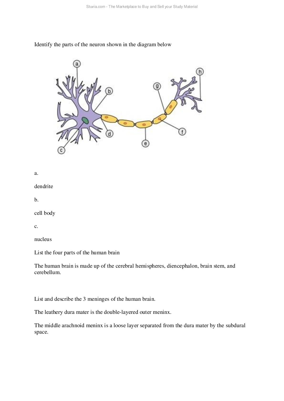
All of portage modules BIOD 152 Intro to the Nervous System Notice: To optimize your learning in this course, we advise that you complete the labs and modules as indicated in the BIOD 152 Lab Schedule... . The nervous system receives and processes information and sends out signals to the muscles and glands to elicit an appropriate response. In this way, the nervous system integrates and controls the other systems of the body. In the human nervous system, the central nervous system (Figure below) includes the brain and the spinal cord (dorsal nerve cord), which lie in the midline of the body. The skull protects the brain and the vertebrae protect the spinal cord. The central nervous system can send signals or impulses to and receive impulses from the peripheral nervous system. The peripheral nervous system includes all nerves not in the brain or spinal cord which are the cranial nerves that connect directly to the brain and the spinal nerves which project from either side of the spinal cord. The peripheral nervous system connects all parts of the body to the central nervous system and can be divided into a sensory or afferent division and a motor or efferent division. The peripheral nervous system receives impulses from the sensory organs via the afferent division and then relays signals or impulses from the central nervous system to muscles and glands via the motor or efferent division. The efferent division can be further divided into the somatic system and the autonomic system. The somatic system nerves control skeletal muscles, skin, and joints. The autonomic system nerves control the glands and smooth muscles of the internal organs and are not generally under conscious control and can be divided into two systems: the sympathetic system which activates and prepares the body for vigorous muscular activity, stress, and emergencies and the parasympathetic system which lowers activity, operates during normal situations, permits digestion, and conserves energy. Problem Set 1: 1. The function of the nervous system is to integrate and control the other body systems. Explain how the nervous system does this. the nervous system is divided into the central and peripheral nervous system. Within the peripheral nervous system their is the autonomic system which communicated with smooth muscle and internal organs and glands. Also in the peripheral system is somatic system which communicates with sense organs and voluntary muscles. 2. List the 2 parts of the central nervous system. Brain and spinal cord 3. How are the parts of the central nervous system protected? brain and the vertebrae 4. How do the central nervous system and the peripheral nervous system interact? through signals and impulses 5. What is included in the peripheral nervous system? all the nerves not in the brain or spinal cord 6. What are the 2 divisions of the peripheral nervous system? sensory (afferent) and motor (efferent) 7. Describe the movement of nerve impulses in the peripheral nervous system. Within the peripheral nervous system their is the autonomic system which communicated with smooth muscle and internal organs and glands. Also in the peripheral system is somatic system which communicates with sense organs and voluntary muscles. 8. What are the 2 divisions of the efferent division of the peripheral nervous system? autonomic and somatic systems 9. What is controlled by the somatic and autonomic nervous systems? automatic communicates with smooth muscles, internal organs and glands. somatic communicates with skeletal muscles, skin and joints 10. What are the 2 divisions of the autonomic nervous system? sympathetic and parasympathetic 11. What is the function of the sympathetic nervous system? to arouse and activate muscles for activity, stress and emergency 12. What is the function of the parasympathetic nervous system? To calm and lower activity. Permits digestion and conserves energy Neurons Neurons (Figure below) are nerve cells that vary in size and shape. They do not undergo mitosis (cell division), require enormous amounts of fuel, are able to survive just minutes without oxygen, and can last an entire human lifetime. Neurons all have three parts: the dendrites, the cell body, and the axon. The neuron cell body, which synthesizes all nerve cell products, consists of a large nucleus with surrounding cytoplasm containing the normal organelles. The dendrites are numerous short extensions that emanate from the cell body which receive information from other neurons conducting those nerve impulses toward the cell body. The single axon, on the other hand, conducts nerve impulses away from the cell body to its axon terminals where it is emitted across a synapse to the dendrite of another neuron. Axons can vary in length being very short or as long as three feet, the length of the axon which extends from the bottom of the spine to the big toe. Axons are composed of cells like the cell body but lack rough endoplasmic reticulum, depending on the cell body for necessary proteins. The peripheral nerve axon is coated in short sections called Schwann cells which are mainly composed of a white fatty layer called the myelin sheath rolled around the axon which insulates the nerve fiber from others and increases the speed of nerve impulses. There are also unmyelinated fibers, which are common in the gray matter of the brain and spinal cord, in which the Schwann cells do not wrap around the axon but are just loosely associated with the axon. The Schwann cell insulating sections are not continuous, having gaps between them called Nodes of Ranvier. At these exposed nodes, the nerve impulse is forced to jump to the next node in a manner called salutatory conduction, greatly increasing the nerve impulse transmission along the axon. The cell body contains the nucleus and other organelles typically found in cells with the exception of centrioles (since it is not capable of mitosis). One of the main functions of the cell body is to manufacture neurotransmitters, which are chemicals stored in secretory vesicles at the end of axon terminals. When neurotransmitters are released by the axon terminal vesicles, they participate in the transmission of the nerve impulse from one neuron to another. Problem Set 2: 13. Identify the parts of the neuron shown in the diagram below. a. Dendrite b. Cell body c. Nucleus d. Axon e. Myelin sheath f. Schwann cell g. Node of Ranvier h. Axon terminal 14. List 3 unusual characteristics of neurons. Do not undergo mitosis. Need oxygen to survive. Require a lot of energy and fuel to survive. 15. List the 3 parts of a neuron. The cell body, dendrites, and axon 16. Describe the structure and function of the neuron cell body. The cell body has a large nucleus with cytoplasm. It synthesizes all nerve cell products. The cell body produces neurotransmitters and chemicals. When the neurotransmitters are released by the axon terminals vesicles out of storage in secretory vesicles, they then help the process of nerve impulses form one neuron to another. 17. Describe the structure and function of the dendrite. The dendrite has numerous short extensions that receives information from other neurons conducting them toward the cell body. 18. Describe the axon, including the number in each neuron, function, structure and organelles. The axon can very in length, can be short or up to three feet tall. conducts nerve impulses away from the cell body towards the axon terminal to be able to communicate from one neuron to another. Axons are composed of cells like the cell body but lack rough endoplasmic reticulum. 19. Describe the composition and function of Schwann cells. Schwann cells are composed of white fatty layer called myelin sheath which insulates nerve fibers from other and increases nerve impulse speed. 20. Describe the location and function of the Nodes of Ranvier. Nodes of Ranvier are located in the gaps between the non continuous shwann cells. These gaps force impulses to jump to the next node which is called salutatory conduction which increases the transmission of nerve impulses down the axon. 21. What important organelle is absent from the neuron cell body and what does the absence of this organelle indicate about activity of the cell body? Centrioles are absent in the cell body resulting in no mitosis in the cell body. 22. Describe the function and site of synthesis and storage of neurotransmitters. The cell body produces neurotransmitters and chemicals. When the neurotransmitters are released by the axon terminals vesicles out of storage in secretory vesicles, they then help the process of nerve impulses form one neuron to another. Neuroglial Cells A nerve consists of hundreds of thousands of axons (#3) wrapped together in a connective tissue. In the peripheral nervous system the cell bodies of neurons (#2) are grouped together in masses called ganglia which are part of a single nerve. The neurons are also accompanied by non-nerve "supporting" cells known collectively as neuroglial cells which include (as shown in the diagram below) ependymal cells (#1), oligodendrocytes (#4), astrocytes (#5) and microglial cells (#7). The functions of these supporting cells are as follows: ependymal cells (circulate cerebrospinal fluid and allow fluid exchange between brain, spinal cord and CSF), oligodendrocytes (insulation of central nervous system axons), astrocytes (control chemical environment of neurons) and microglial cells (protect CNS by scavenging dead cells and infectious microoganisms). Neurons can be classified as to their structure and function. Structurally, neurons are classified according to the number of extension from their cell body, as multipolar, bipolar and unipolar neurons. Multipolar neurons, the most common type in humans found as motor neurons or interneurons within the CNS, have three or more extensions, one axon and many dendrites. Bipolar neurons, found as receptors cells in the visual and olfactory systems, have two extensions, one axon and one dendrite. Unipolar neurons, found as sensory neurons in the peripheral nervous system, have one extension which branches into two, one central process running to the CNS and another peripheral process running to the sensory receptor. Functionally, neurons are classified as sensory or afferent neurons, motor or efferent neurons and association or interneurons. Most sensory neurons are unipolar and carry impulses from receptors in the skin or internal organs toward the CNS. Most motor neurons are multipolar and carry impulses from the central nervous system to muscle fibers or glands. Interneurons are usually multipolar and found within the central nervous system only. They transmit impulses between sensory and motors neurons conveying messages between various parts of the central nervous system, such as from one side of the brain or spinal cord to the other, or from the brain to the spinal cord, and vice versa. Problem Set 3: 23. List the four types of support neuroglial cells and a function of each. Ependymal cells, circulate cerebrospinal fluid and allow fluid exchange between the brain, spinal cord and CSF. Oligodendrocytes, insulate CNS axons Astrocytes, control chemical environment of neurons Microglial cells, protect CNS by scavenging dead cells and infectious microorganisms 24. List the 3 structural classes of neurons, and describe the structure and an example of each. Multipolar, which is most common in humans have three or more extensions, one axon and many dendrites. They are found as motor neurons or interneurons. Bipolar, have two extensions, one axon and one dendrite. They are found as receptor cells in the visual and olfactory systems. Unipolar, have one extension which branches into two. One central process running to the CNS and another to PNS running to the sensory receptors. Are found as sensory neurons in the PNS. 25. List the 3 functional classes of neurons, and describe the structure and an example of each. Sensory neurons, are unipolar and carry impulses from receptors in the skin or internal organs towards CNS. Motor neurons, multipolar and carry impulses from CNS toward muscle fibers and glands. Interneurons, are multipolar and found within CNS only and transmit impulses between sensory and motor neurons conveying messages between various parts of the CNS, such as from one side of the brain or spinal cord to the other side. Action Potentials Neurons are specialized to conduct electric impulses called action potentials. The nerve impulse is an electrochemical charge moving along an axon created by the movement of unequally distributed ions on either side of an axon’s plasma membrane. At rest the plasma membrane is said to be polarized, meaning that one side has a different charge than the other side. When the axon is not conducting an impulse, this difference in electrical charge is called resting potential and is equal to about -70mV (millivolts). This means that the charge on the inside of the axon's cell membrane is 70 millivolts less than the outside of the membrane. This is maintained by a sodium-potassium pump which uses active transport to carry ions across the plasma membrane. The pump works by using an integral carrier protein that, for every three Na+ ions that are pumped out, two K+ ions are pumped in. The pump must keep in constant operation, because the Na+ and K+ ions will naturally diffuse back to where they originated. Because the plasma membrane is more permeable to K+ diffusing outward and because more Na+ ions are being pumped outward than K+ pumped inward, a relative positive charge develops and is maintained on the outside of the membrane. If the axon is stimulated to conduct a nerve impulse, there is a rapid change in the polarity. This change in polarity is called the action potential. First, the membrane potential becomes more positive (called depolarization), indicating that the inside of the membrane is now more positive than the outside. Then the potential returns to normal (called re-polarization), indicating that the inside of the axon is negative again. The action potential is due to special protein-lined channels in the membrane, which can open to allow either sodium or potassium ions to pass through. These channels have gates, called sodium gates and potassium gates. During the resting phase both sodium and potassium gates are closed. The sodium gates open and sodium rushes into the axon during the depolarization phase of the action potential. Voltage travels to zero and then on up to +40mV. Once this phase is complete, re-polarization occurs. The sodium gates close and potassium gates open allowing potassium to rush out of the axon. This returns a negative voltage to the inside of the axon but these gates are slow to close and there is generally an afterpolarization undershoot of the potential. These channels and their gates are voltage activated, as proteins respond to changes in voltage with changes in shape. The action potential travels along the length of an axon like a wave. It is self-propagating because the ion channels are prompted to open whenever the membrane potential decreases (depolarizes) in an adjacent area. An action potential is an all-or-nothing response either occurring or not. Since no variation exists in the strength of a single impulse, we distinguish the difference in intensity of a sensation (minor pain/major pain) by the number of neurons stimulated and the frequency with which they are activated. An impulse passing from one vertebrate nerve cell to another always moves in only one direction and there is a very short delay in transmission of the nerve impulse from one neuron to another. Neurons do not touch. There is a minute fluid-filled space, called a synapse, between the axon terminal of the sending (presynaptic) neuron and the dendrite of the receiving (postsynaptic) neuron. [Show More]
Last updated: 1 year ago
Preview 1 out of 207 pages
Instant download
Buy this document to get the full access instantly
Instant Download Access after purchase
Add to cartInstant download
Reviews( 0 )
Document information
Connected school, study & course
About the document
Uploaded On
Jun 29, 2021
Number of pages
207
Written in

Additional information
This document has been written for:
Uploaded
Jun 29, 2021
Downloads
0
Views
47
.png)