Biology > Study Notes > Midterm 2A Study Guide and Notes (All)
Midterm 2A Study Guide and Notes
Document Content and Description Below
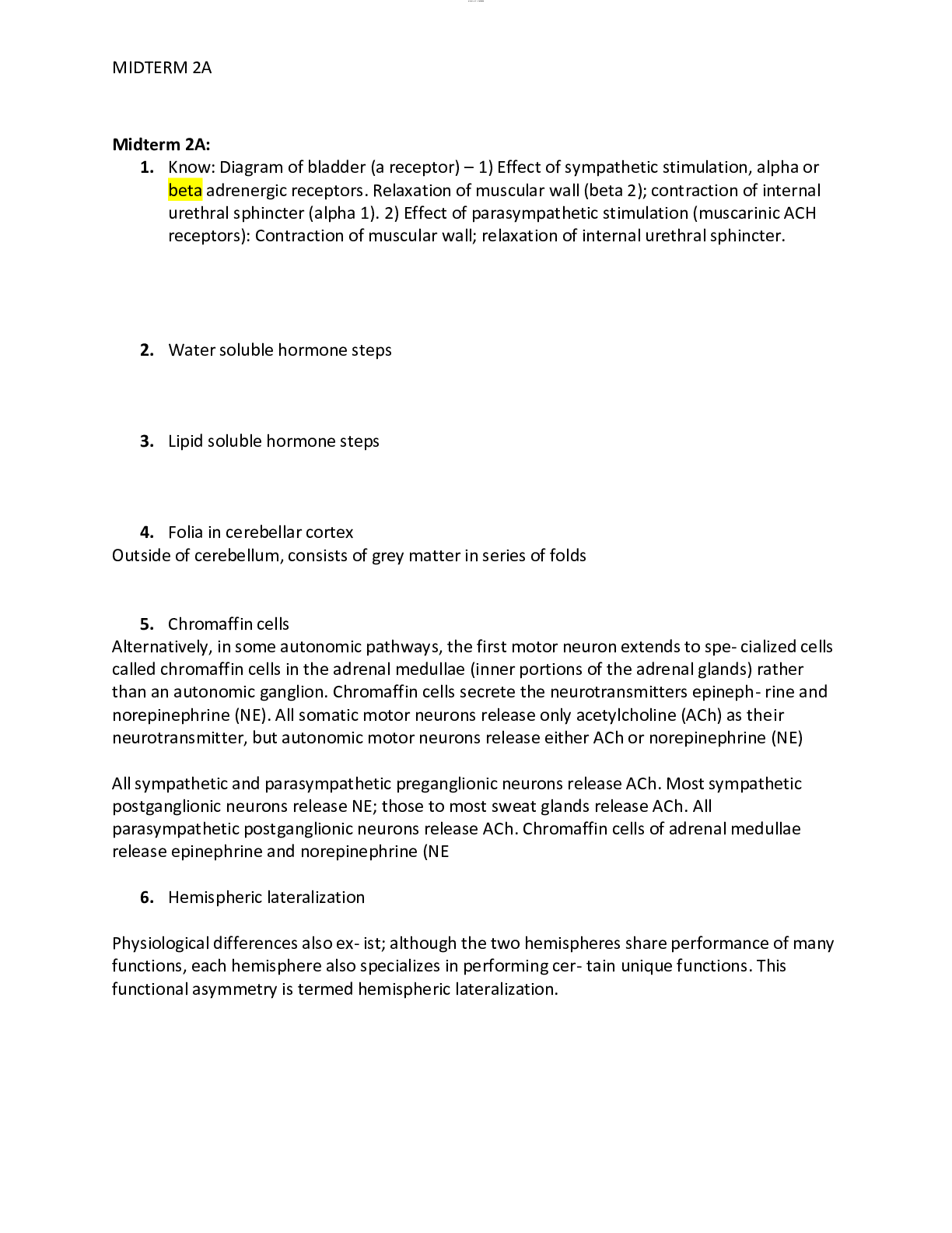
Midterm 2A: 1. Know: Diagram of bladder (a receptor) – 1) Effect of sympathetic stimulation, alpha or beta adrenergic receptors. Relaxation of muscular wall (beta 2); contraction of internal uret... hral sphincter (alpha 1). 2) Effect of parasympathetic stimulation (muscarinic ACH receptors): Contraction of muscular wall; relaxation of internal urethral sphincter. 2. Watersoluble hormone steps 3. Lipid soluble hormone steps 4. Folia in cerebellar cortex Outside of cerebellum, consists of grey matter in series of folds 5. Chromaffin cells Alternatively, in some autonomic pathways, the first motor neuron extends to spe- cialized cells called chromaffin cells in the adrenal medullae (inner portions of the adrenal glands) rather than an autonomic ganglion. Chromaffin cells secrete the neurotransmitters epineph- rine and norepinephrine (NE). All somatic motor neurons release only acetylcholine (ACh) as their neurotransmitter, but autonomic motor neurons release either ACh or norepinephrine (NE) Allsympathetic and parasympathetic preganglionic neurons release ACh. Mostsympathetic postganglionic neurons release NE; those to most sweat glands release ACh. All parasympathetic postganglionic neurons release ACh. Chromaffin cells of adrenal medullae release epinephrine and norepinephrine (NE 6. Hemispheric lateralization Physiological differences also ex- ist; although the two hemispheres share performance of many functions, each hemisphere also specializes in performing cer- tain unique functions. This functional asymmetry is termed hemispheric lateralization. lOMoARcPSD|549 808 2 MIDTERM 2A Despite some dramatic differences in functions of the two hemispheres, there is considerable variation from one person to another. Also, lateralization seems less pronounced in fe- males than in males, both for language (left hemisphere) and for visual and spatial skills (right hemisphere). For instance, females are less likely than males to suffer aphasia after damage 7. Voltage gated channels -- specifically ones that use sodium ions Gated channels that open in response to voltage stimulus (change in membrane potential). Axons of all types of neurons. Ie threshhod stimulus causes voltage gated na+ channels to open causing depolarization of cell and propagation of action potential down axon lOMoARcPSD|549 808 2 MIDTERM 2A 8. astigmatism, nearsightedness and farsightedness astigmatism is when they eye does no refract light evenly on the retina, leads to blurred vision at any distance nearsightedness: Can see close, cant see far. AKA Myopia. Due to issue with refracting the light, in near sightednees image gets focused in front of retina not on it farsightedness: Can see far things, but not close things. AKA hyperopia. Visual image gets focused behinf retina 9. CNS & PNS 10. Resting membrane potential voltage 11. Calcium channels Muscle action potential along ttubes causes calcium to be pumped out ofsarcolemma and calcium c hannels to open 12. triggerzones on neurons axon hillock ( basically the verey beginning of an axon, last space where ESPS and ISPS are summated to see if a threshold potential is met lOMoARcPSD|549 808 2 MIDTERM 2A 13. dermatomes A dermatome is an area of skin in which sensory nerves derive from a single spinal nerve root (see the following image). Dermatomes of the head, face, and neck. 14. diverging circuits In a diverging circuit, the nerve impulse from a single presynap- tic neuron causes the stimulation of increasing numbers of cells along the circuit. Allows signals to be amplified lOMoARcPSD|549 808 2 MIDTERM 2A 15. lumbar and sacral plexus The roots (anterior rami) of spinal nerves L1–L4 form the lum- bar plexus (LUM-bar) (Figure 13.9). Unlike the brachial plexus, there is minimal intermingling of fibers in the lumbar plexus. The lumbar plexus supplies the anterolateral abdominal wall, external genitals, and part of the lower limbs The roots (anterior rami) of spinal nerves L4–L5 and S1–S4 form the sacral plexus (SAˉ-kral) (Figure 13.10). This plexus is situated largely anterior to the sacrum. The sacral plexus supplies the buttocks, perineum, and lower limbs. The largest nerve in the body—the sciatic nerve— arises from the sacral plexus. 16. muscles that flex thigh quadriceps femoris, rectus femoris 17. central sulcus diagram - where it is it is in the cerebrum and it seperates the frontal lobe from the parietal lobe 18. agonist/prime mover is the prime mover that exerts the force to move the load lOMoARcPSD|549 808 2 MIDTERM 2A 19. process of hearing Bending of the stereocilia of the hair cells of the spiral organ causes the release of a neurotransmitter (probably glutamate), which generates nerve impulses in the sensory neurons that innervate the hair cells. The cell bodies of the sensory neurons are located in the spiral ganglia. Nerve impulses pass along the axons of these neurons, which form the cochlear branch of the vestibulocochlear (VIII) nerve (Figure 17.23). These axons synapse with neurons in the cochlear nuclei in the medulla oblongata on the same side. Some of the axons from the cochlear nuclei decussate (cross over) in the medulla, ascend in a tract called the lateral lemniscus on the opposite side, and terminate in the inferior colliculus in the midbrain. 20. descriptive word forsize of muscle – vastus? Vastus = huge 21. presynaptic and postsynaptic excitations presynaptic neurons sends the information and postsynaptic receive the info secretory vesicles get released at the synaptic cleft and can have either an ISPS or ESPS effect on that neron to inhib or excite the postsynaptic neuron 22. hormone functions Help regulate: extracellular fluid metabolism biological clock contraction of cardiac & smooth muscle glandular secretion some immune functions growth and development reproduction lOMoARcPSD|549 808 2 MIDTERM 2A 23. diagram ofzygomaticus The zygomaticus major directs the motion of upper lip outward and superiorly, and it especially controls smiling......The zygomaticus minor is located anterior towards the zygomaticus major as well as supports the larger muscle in order to shifts the upper lip upwards and outwards and backward as wel 24. tongue (what is the taste bud that disappears when you are an adult) Foliate pappilae It is located on the back lateral aspect of the tongue 25. sphenoid bone is the one that houses pituitary gland is in contact with all other bones of the cranium houses the pituitatry gland lOMoARcPSD|549 808 2 MIDTERM 2A 26. the stretch reflex A stretch reflex operates as follows (Figure 13.14): 1 Slight stretching of a muscle stimulates sensory receptors in the muscle called muscle spindles (shown in more detail in Figure 16.4). The spindles monitor changes in the length of the muscle. 2 In response to being stretched, a muscle spindle generates one or more nerve impulses that propagate along a somatic sensory neuron through the posterior root of the spinal nerve and into the spinal cord. 3 In the spinal cord (integrating center), the sensory neuron makes an excitatory synapse with, and thereby activates, a motor neuron in the anterior gray horn. 4 If the excitation is strong enough, one or more nerve impulses arises in the motor neuron and propagates, along its axon, which extends from the spinal cord into the anterior root and through peripheral nerves to the stimulated muscle. The axon terminals of the motor neuron form neuromuscu- lar junctions (NMJs) with skeletal muscle fibers of the stretched muscle. 5 Acetylcholine released by nerve impulses at the NMJs trig- gers one or more muscle action potentials in the stretched muscle (effector), and the muscle contracts. Thus, muscle stretch is followed by muscle contraction, which relieves the stretching. In the reflex arc just described, sensory nerve impulses enter the spinal cord on the same side from which motor nerve im- pulses leave it. This arrangement is called an ipsilateral reflex (ipsi-LAT-er-al same side). All monosynaptic reflexes are ip- silateral. 27. medulla (adrenal medulla?) The adrenal medulla, the inner part of an adrenal gland, controls hormones that initiate the flight or fight response. The main hormones secreted by the adrenal medulla include epinephrine (adrenaline) and norepinephrine (noradrenaline), which have similar functions.cranial nerve for vision/location 28. retina a layer at the back of the eyeball containing cells that are sensitive to light and that trigger nerve impulses that pass via the optic nerve to the brain, where a visual image is formed. 29. cochlea The cochlea is a hollow, spiral-shaped bone found in the inner ear that plays a key role in the sense of hearing and participates in the process of auditory transduction. Sound waves are transduced into electrical impulses that can be interpreted by the brain as individual frequencies of sound. [Show More]
Last updated: 1 year ago
Preview 1 out of 30 pages
Reviews( 0 )
Document information
Connected school, study & course
About the document
Uploaded On
Jul 09, 2021
Number of pages
30
Written in
Additional information
This document has been written for:
Uploaded
Jul 09, 2021
Downloads
0
Views
65

.png)
.png)


.png)






.png)



.png)
How Do Geographically Dispersed Teams Collaborate Effectively Paper.png)


