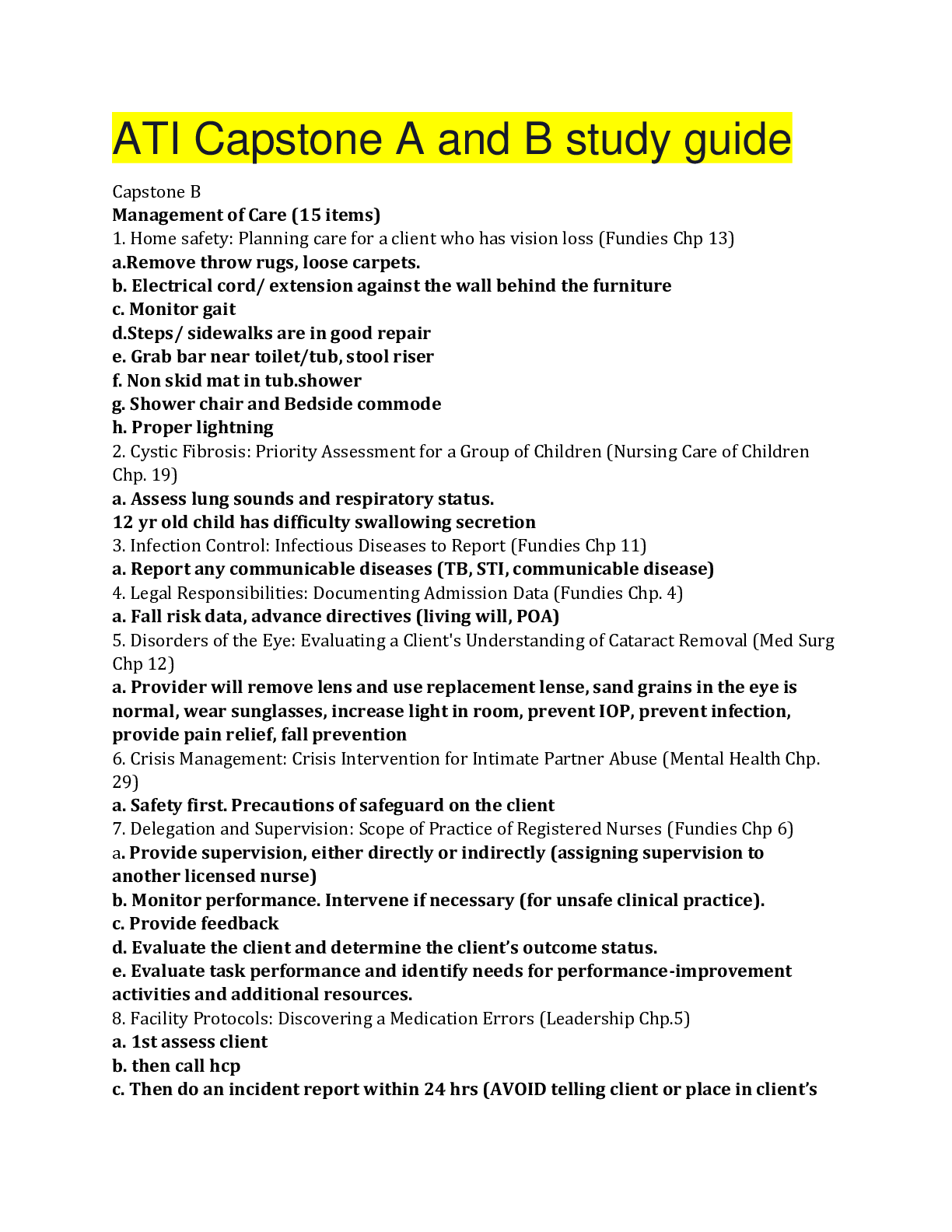
Pathophysiology > QUESTIONS and ANSWERS > PATHOPHYSIOLOGY NR 507WK4TD2 (All)
PATHOPHYSIOLOGY NR 507WK4TD2
Document Content and Description Below
PATHOPHYSIOLOGY NR 507WK4TD2 Week 4: Alterations in Renal Function - Discussion Part Two Loading... Discussion This week's graded topics relate to the following Course Outcomes (COs). 1 Analyze pa... thophysiologic mechanisms associated with selected disease states. (PO 1) 2 Differentiate the epidemiology, etiology, developmental considerations, pathogenesis, and clinical and laboratory manifestations of specific disease processes. (PO 1) 3 Examine the way in which homeostatic, adaptive, and compensatory physiological mechanisms can be supported and/or altered through specific therapeutic interventions. (PO 1, 7) 4 Distinguish risk factors associated with selected disease states. (PO 1) 5 Describe outcomes of disruptive or alterations in specific physiologic processes. (PO 1) 6 Distinguish risk factors associated with selected disease states. (PO 1) 7 Explore age-specific and developmental alterations in physiologic and disease states. (PO 1, 4) Discussion Part Two (graded) A 56-year-old female comes into the clinic complaining of intermittent severe pain that radiates from the flank to the groin and sometimes to the inner thigh. Upon further questioning she tell you that she has an urge to always go to the restroom and that she sometime sweats and feels nauseous. A urinalysis provides traces of blood, a few white blood cells and no bacteria. • What is your differential diagnosis? Discuss in detail the pathophysiology of each item in your differential and how it might fit in describing this case. • The x-ray comes back and there is nothing abnormal except a slightly dilated ureter. Does this change your differential or narrow it? • How would you treat the pain in this case? Responses Lorna Durfee Discussion Part Two Dr. Brown and Class: What is your differential diagnosis? Urinary Calculi. Nephrolithiasis A 56-year-old female comes into the clinic complaining of intermittent severe pain that radiates from the flank to the groin and sometimes to the inner thigh. Upon further questioning, she tells you that she has an urge always to go to the restroom and that she sometimes sweats and feels nauseous. A urinalysis provides traces of blood, a few white blood cells, and no bacteria. Tests: Urinalysis shows few white blood cells, trace blood = hematuria or microhematuria and no bacteria. Patients with nephrolithiasis can have macroscopic or microscopic hematuria, but the urine may be normal. There can be pyuria without bacteria. If there is pyuria and foul-smelling urine and fever, there can be an infection. If there is suspicion of crystalline substances in the sediment, further testing will be necessary (Preminger, 2014). X-ray: Slightly dilated ureter. Discuss in detail the pathophysiology of each item in your differential and how it might fit in describing this case. Pathophysiology of following items: Flank to groin pain into the thigh, severe and intermittent, urgency, sweats, nausea. 5/23/2016 1:47:48 PMPreminger (2014) relates that calculi can remain in the renal parenchyma or renal pelvis or passed to the ureter or bladder. An abrasion from a stone passing and trauma in the ureter and bladder walls can cause pain. Calculi, when lodged in the ureter, can cause obstruction and decreased urine flow. They can cause hydroureter or dilation of the ureter, and hydronephrosis, or a swelling in the kidney due to the build-up of urine (Preminger, 2014). Common areas of that stones get lodged are in the ureteropelvic junction and the distal ureter. Calculi that have a diameter of greater than 5 mm can be lodged in the ureteropelvic junction. Any calculi that are smaller are likely to pass through the system. When there is an obstruction, there is a decrease in the filtration of the glomeruli. With swelling in the kidney there is an increased glomerular pressure and decline in blood flow and then renal function (Preminger, 2014). Preminger (2014) tells us that stone remaining in the renal pelvis are not symptomatic until they cause obstruction and/or infection. The signs and symptoms that accompany this condition are severe pain that is accompanied by nausea. There can also be vomiting that usually occurs when calculi pass into the ureter or cause obstruction. The feeling of pain is due to renal colic that is excruciating and is intermittent. The pain continues in a cycle. When there is pain in the flank or kidney and radiates across the abdomen, it is suggestive of a ureteral or renal pelvic obstruction. If the pain radiates into the groin along the ureter, there can be a lower ureteral obstruction. If the pain is suprapubic, it can be a distal ureteral, ureterovesical calculus. It could also be a bladder calculus (Preminger, 2014). Nephrolithiasis: Curhan, Aronson & Preminger (2015) relate that renal and ureteral stones are seen most commonly seen in primary care. Patients usually present with renal colic and hematuria. Other patients may present with vague abdominal pain, acute abdominal flank pain, nausea. There is also urinary urgency and frequency and difficulty urinating. Approximately 80 percent of patient with this condition have calcium stones. Other stone types include calcium phosphate, uric acid, struvite, and cystine. The theory is that stone formation happens when the soluble material (calcium oxalate) supersaturates the urine, and this begins the crystal formation process. These crystals are anchored to collection ducts, and their size will increase with time. This process happens at sites of epithelial injury, and the crystals may cause this. Another theory is that stones and their formation begin at the renal medullary interstitium. Calcium phosphate crystals form, in the interstitium and then extruded from the renal papilla. The crystals of calcium oxalate and phosphate deposit on top of this place and then remain attached to the papilla (Curhan et al., 2015). Preminger and Curhan (2015) tell us that approximately 8 percent of women will have at least one stone by the age of 70. These stones will contain calcium oxalate. They also suggest that obesity among women has a part to play in the formation of the stones (Preminger & Curhan, 2015). Gould and Dyer (2011) tell us that stones in the kidney or bladder are asymptomatic frequently. Sometimes flank pain occurs because of distention in the renal capsule. With obstruction of the ureter, there are intense spasms of pain in the flank that radiates into the groin that can last until the stone can be removed. This pain comes from the ureter contractions which is trying to force the stone out. There can be a rapid pulse, nausea, and vomiting (Gould and Dyer, 2011, p. 455). The x-ray comes back, and there is nothing abnormal except a slightly dilated ureter. Does this change your differential or narrow it? I would not change the differential. The dilated ureter gives a definitive symptom that is related to urinary calculi. This patient needs further investigation. The use of a non-contrast helical CT can detect location and degree of obstruction. However, if this patient has had many CT scans, there is a concern for radiation exposure (Preminger, 2014). How would you treat the pain in this case? As the Centers for Disease Control and Prevention have Guidelines for Providers when it comes to opioids, I would try the recommendation of starting low and going slow when it comes to opioid medication. I would prefer not to use opioids and prefer anti-inflammatories. If opioids are to be used, they need to be at the lowest possible dose and the use of immediate-release opioids. I would only want to provide quantity needed for the duration of pain (The Centers for Disease Control and Prevention, 2016). Preminger (2014) states that treatment of renal colic is with the use of opioids such as morphine, or for rapid onset, fentanyl. He states both are used for pain relief and are appropriate. An antiemetic could also be used to stop the vomiting. Although increasing fluids has been traditionally used it has not proven to speed the passage of calculi. If the calculi do not pass within 6 to 8 weeks, then removal is necessary. When there is an immediate need for obstruction a ureteral stent can be used and then removal of the calculi (Preminger, 2014). References Curhan, G. C., Aronson, M. D., & Preminger, G. M. (2015). Diagnosis and acute management of suspected nephrolithiasis in adults. Retrieved from http://www.uptodate.com/contents/diagnosis-and-acute-management-of-suspected-nephrolithiasis-in-adults Gould, B. E., & Dyer, R. M. (2011). Urinary System Disorders. In Pathophysiology for the health professions (4th ed., p. 450-455). Preminger, G. M. (2014). Urinary Calculi (Nephrolithiasis). In Merck Manual online. Retrieved from http://www.merckmanuals.com/professional/genitourinary-disorders/urinary-calculi/urinary-calculi The Centers for Disease Control and Prevention. (2016). Guideline Information for Providers. Retrieved from http://www.cdc.gov/drugoverdose/prescribing/providers.html Qaseem, A., Dallas, P., Forciea, M. A., Starkey, M., & Denberg, T. D. (2014). Dietary and pharmacologic management to prevent recurrent nephrolithiasis in adults: a clinical practice guideline from the American College of Physicians. Annals Of Internal Medicine, 161(9), 659-667. doi:10.7326/M13-2908 Liberty Neoh Discussion 2 5/24/2016 7:35:48 PM Dr. Brown and Class, Based from the symptoms presented, the patient has kidney stones. Kidney stones can cause flank pain, typically there are acute spasms of severe pain, radiation of the pain to the groin, and then testicles in men and labia in women, nausea, and vomiting. The dilated ureter from the x ray result may be due to the obstruction caused by kidneys stones (Skolarikos, Dellis, & Knoll, 2015).Other differentials may include gallstones, irritable bowel syndromes, and other gastrointestinal disturbances. Most common complaints are abdominal pain. Patients’ health histories must be obtained in detail and needs to include accompanying signs such as abdominal pain relieved by defecation, pain associated with looser stools, pain associated with more frequent stools, sensation of incomplete rectal emptying, passage of mucous, and visible abdominal distension (Kirk et al, 2011). According to Resorlu and his colleagues (2013), “shock wave lithotripsy (SWL), percutaneous nephrolithotomy (PNL), and retrograde intrarenal surgery (RIRS) are the three main modalities for the management of upper urinary stone disease”. Treatments are usually performed in outpatient settings. References Kirk, G., Kennedy, G., McKie, L., Diamond, T., & Clements, B. (2011). Preoperative symptoms of irritable bowel syndrome predict poor outcome after laparoscopic cholecystectomy. Surgical Endoscopy, 53. doi: 10.1007/s00464-011-1729-7 Resorlu, B., Unsal, A., Ziypak, T., Diri, A., Atis, G., Guven, S.,…Oztuna, D. (2013). Comparison of retrograde intrarenal surgery, shockwave lithotripsy, and percutaneous ephrolithotomy for treatment of medium-sized radiolucent renal stones. World Journal of Urology, 31. doi: 10.1007/s00345-012-0991-1 Skolarikos , A., Dellis, A., & Knoll, T (2015). Ureteropelvic obstruction and renal stones: etiology and treatment. Urolithiasis, 43. doi: 10.1007/s00240-014-0736-2 Brooke Lobianco reply to Liberty Neoh RE: Discussion 2 5/29/2016 3:33:42 PM Liberty, Thank you for your post! Just to talk a little bit more in depth regarding the types of stones; most kidney stones are radiopaque these are the calcium containing stones; (calcium oxalate and calcium phosphate) with the most common being the calcium oxalate stones. "Calcium oxalate stones are found in 56 - 61% of adults, calcium phosphate in 8 - 18%", (Frassetto and Kohlstadt, 2011). Uric acid stones, crystine, and sturvite, are the stones that are radiolucent. "Uric acid stones are found in 9 - 17% of adults, sturvite in 2 - 4% and crystine only in about 1% of all adults", (Frassetto and Kohlstadt, 2011). These stones are not very common, they develop when there is to much uric acid in the urine. People who are known to have to much acid in their urine are those who are obese, have diabetes and people with gout. As we know gout is a result of too much uric acid in the blood. It is not uncommon for a diabetic to have gout or kidney stones. Frassetto and Kohlstadt (2011) wrote, "Contributing risk factors for kidney stones are obesity, insulin resistance, gastrointestinal pathology, living in warmer climates, and certain dietary patterns and medications". It was also stated that "patients with kidney stones should increase fluid intake to at least 2 L per 24 hours", (Frassetto and Kohlstadt, 2011). Frassstto, L. & Kohlstadt, I. (2011). Treatment and prevention of kidney stones:an update. American Family Physician 84(11). Retrieved from http://www.aafp.org/afp/2011/1201/p1234.html Rechel DelAntar Differential Diagnoses Hello Professor and Class, Differential Diagnoses This is a case of a 56 year old female complaining of intermittent severe pain radiating to the flank area extending to the groin accompanied by urgency in urination, nausea and periods of sweating. Further testing shows a Urinalysis result of trace blood, few white cells but no bacteria and an x-ray result, which shows a slightly dilated ureter otherwise, nothing significant. Based on these symptoms the patient may have: 1. Renal Stones = also known as a renal calculus or nephrolith, is a solid piece of material which is formed in the kidneys from minerals in urine. Kidney stones form when your urine contains more crystal-forming substances such as calcium, oxalate and uric acid than the fluid in your urine can dilute. At the same time, your urine may lack substances that prevent crystals from sticking together, creating an ideal environment for kidney stones to form. A stone may stay in the kidney or travel down the urinary tract and they vary in sizes. Small stone may pass on its own, causing little or no pain while larger stone may get stuck along the urinary tract and can block the flow of urine, causing severe pain or bleeding. Other associated symptoms include: nausea, vomiting, fever, blood in the urine, and painful urination. Blockage of the ureter can cause decreased kidney function and dilation of the kidney, which can be seen in an x-ray (Worcester, E., et. al., 2014). This diagnosis fits the symptomatology of the patient and is most likely what the patient is experiencing. 2. Interstitial Cystitis = an inflammation of the bladder. Otherwise known as Painful bladder syndrome, Interstitial cystitis (IC) is a condition that results in recurring discomfort or pain in the bladder and the surrounding pelvic region. The common denominator in interstitial cystitis/painful bladder syndrome is damage to the urothelium, which normally acts as a barrier against insults to the bladder. Damaged urothelium produces cytokines that activate mast cells in the interstitium. The diffusion of excess potassium into the bladder interstitium through a defective urothelium also triggers mast cell activation. The activation of mast cells results in a cycle of neuronal hyperexcitability leading to secretion of neurotransmitters and triggering further mast cell stimulation and degranulation. This process appears to contribute to the chronic pain, urgency, and frequency experienced by patients. People may experience mild discomfort, pressure, tenderness, or intense pain in the bladder and pelvic area. Symptoms may include an urgent need to urinate, a frequent need to urinate, or a combination of these symptoms. Pain may change in intensity as the bladder fills with urine or as it empties (National Institute of Diabetes and Digestive and kidney Disease, 2013). In PBS/IC, the patient does not exhibit fever and chills or signs of infection however the pain is concentrated more in the abdominal and pubic area. It doe not create ureteral dilatation as seen in the patient’s x-ray eliminating it as a possible diagnosis. 5/24/2016 8:28:03 PM3. Pyelonephritis = Pyelonephritis results when a UTI progresses to involve the upper urinary system (the kidneys and ureters) and is common among females. Symptoms include fever, chills, abdominal pain, nausea and vomiting, painful urination and frequent urination. In this case study however, the patient although experiencing nausea, painful urination and urgency, the patient is not having any fever and chills. The flank pain experienced with pyelonephritis is dull and achy compared to the severe pain experienced by the patient (Imam, T., 2016). Also, pyelonephritis does not use dilation in the ureters as shown in the patient’s x-rays. 4. Urinary Tract Infection = A urinary tract infection (UTI) is an infection in any part of the urinary system; kidneys, ureters, bladder and urethra. Most infections involve the lower urinary tract, the bladder and the urethra and occurs mostly in women. Bacteria are the most common cause of UTIs. Normally, bacteria that enter the urinary tract are rapidly removed by the body before they cause symptoms. However, sometimes bacteria overcome the body’s natural defenses and cause infection. Signs and symptoms include a strong, persistent urge to urinate, burning and frequent urination, cloudy urine to blood tinged urine, foul smelling urine and pelvic pain in women from the pelvis to the pubic area. (Lane, D.R. and Takhar, S.S., 2011). The patient although experiencing frequent urination is not experiencing cloudy and foul smelling urine. Also, the type of pain described by the patient does not fit the pain symptoms of a UTI making this diagnosis unlikely. Pain experienced with renal calculi is often described as the strongest sensation ever felt and known as renal colic. It typically comes in waves lasting 20 to 60 minutes caused by peristaltic contractions of the ureter as it attempts to expel the stone. Oral hydration and pain management using NSAIDs are part of the acute treatment of renal calculi. Severe pain is treated by IV pain meds, antispasmodics and opioids to relieve symptoms until other interventions are done to remove renal calculi (Lane D.R. and Takhar, S.S., 2011). References: Imam, T. (2016). Kidney Infection. Retrieved from http://www.merckmanuals.com/ home/kidney-and-urinary-tract-disorders/urinary-tract-infections-uti/kidney-infection. Lane, D.R. and Takhar, S.S. (2011). Diagnosis and management of urinary tract Infection and pyelonephritis. Emergency Medicine Clinics of North America.29(3). 539-552. National Institute of Diabetes and Digestive and Kidney Disease. (2013). Insterstitial Cystitis/Painful Bladder Syndrome. Retrieved from http://www.niddk.nih.gov/health-information/health-topics/urologic- disease/interstitial-cystitis-painful-bladder-syndrome/Pages/facts.aspx. Worcester, E., Goldfarb, S. and Lam, A. (2014). Cystine Stones. Retrieved from http://www.uptodate.com/contents/cystine-stones. Sarah Boulware reply to Rechel DelAntar RE: Differential Diagnoses Rechel, I found your post very informative. During my research I found some information on the treatment of kidney stones. The National Institute of Diabetes and Digestive Kidney Disease (2013) found that if a person is experiencing severe pain from a larger stone shock wave lithotripsy might be needed. The lithotripter machine generates shock waves that pass through the person’s body and break the kidney stone into smaller pieces that can readily pass through the urinary tract. A ureterscope can also be used to perform a Ureteroscopy to enter the bladder up into the urethra and retrieve the stone. These are more extreme treatments. Like you mentioned in your post pain medication and fluids are often used. If possible the patient should drink a lot of fluid to help move the stone along the ureter and out of the body. If the patient is dehydrated from vomiting and nausea IV fluids will be necessary. Many patients don’t seek treatment until they are having pain caused by the stone. At this point they are in a lot of pain requiring medication and fluid. Reference The National Institute of Diabetes and Digestive and Kidney Diseases. (2013). Kidney stones in adults. Retrieved from http://www.niddk.nih.gov/health-information/health-topics/urologic- disease/kidney-stones-in-adults/Pages/facts.aspx#treated Rechel DelAntar reply to Sarah Boulware RE: Differential Diagnoses 5/26/2016 6:58:50 PM Hello Sarah, Im glad you find my post informative. You are right, lithotripsy is used as non-invasive way of removing stones that are too big to pass and at this point is pressing on the renal calyx causing pain. Most renal stones that are too big remain on calyx since they are unable to pass to the ureters. These stones are so big they cause hydronephrosis and getting them out becomes urgent. Treatment for renal calculi starts with pain medications until the size of the calculi is determined via ultrasound or x-ray. Once it is determined it is too big to pass then a lithotripsy will then be performed to break up the larger stones to smaller one allowing the patient to pass it through the urinary tract. Patient's are encouraged to strain urine in order to determine if they have passed the stones (Cecen, K. et.al., 2014). And you are right, increase in fluid intake is necessary to flush out the calculi. If this is not effective then more invasive means have to be done to extract the renal calculi. Reference: Cecen, K., Karadag, M., Demir, A., Bagcioglu, M., Kocaasia, R. and Sofikerim, M. (2014). Flexible Ureteroscopy versus Extracorporeal Shockwave Lithotripsy for the treatment of upper/middle calyx kidney stones of 10-20mm: a retrospective analysis of 174 patients. Retrieved from http://springerplus.springeropen.com/articles/10.1186/2193-1801-3-557. 5/26/2016 11:34:54 AM Deborah Matheny reply to Sarah Boulware RE: Differential Diagnoses 5/29/2016 5:56:29 PM Hello Sarah and Rechel: I found both of your posts informative and I can add to the conversation through personal experience. In 1994 I had my first encounter with kidney stones and I can confirm that the pain is excruciating especially when a blockage has occurred. I had a complete blockage and had to have a stent placed and removal with what they had called a basket then I had lithotripsy to break up remaining stones for ease of passage. Lithotripsy is when they use shock waves to break up the stones (crush them almost into a sandy material) and afterwards I can tell you that you feel as if you had been hit by a truck as you ache for several days afterwards. Several years later I had a recurrence of stones but no stent was needed at this time but lithotripsy was still called for as several stones were too large to pass on their own and a third and final experience occurred in 2000 where blockage again occurred, a stent was placed and lithotripsy was also used. In two cases the stones were calcium oxalate while the third and final time the stones were calcium phosphate. I can honestly say that it took three episodes for me to finally believe that drinking more fluids (not soda and coffee) was needed to prevent any further episodes (I admit to being hard-headed at times). My urologist encouraged me to drink one 6-oz glass of cranberry juice daily as this would help flush any sediment out of my kidneys before any stones formed again. I have not had another episode since 2000, I drink 4 to 6 bottles of water daily, and I still drink that one glass of cranberry juice daily. Whether it works or not I believe it does and will continue to drink it. I will admit that all three experiences were very painful with the stones and with the lithotripsy.As a RN I have discussed my experience with several urologists and have found that those that have been in practice for a longer period recommend use of cranberry juice while those who have less time in this field encourage more fluid intake but are on the fence about whether cranberry juice works or not. If I am asked I tell about my experience only but as a nurse practitioner I believe I will encourage drinking cranberry juice to help in preventing reoccurrence. Again let me thank you both for an informative and interesting read. Debbie Jonathan Bidey reply to Rechel DelAntar RE: Differential Diagnoses Rachel, Excellent post! You did a wonderful job describing the differential diagnoses for this patient. I definitely agree that this patient is most likely experiencing kidney stones. You mentioned pain management until the stone is able to be removed. Under normal circumstances, the stone should be allowed to pass without retrieval. However, sometimes the stone is too large or becomes lodged in the kidney or ureter. In order to remove these stones, intervention is required. Surgery can be performed, but the most widely used intervention is lithotripsy. Lithotripsy uses sound waves to break up the stone. The stone is then able to be passed by the patient since it is now smaller. Lithotripsy can be performed in three different ways. These are electrohydraulic, piezoelectric, or electromagnetic (Neisius et al., 2015). The electrohydraulic method creates a shock wave through an electric spark gap in water (Neisius et al., 2015). The piezoelectric method creates a shock wave through high voltage pulsed energy focused on ceramic elements which elicit an abrupt expansion (Neisius et al., 2015). The electromagnetic method creates shock waves through a metallic membrane which is propelled with an electrical impulse through a shock tube (Neisius et al., 2015). -Jonathan Bidey Reference: Neisius, A. (2015). Shock wave lithotripsy: The new phoenix? World Journal of Urology, 33(2), 213-221. http://dx.doi.org/10.1007/s00345-014-1369-3 5/29/2016 10:23:38 AM Sarah Boulware Part Two 5/25/2016 2:45:45 PM Dr. Brown and Class, 1. Perirenal abscess secondary to urolithiasis A perirenal abscess is a pocket of pus around one or both of the kidneys. Pus accumulates in the space between the renal capsule and Gerota’s fascia. They are most often caused by urinary tract infections that begin in the bladder and spread to the kidney. Surgery in the urinary tract or reproductive system or a bloodstream infection can also lead to a perirenal abscess. The biggest risk factor for a perirenal abscess is kidney stones. When kidney stones block urine flow it provides a place for infection to grow. Bacteria tend to stick to the stones and antibiotics cannot kill the bacteria there. Kidney stones are found in 20 to 60 percent of patients with perirenal abscess. Symptoms include pain in the flank that may extend to the groin or down the leg, sweating, fever, and chills (U.S. National Library of Medicine, 2015). The patient’s symptoms are concurrent with a perirenal abscess. She is not exhibiting fever or chills but does have radiating pain from her flank down to her groin and leg as well as sweating. Part Two: The x-ray came back with only a sign of a slightly dilated ureter. This indicates there may have been a urolithiasis obstruction, which could have provided the perfect place for bacteria to colonize. The stone may have passed leaving no signs other than the slightly dilated ureter. The pus from the abscess can be drained through a catheter that is placed through the skin or during surgery. Antibiotics are also indicated. Pain should resolve with treatment (U.S. National Library of Medicine, 2015). 2. Obstructive Uropathy secondary to Urolithiasis The patient is exhibiting what is commonly referred to as renal colic symptoms including flank pain, nausea, and hematuria. Renal colic pain is described as waves of severe pain that is first felt in the flank and radiates toward the groin. This type of pain does not change with position and can last from 20 to 60 minutes. The location of the pain may correlate with the location of the stone. If the stone is at the ureteropelvic junction it can cause acute flank pain. If a stone obstructs the urtero-vesical junction symptoms will include dysuria, urinary frequency, and urgency (Hochwind & Ashcroft, 2012). Urolithiasis is often easily diagnosed due its classic presentation. Kidney stones, or calculi, form when various minerals combine in the urine to form crystals. The most common minerals are calcium, oxalate, and phosphate. The crystals precipitate to form a stone. The stone can be formed from many different causes including low total urine volume, alterations in urine pH balance, or when there is an overabundance of stone-forming mineral salts in the urine causing it to become super-saturated (Hochwind.& Ashcroft, 2012). Saturation is often described as the concentration ratios of calcium oxalate or calcium phosphate to its solubility. The majority of kidney stones contain calcium.The calcium-based stones are composed of calcium oxalate, calcium phosphate or both. Symptoms occur when there is an obstruction and the type of pain usually correlates with the location of the stone (Chung, Stern, & Dufton, 2013). 3. Bladder Cancer Most cancers involving the bladder, renal pelvises, ureters, and the proximal urethra are transitional cell carcinomas that derive from the transitional epithelium. Normally the transitional epithelium consists of a specialized mucous membrane that lines these structures. Bladder cancer can be low-grade, which is a mild version of the cancer, or high-grade that has a strong tendency to invade the muscular wall of the bladder and spread. This is classified as muscle-invasive bladder cancer and is likely to spread to other parts of the body. Nonmuscle-invasive disease is confined to the bladder. Transitional cell carcinomas represent the majority of bladder cancer occurrences. Bladder cancer typically presents with gross or microscopic hematuria. Patients can experience urinary frequency, nocturia, and dysuria but this is more common in patients with carcinoma in situ. Patients that present with pain may have an upper urinary tract urothelial carcinoma with tumor obstruction. The patient does have symptoms associated with tumor obstruction related to upper urinary tract urothelial carcinoma. She also presents with traces of blood in her urine, however it is less than is often seen with bladder cancer. Further diagnostic testing is needed to rule out this differential diagnosis (National Cancer Institute, 2016). 4. Painful Bladder Syndrome (PBS)/ Interstitial cystitis (IC) PBS/IC is defined as pain and urinary discomfort in the absence of infection or other identifiable causes. It is often characterized by bladder pain, urinary frequency, urgency, and nocturia. Pain is often located suprapubicaly and can sometimes radiate to the groin. Pain is relieved by voiding but returns quickly. The mucous layer of the bladder that provides the primary protection is defective or damaged, which results in direct exposure of the bladder mucosa and submucosal nerve endings to urine. The high content of potassium and other irritants that are found in urine cause an inflammatory reaction in the bladder wall. The patient’s urinalysis showed no major causes for infection or other reason for pain and frequency. The pain is not suprapubic but does radiate to her groin (Flander, 2013). References Chung, C., Stern, P., & Dufton, J. (2013). Urolithiasis presenting as right flank pain: a case report. Journal of Canadian Chiropractic Association, 57(1), 69-75.Flander, N. (2013). Painful bladder syndrome and interstitial cystitis: treatment options. British Journal of Nursing, 22, 20-27. Hochwind, C. & Ashcroft, K. (2012). Tamsulosin for ureteral stones – use in a pediatric population? Urologic Nursing, 32(2), 88-92. Liu, X., Wang, C., Liu, Y., Lui, K. (2016). Renal and perinephric abscesses in West China hospital: 10-year retrospective-descriptive study. World Journal of Nephrology, 5(1), 108-114. doi: 10.5527/wjn.v5.i1.108 National Cancer Institute. Bladder cancer treatment – health professional version. Retrieved from http://www.cancer.gov/types/bladder/hp/bladder-treatment-pdq U.S. National Library of Medicine. (2015). Perirenal abscess. Retrieved from https://www.nlm.nih.gov/medlineplus/ency/article/001274.htm Lanre Abawonse Discussion Part Two 5/25/2016 7:42:19 PM Kidney Stones are stones that form in the kidneys from the crystallization ofminerals and other substances that are normally dissolved in the urine. Stones are solutes that grow in supersaturated urine. The more common forms of stones are made up of calcium, oxalate, sodium, phosphorus, cystine and uric acid (Dawson& Tomson, 2012). The precise cause of kidney stones is unknown; it is thought they are associated with dehydration, urinary obstruction, and calcium levels. The symptoms of kidney stones do not appear until the stone dislodges and begins to travel down the urinary tract and enters the ureter. The pain that the patient is experiencing begins in the flank and radiate into the lower groin. One of the classic signs of a kidney stone is pain from the flank to the groin and traces of blood found on the urinalysis (Kidney Stones, 2012). Urinary Tract Obstruction Obstructive disorder of the urinary tract interferes with the flow of urine. It may be congenital or acquired intraluminal (renal calculi) or secondary to extrinsic compression (tumor). In complete or significant partial obstruction, hydrostatic pressure increases proximal to the obstruction as a consequence of continued glomerular filtration and simultaneous obstruction to the flow of urine. Obstruction can be complete or partial and it can lead to kidney damage (McCance, Huether, Brashers, & Rote, 2013). Symptoms include flank pain, which can be intermittent, and nausea and vomiting. These symptoms are consistent with the patient’s symptoms. Partial obstructions can be treated with medications. Complete obstructions may require surgery (Aleksic, 2015). Pyelonephritis is a type of urinary tract infection of the renal pelvis and renal tissue; it is caused by an invasion of microorganisms. This is usually unilateral, involving the right kidney 50% of the time and left kidney 25% of the time. In acute cases, it originates from an ascending infection but may arrive at the kidney via the bloodstream. Once in the kidney, bacteria bind to epithelial cell receptors, initiating an inflammatory response. Inflammatory mediators and bacterial toxins are responsible for the parenchymal damage to the kidney. Symptoms include flank pain, hematuria, polyuria, dysuria, urgency, malodorous urine and fever. (Bethel, 2012). The x-ray comes back and there is nothing abnormal except a slightly dilated ureter. Does this change your differential or narrow it? With the x-ray result confirming the dilation of the ureter, this helps to narrow down the diagnosis, as obstruction in a ureter causes dilation of the ureter (megaureter). Kidney stones are a common cause of urinary tract obstructions (McCance, Huether, Brashers, & Rote, 2013). How would you treat the pain in this case? Different modalities can be used to treat the pain associated with the kidney stone. The use of narcotics and nonsteroidal anti- inflammatory agents is considered useful as it helps to relax the ureter and facilitate passage of the stone. The drugs of choice are morphine sulfate and ketorolac. The use of alpha blockers can be used to relax the smooth muscle in the ureter to help the stone pass and relieve the pain (Kidney Stones, 2012). Reference: Aleksic, D. (2015). Acute urinary tract obstruction. Serbian Journal of Experimental & Clinical Research, 16(3), 249. doi:10.1515/sjecr-2015-0033 Bethel, J. (2012). Acute pyelonephritis: Risk factors, diagnosis and treatment. Nursing Standard, 27(5), 51-56 6p. Dawson, C. H., & Tomson, C. V. (2012). Kidney stone disease: pathophysiology, investigation and medical treatment. Clinical Medicine (London, England), 12(5), 467-471. Kidney stones: Common, painful, preventable. (2012). Harvard Men's Health Watch, 16(6), 1-5 McCance, K. L., Huether, S. E., Brashers, V. L., & Rote, N. S. (2013). Pathophysiology: The biologic basis for disease in adults and children (7th ed.). St. Louis, MO: Mosby. Brittany Heller Kidney stone 5/26/2016 7:40:59 AM My initial diagnosis will be a kidney stone due to all the patient’s reporting symptoms. Kidney Stones: “A Calculi or urinary stone are masses of crystals, protein, or other substances, that are common cause of urinary tract obstruction in adults” (McCance & Huether, 2014, p. 1731). Kidney stones are more common in men than women. Risk factors associated with kidney stones are: age, gender, race, geographic location, seasonal factors, fluid intake, diet, occupation, genetic predisposition, UTI, hypertension, atherosclerosis, metabolic syndrome, obesity anddiabetes (McCance & Huether, 2014, p. 1343). Urinary stones are classified according to what they are compromised of. Up to 80% of the kidney stone types are composed of calcium oxalate or phosphate (McCance & Huether, 2014, p. 1343). Clinical presentation of kidney stones include renal colic. Renal colic is moderate to severe pain originating from the flank to the groin. This type of pain indicates there is an obstruction in the midureter and will have lower urinary tract symptoms such as urgency, frequency, and incontinence (McCance & Huether, 2014, p. 1344). The severe pain can also be associated with nausea and vomiting. These are all symptoms that the patient is presenting with and why this is my leading diagnosis. Other signs and symptoms include gross or microscopic hematuria, which the patient has as well. The formation of renal calculi depends on the formation or presence of four processes. The first step would be he supersaturation of one or more salts in the urine (McCance & Huehter, 2014, p. 1343). The second step would include the precipitation of the salts from a liquid to a solid state to form crystals. The third step would be the growth of the crystals through crystallization or agglomeration (McCance & Huether, 2014, p. 1343). The fourth step would also include the presence or absence of the stones inhibitors. Due to the patient’s presenting factors, the dilated ureter means that there is an obstruction and that means I would not change my diagnosis. Treatment of kidney stones include managing the acute pain, promote the passage of the stone, possibly reduce the size of the stones that have formed, and prevention of new stones forming (McCance & Huether, 2014, p. 1344). Oral or parenteral analgesics would be ordered and given. McCance, K. & Huether, S. (2014) Pathophysiology: The Biologic Basis for Disease in Adults and Children(7 ed). St. Lois: Elsevier. th Lorna Durfee reply to Brittany Heller RE: Kidney stone Brittany: I found something interesting related to children and kidney stones. Smith and Stapleton (2016) inform us that stones are less common in children. However, most who do develop kidney stones have an underlying condition, or they develop them for unknown reasons. The most common symptoms of kidney stones are a pain in the belly, hematuria, nausea or vomiting and an urgency to go to the bathroom. Sometimes young children do not have any symptoms, and usually, a stone is found on the x-ray done for another medical issue (Smith & Stapleton, 2016). There are certain factors that increase a child’s risk. If a child has a prior history of stones in the past, they are more likely to have another stone in the future. Children sometimes do not drink enough fluid, and this affects the amount of urine the body makes. As we know, when drinking small amounts of liquid it concentrates the stone-forming substances in the urine. To prevent that from happening drinking more fluids can reduce the risk of recurrence of stones by making the urine less concentrated. Also, other conditions can increase the risk of stone formation. For example, children with epilepsy, who use the ketogenic diet can increase the risk of development of kidney stones. This diet is used to treat seizures. Children with cystic fibrosis are also at high risk for development of stones. If a child has a urinary tract abnormality of the kidneys or other abnormalities in the urinary tract that can increase the risk for problems also. There are also inherited disorders that can increase the risk of stone development (Smith & Stapleton, 2016). Smith and Stapleton (2016) suggest that if a child has symptoms of a kidney stone, they should see a doctor or nurse as soon as they can. Sometimes trying home treatment can help, if the pain is manageable and the child has no other health issues. They recommend the use of Advil. They recommend pushing more fluids to help push out the stone. Treatment at home also consists of straining the urine of the child for a few days. If there is no resolution, then the child may need to be hospitalized, particularly if there is severe pain and vomiting. Stones can cause permanent damage if not treated promptly. Treatments then can progress to shock wave lithotripsy, percutaneous nephrolithotomy, and ureteroscopy. For prevention or recurrence of stones blood and urine tests can be done. Also, if a stone has passed it is vital to know what type of stone it was. Future management and kidney stone prevention is based on kidney stone composition. To prevent recurrence, we have the patient utilize dietary and fluid management according to stone type. References Smith, J., & Stapleton, F. B. (2016). In T. W. Post (Ed.), UpToDate. Kidney stones in children. Retrieved from http://www.uptodate.com/contents/kidney-stones-in-children-beyond-the-basics Brooke Lobianco Kidney Stones Patient: Betty Doe- 56-year-old female Chief Complaint: Intermittent severe pain that radiates from the flank to the groin and sometimes to the inner thigh, urinary frequency, sweating with associated nausea Medical History Medications Allergies Surgical History Social History -N/A -N/A - N/A - N/A - N/A Vitals: N/A Physical Exam: Unremarkable Urinalysis: Traces of blood, a few white blood cells and no bacteria. Kidney Stones (Nephrolithiasis) Per Goroll and Mulley (2014), 1-3% of the population will experience a renal calculus (stone) at some point in their lives. Due to the patient’s current complaints, she may be experiencing a kidney stone, also referred to as nephrolithiasis or renal calculi. 75% of kidney stones are composed of calcium salts like calcium oxalate, and less commonly of calcium phosphate, uric acid, struvite or apatite, and cysteine (Goroll & Mulley, 2014). The two main factors that are key in the pathogenesis of stone formation include changes in urinary concentration, and physiochemical changes (Goroll & Mulley, 2014). Hypercalciuric and hperoxaluric states promote stone formation via several methods (Goroll & Mulley, 2014). The main methods include increased gut absorption of dietary calcium, increased resorption of calcium from the bone, and presence of a renal calcium leak (Goroll & Mulley, 2014). 50% of patients with calculus are hypercalciuric due to genetic factors that increase renal calcium excretion, however, potentially contributing gene mutations have yet to be discovered (Goroll & Mulley, 2014). 30% of patients are 5/26/2016 9:59:51 AM 5/26/2016 9:49:39 AMhyperoxaluric, which may be secondary to an increased absorption of dietary oxalate (Goroll & Mulley, 2014). Physiochemical changes refer to changes in the urinary PH and concentration of potential inhibitors of stone formation, such as magnesium, citrate, sulfate, and pyrophosphate) (Goroll & Mulley, 2014). Prevention is largely dietary. Per Gul and Munga (2014), those at high risk should be drinking enough fluid to produce 2.5 liters of urine daily. Drinking fruit juices reduces stone formation by increasing urine volume, and it is also high in potassium and citric acid. Citrate, such as that found in the recommended 4 ounces of lemon juice daily, binds with urinary calcium and reduces super saturation of urine, while binding to calcium oxalate crystals to prevent crystal growth (Gul & Munga, 2014). Symptoms of nephrolithiasis range from asymptomatic to severe (Goroll & Mulley, 2014). A common characteristic is renal colic, which refers to a ureteral obstruction presenting as an abrupt onset of persistent, waxing and waning pain, that localizes in one flank as the stone wedges in the uretero-pelvic junction, and radiates to the genitalia and groin as the stone wedges lower in the urethra (Goroll & Mulley, 2014). Nausea and vomiting is also not uncommon (McCance, Huether, Brashers, & Rote, 2013). This is caused by the anatomical localization of the autonomic nervous system, which is in close proximity of the visceral nerves, allowing interaction between nerves. The pain sensation in renal colic is due to the obstruction of urinary flow with subsequent increasing wall tension in the urinary tract (Jung & Osther, 2015). Urinary frequency can be explained based on the location of the stone. Bothersome lower urinary tract symptoms indicate obstruction of the lower ureter or ureterovesical junction (McCance et al., 2013). A dilated ureter on x-ray can be interpreted as hydroureter (an accumulation of urine in the ureter). This dilation is an indication of obstruction such as that caused by a stone (McCance et al., 2013). Pain management is a main component of kidney stone management. According to Jung and Osther (2015) the use of NSAIDs is first-choice analgesia due to their direct effect on prostaglandin release thereby decreasing the renal pressure resulting in pain relief. Goroll, A.H., & Mulley, A.G. (2014). Approach to the patient with nephrolithiasis. In A.H. Goroll & A.H. Mulley (Eds.) Primary Care Medicine: Office Evaluation and Management of the Adult Patient (7 ed.) (pp.981-982). Philadelphia, PA: Wolters Kluwer. th Gul, Z., & Munga, M. (2014). Medical and Dietary Therapy for Kidney Stone Prevention. Korean Journal Of Urology, 55(12), 775-779. Jung, H., & Osther, P. (2015). Acute management of stones: When to treat or not to treat?. World Journal Of Urology, 33(2), 203. doi:10.1007/s00345-014- 1353-y McCance, K. L., Huether, S. E., Brashers, V. L., & Rote, N. S. (2013). Pathophysiology: The biologic basis for disease in adults and children (7th ed.). St. Louis, MO: Mosby. Michelle Demey Discussion Part Two 5/26/2016 4:29:02 PM A 56-year-old female presents with complaints of intermittent severe pain that radiates from the flank to the groin and sometimes inner thigh. She also has frequent urination and sometimes sweats and feels nauseous. Urinalysis provides traces of blood, and a few white blood cells, negative for bacteria. X-ray impression notes a dilated ureter. The differential diagnosis of anatomical obstruction secondary to kidney stones (urolithiasis) is likely the cause of this patients signs and symptoms. Radiology findings, and microscopic hematuria in the presence of acute flank, groin, and thigh pain is suggestive of renal colic. The pathophysiology is that urine is a metastable solution that contains many compounds and salts including calcium, oxalate, phosphate, and uric acid. Kidney stones form when urine becomes supersaturated with stone-forming salts (Carter et al., 2011). These salts precipitate out of solution and form crystals, which accumulate at anchoring sites to form stones (Carter et al., 2011). Acute flank pain is a common presenting complain to the emergency department, requiring a broad differential diagnosis and work-up. Nephrolithiasis appears to be the most frequent cause of flank pain, affecting 3% to 5% of the population. The stone is a calculus of mineral or organic solids that can form anywhere in the urinary tract or more specifically in the ureter (Carter et al., 2011). Precipitates and stones less than 5 millimeters are often passed through the urinary tract with normal micturition. Larger stones that are unable to pass may create a partial or complete urinary tract obstruction with subsequent flank pain, nausea, and vomiting (Chamberlain College of Nursing [CCN], 2015). Obstructions, partial or complete, often result in inflammation and infection. Each ureter is made up of smooth muscle close in proximity allowing the direction of transmission of electrical stimulation from one cell to another. This allows for peristalsis even though the ureter is denervated. Sensory innervation for the upper part of the ureter arises from the tenth thoracic nerve roots, with referred pain to the umbilicus. The innervation of lower segments arises from the sacral nerve with referred pain to the vulva or penis (McCance & Huether, 2014). Renal colic is defined as severe intermittent flank pain that radiates to the groin, lower abdomen, or genitalia due to the passage of stone through the urinary system. Pain is often accompanied by nausea, vomiting, dysuria, and hematuria (Carter et al., 2014). The innervation of the bladder and internal urethral sphincter is supplied by parasympathetic fibers of the autonomic nervous system that arise from the sacral levels of the spinal cord, S2 and S4. Skeletal motor neurons in the pudendal nerve innervate the external urethral sphincter. The reflex arc required for micturition is stimulated by mechanoreceptors that respond to stretching of tissue. When the bladder accumulates 250 to 300 ml of urine, the bladders contracts and the internal sphincter relaxes through activation of the spinal reflex arc. It is at this time a person feels the urge to void (McCance & Huether, 2014). If a person is unable to empty the bladder or empties small amounts, secondary to partial or complete obstruction, the tissue continues to stretch and stimulates the urge to urinate. Injury causes an immune response that includes type II, III, or IV hypersensitivity reactions. The inflammation and accumulation of cells places pressure on the capillaries and decreases blood flow. The damage to capillary membrane can cause increased permeability of the membrane and loss of proteins and red blood cells into the urine (CCN, 2015). Also the bladder has a profuse blood supply, accounting for the bleeding that readily occurs with trauma, surgery, or inflammation (McCance & Huether, 2014). The acute treatment of kidney stones addresses pain management and focuses on the morbidity associated with an obstructed renal system. The diagnostic modality of choice is a non-contrast computed tomography (CT); ultrasonography is preferred in pregnant patients and children. Combining opiods with non-steroidal anti-inflammatory drugs (NSAIDS) is the optimal evidence-based regimen to treat severe symptoms (Carter et al., 2011). References Carter, M., Green, B., Ban, K., & Shah, K. (2011, July). Renal calculi: emergency department diagnosis and treatment.EBMEDICINE.NET, 13(7). Retrieved from http://www.ncbi.nlm.nih.gov/pubmed Alterations in renal function [lesson week 4] (2015, November). In Chamberlain College of Nursing. Retrieved May 26, 2016, from https://devry.equella.ecollege.com/file/2f71861b- ba05-455f-be03-59d157f82e5c/2/NR507McCance, K., & Huether, S. (2014). Pathophysiology: The biological basis for disease in adults and children vol. 2 (7th ed.). St. Louis, MO: Elsevier Mosby. Heather Orr Discussion Part 2 5/26/2016 5:17:36 PM A 56-year-old female comes into the clinic complaining of intermittent severe pain that radiates from the flank to the groin and sometimes to the inner thigh. Upon further questioning she tell you that she has an urge to always go to the restroom and that she sometime sweats and feels nauseous. A urinalysis provides traces of blood, a few white blood cells and no bacteria. • What is your differential diagnosis? Discuss in detail the pathophysiology of each item in your differential and how it might fit in describing this case. 1. Kidney stone – Kidney stones occur when substances found in the urine such as salts become concentrated and form solid masses. This typically occurs in periods of dehydration or when dietary changes happen which promote stone formation. When the salts found in the urine become higher in concentration than the fluid supersaturation occurs and the fluid is no longer able to dissolve all of the salts. When this happens in the urine it promotes stone formation because the salts begin to form small crystals and then these small crystals continue to grow eventually forming the stones. When kidney stones are present patients will complain of flank pain that is severe in nature often causing nausea and vomiting and that radiates to the groin exactly as our patient describes. Also present due to the microtrauma the stones inflict will be either microscopic or gross hematuria as found in our patient’s urinalysis (McCance and Huether, 2014). 2. Pyelonephritis – Pyelonephritis is an infection occurring in the kidneys and usually occurs as a result of pathogens making their way from the bladder to the ureters and finally the kidneys. Because of the inflammation in the kidneys secondary to the infection flank pain can occur and be accompanied by nausea, vomiting, alternating chills and sweats, and fever. We are not given information on our patient’s temperature however she does report nausea, flank pain, and periodic sweating. Urinalysis results also support pyelonephritis as pyuria is present however most often pyuria in pyelonephritis is significant and microscopic hematuria is not always present (Colgan, Williams, and Johnson, 2011). 3. Glomerulonephritis – The glomerulus in the kidney are susceptible to primary injury due to drugs, infection, immunologic responses, and ischemia or secondary injury as a result of systemic diseases. When the glomerulus experience injury they become inflamed and the condition is called glomerulonephritis. When injury and inflammation occur to the cellular components of the glomerulus red blood cells are released and the normal filtration barrier is disrupted. This disruption leads to protein in the urine which is not mentioned in our case study making this diagnosis less likely (Lewis and Maxwell, 2015). 4. Renal Tumor – Renal tumors rarely benign and renal malignancies currently account for 2.3% of deaths attributable to cancer. As tumors grow in the kidney ruptures of blood vessels in the kidney occur which cause either gross or microscopic hematuria while the inflammatory response of the body to the tumor causes pyuria both seen in our case. Also, flank pain which may radiate to other areas of the abdomen is often one of the first symptoms and what brings patients in to be examined. While general episodes of nausea accompanied by sweating are not typically reported with renal tumors there is an incidence of reported night sweats and a further description of our patients sweat experience should be garnered. (Furlow, 2016; McCance and Huether, 2014). • The x-ray comes back and there is nothing abnormal except a slightly dilated ureter. Does this change your differential or narrow it? ◦ This narrows my differential to the most likely diagnosis of kidney stone. When there is an obstruction of the upper urinary tract such as in the case of kidney stones hydroureter can result. This is visualized as dilation of the ureter. • How would you treat the pain in this case? ◦ Pain in kidney stones is reported to be incredibly severe. If the patient was going to be admitted to the hospital I would use IV pain medication such as Dilaudid 0.5mg q4 hr or Morphine 4mg IV q4h. If the patient were to be sent home I would prescribe Norco 10/325 q4 hr. References: Colgan, R., Williams, M., Johnson, J.R. (2011). Diagnosis and treatment of acute pyelonephritis in women. American Family Physician, 84(5), 519-526. Furlow, B. (2016). Computed tomography of renal masses in adults. Radiologic Technology, 87(3), 305-329. Lewis, G. & Maxwell, A. P. (2015). Timely diagnosis and treatment essential in glomerulonephritis. The Practitioner, 259(1779), 13. McCance, K. L., & Huether, S. E. (2014). Pathophysiology: The biologic basis for disease in adults & children (7th ed.). St. Louis: Mosby. Jennifer Roth Part 2 5/26/2016 5:42:31 PM Hello Dr. Brown and Classmates, The differential diagnosis is urolithiasis, or a kidney stone. Approximately 80% of calcium kidney stones are calcium oxalate (CaOx), with a small percentage (15%) of calcium phosphate (CaP) (Sakhaee, Maalouf, & Sinnott, 2012). The pathophysiological mechanisms for calcium kidney stone formation are complex and diverse and include low urine volume, hypercalciuria, hyperuricosuria, hypocitraturia, hyperoxaluria, and abnormalities in urine pH (Sakhaee, Maalouf,& Sinnott, 2012). The pathophysiological mechanisms for hypercalciuria are numerous and may involve increased intestinal calcium absorption, decreased renal calcium reabsorption, and enhanced calcium mobilization from bone (Sakhaee, Maalouf, & Sinnott, 2012). However, intestinal calcium hyperabsorption is the most common abnormality in this population. “Hypercalciuria is a heterogeneous disorder in which intestinal calcium hyperabsorption may be dependent or independent of 1,25-dihydroxyvitamin D” (Sakhaee, Maalouf, & Sinnott, 2012, p. 1850). The most severe variant is characterized by normocalcemia, hypercalciuria, intestinal hyperabsorption of calcium, and normal or suppressed serum PTH and/or urinary cAMP (Sakhaee, Maalouf, & Sinnott, 2012). However, a less severe form shares many of the same biochemical characteristics, but hypercalciuria normalizes after a restricted calcium diet of <400 mg/d (Sakhaee, Maalouf, & Sinnott, 2012). The pathophysiological mechanism underlying hyperuricosuria is attributed to a high purine diet. The physicochemical basis involved in this process has not been well established. One study has attributed the physicochemical process to urinary supersaturation with colloidal monosodium urate-induced CaOx crystallization, another study has shown a lack of effect of monosodium urate and attributes CaOx stone formation to decreased solubility of CaOx in solution (Sakhaee, Maalouf, & Sinnott, 2012). Hypocitraturia commonly occurs with metabolic acidosis or acid loading mediated through up- regulation of proximal renal tubular reabsorption of citrate (Sakhaee, Maalouf, & Sinnott, 2012). “The physicochemical basis for the inhibitory role of citrate involves the formation of soluble complexes and reduction of urinary saturation with respect to calcium salts in addition to direct inhibition of CaOx crystallization processes” (Sakhaee, Maalouf, & Sinnott, 2012, p. 1853). The underlying mechanisms of hyperoxaluria can be divided into: oxalate overproduction as a result of an error in metabolism, increased dietary intake, and increased intestinal oxalate absorption. “Inborn errors in metabolism include type I hyperoxaluria resulting from a deficiency or mistargeting of hepatic alanine glyoxylate transferase, type II primaryhyperoxaluria due to a deficiency in glyoxylate reductase/hydroxypyruvate reductase, and the rare type III hyperoxaluria as a result of the gain of function of hepatic or renal mitochondrial 4-hydroxy-2-oxoglutarate aldolase” (Sakhaee, Maalouf, & Sinnott, 2012, p. 1854). Both highly acidic urine (pH ≤ 5.5) and highly alkaline urine (pH ≥ 6.7) predispose patients to calcium kidney stone formation. With acidic pH, urine becomes supersaturated with uric acid that participates in CaOx crystallization. The mechanism for the formation of calcium stones is increased urinary supersaturation of stone-forming salts, which leads to homogeneous nucleation in the lumen of the nephron, followed by crystal growth and consequent obstruction in the distal nephron. Since the x-ray was negative for calculi, I would narrow the diagnosis down to renal colic. The calculi has likely already passed out of the body. I would treat renal colic with an NSAID and an antiemetic if nausea continues, as well as educate the individual on the necessity for high fluid intake and the benefits of using lemons to prevent calculi formation. Non-steroidal anti-inflammatory drugs offer the best initial analgesia, with opiates as a second line treatment (Bultitude & Rees, 2012). Sometimes the pain can be excruciating and a narcotic pain reliever may be warranted. Reference Bultitude, M. & Rees, J. (2012). Management of renal colic. British Medical Journal, 345:e5499. doi: 10.1136/bmj.e5499 Sakhaee, K., Maalouf, N.M., & Sinnott, B. (2012). Kidney stones 2012: Pathogenesis, diagnosis, and management. Journal of Clinical Endocrinology Metabolism 97(6), 1847-1860. doi: 10.1210/jc.2011-3492 Matthew Dove Week 4, Case study 2 1) Kidney stones/nephrolithiasis [Show More]
Last updated: 1 year ago
Preview 1 out of 19 pages
Instant download
.png)
Instant download
Reviews( 0 )
Document information
Connected school, study & course
About the document
Uploaded On
Sep 08, 2021
Number of pages
19
Written in

Additional information
This document has been written for:
Uploaded
Sep 08, 2021
Downloads
0
Views
87
.png)
.png)
.png)
.png)
.png)
.png)
.png)
.png)
.png)
.png)
.png)
.png)
.png)
.png)