BIOLOGY C405 - Anatomy and Physiology Task 1 Dissection
Document Content and Description Below
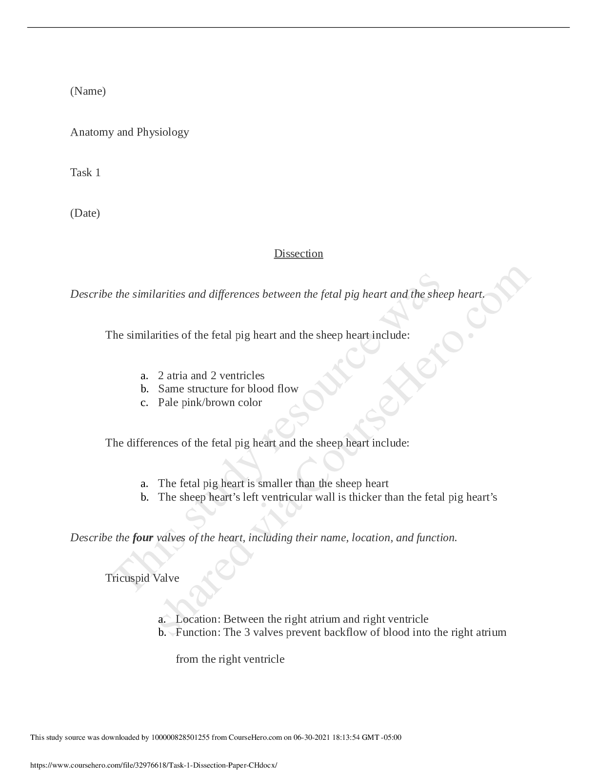
A&P II Task 1; Dissections 1. Discuss your dissection of the sheep heart and the cardiovascular system of the fetal pig by doing the following: a. Describe the similarities and differences between the... fetal pig heart and the sheep heart. The sheep’s heart and the fetal pig’s heart are similar in their anatomical structures. Both of them have 2 atriums and 2 ventricles, 4 valves and a pulmonary vein and artery, aorta, and inferior and superior vena cava. Both have 3 layers epicardium, myocardium and endocardium. Both have the sulcus – a depression on the surface along the separation of the left and the right ventricles. They differ in their size – fetal pig’s heart is very small and the sheep’s heart is much bigger, the size of a small human fist. The fetal pig’s heart has the ductus arteriosus between the aortic arch and the pulmonary artery. b. Describe the four valves of the heart, including their name, location, and function. The heart has 2 atrioventricular valves and 2 semilunar valves. Atrioventricular valves are between the atriums and the ventricles. They are open during diastole and closed during systole. The tricuspid valve is between the right atrium and the right ventricle. Its function is to allow blood flow from the atrium into the ventricle during diastole and prevent the blood from flowing back into the atrium during systole. The bicuspid valve, also known as mitral valve is between the left atrium and the left ventricle. When it’s open the blood flows from the atrium to the ventricle and then closes to prevent backflow. The semilunar valves are between the ventricles and the blood vessels. They are open during systole and closed during diastole. The aortic valve is located between the left ventricle and the entrance to the aorta. Its role is to let the oxygenated blood flow through the aorta and get distributed throughout the body. The pulmonary valve can be found between the right ventricle and the pulmonary artery. It allows oxygen-depleted blood to be transported from the heart to the lungs and prevents back flow when closed. c. Discuss the similarities and differences between the left and right sides of the heart. The left and right sides of the heart have similarities and differences. They are similar in that they both have an atrium and a ventricle. Both of them have an atrioventricular and a semilunar valves and chordae tendineae that keep the valves in place. The left ventricle is bigger and thicker than the right ventricle as the blood pumped from it needs to travel much greater distance through the body. The atrioventricular valves are different as well with the tricuspid valve having 3 cusps and the mitral valve having 2 cusps. The left and right sides of the heart are different in their function also. The right side accepts the blood coming back from the body and transports it to the lungs, and the left side accepts the oxygenated blood from the lungs and transports it to the body. d. Compare the structure of the atrioventricular valves to the structure of the semilunar valves. The AV valves are different in structure from the semilunar valves as the AV valves are attached to the papillary muscles by the chordae tendineae to prevent them from turning inside-out. The semilunar valves are self-supporting and are not connected to papillary muscles. e. Describe the appearance of the papillary muscles. The papillary muscles are small thick muscles at the base of the ventricle and are connected via the chordae tendineae to the AV valves. f. Describe the path that blood takes starting in the right atrium and ending in the superior/inferior vena cava. Deoxygenated blood starts from the right atrium, moves through the tricuspid valve to right ventricle and through the pulmonary valve to the pulmonary artery and the lungs, where it gets oxygenated. From the lungs through the pulmonary veins enters the left atrium, flowing through the mitral valve to the left ventricle and enters the aorta via the aortic valve. The oxygenated blood is then distributed through the body via the major and minor blood vessels, supplying oxygen to the tissues and organs and exchanging for carbon dioxide, eventually ending up in the superior and inferior vena cava and back in the right ventricle. 2. Discuss your dissection of the respiratory system of the fetal pig by doing the following: a. Compare the structure of the trachea to the structure of the esophagus. The trachea is a stiff tube connecting the larynx and the lungs and containing several semi-circular cartilage rings. They keep it open for the air passage, but allow for flexibility as well. The trachea is straight for the most part and splits into left and right bronchi at its lower part. The esophagus is located behind the trachea and is a long, muscular tube. It is more flexible than the trachea as it needs to be able to transport the food from the mouth to the stomach. At the time of the dissection is collapsed. b. Describe how the structures of the respiratory system (i.e., trachea, bronchi, and lungs) relate to their functions. The trachea is a stiff, cartilaginous tube connecting the larynx and the lungs. Its structure allows it to stay open. It splits into two main bronchi. The bronchi are similar to the trachea in their structure which keeps them from collapsing. Being open at all times facilitates the transport of air to the lungs with every breath we take. As they are entering deeper into the lungs, the bronchi branch out into smaller structures called bronchioles, which eventually carry the oxygen rich air to the alveoli where the gas exchange takes place. c. Describe the texture of the lungs. The lungs have porous, sponge-like consistency. Because of the air in the alveoli it crepitates when handled. However, since the provided specimen was a fetal pig, the lungs felt stiffer and were darker pink to red in color. d. Describe the similarities and differences between the left lung and the right lung. Both lungs have bronchi supplying them with air, the texture is the same as well as their function. They are similarly cone shaped with an apex and a base. However, they are different in their location and size. The left lung is slightly smaller than the right and consists of three lobes, whereas the right lung consists of four lobes. The reason being that the left lung shares its thoracic space with the heart. 3. Discuss your dissection of the sheep kidneys and the urinary system of the fetal pig by doing the following: a. Compare the structure of the fetal pig kidneys to the structure of the sheep kidneys. Both the sheep and the pig kidneys are bean-shaped and both have the main structures – cortex, medulla and renal pelvis. They were different color; the fetal pig’s was reddish-brown and the sheep’s was greyish-yellowish ( not sure if it was because of the chemicals). b. Describe the location of the kidneys in the fetal pig. Kidneys are located in the retroperitoneum. c. Describe the path that urine takes to exit the body, starting in the kidney. The urine is made in the kidneys through filtration of blood. It then moves through the ureters to the urinary bladder, where is stored. During urination the urine passes through the urethra which is connected to the penis and is discarded outside of the body. 4. Discuss your dissection of the components of the endocrine system of the fetal pig by doing the following: a. Describe the endocrine organs that are located in the throat region (e.g., function and appearance). The two endocrine organs in the throat are the thymus gland and the thyroid gland. The thyroid gland is a dark and solid organ and is located in front of the trachea. It secretes thyroxin, which is important for the metabolic control and calcitonin, which lowers calcium levels in the body. The thymus gland is glandular-looking and is located posteriorly to the larynx and over the thyroid and it extends to partially cover the anterior portion of the heart. The thymus plays an important role in the development and maintenance of the immune system. b. Describe three endocrine organs that are located in the abdominal or pelvic cavities. The three endocrine organs in the abdominal cavity are the pancreas, the adrenal glands and the male gonads. The pancreas is located along the bottom of the stomach. It is yellowish-brown in color with bumpy texture. The adrenal glands are located on the anterior surface of the kidneys. Hey are bean shaped and light in color. My specimen was a male. The testes are located in the scrotal sac in the pelvic cavity. They are small with smooth surface. 5. Discuss your dissection of the digestive system of the fetal pig by doing the following: a. Describe the major digestive organs, including their name, location, relative size, and physical characteristics (e.g., color, shape, texture). The digestive tract has 3 main parts: stomach, small and large intestine. The stomach is located underneath the liver in the left upper quadrant. It is pouch-like that is tan in color with a smooth surface. The small intestine is a thin, long tube which lies in a spiral and that way is able to fit in the tight space in the lower abdominal cavity. The large intestine is also in the lower abdominal cavity and is held together by the mesentery. It is shorter than the small intestine, but larger in diameter. b. Describe the accessory digestive organs, including their name, location, relative size, and physical characteristics (e.g., color, shape, texture) The liver is a large, multi-lobar organ, brownish in color in the right abdominal cavity over the liver. In its posterior side is embedded the gallbladder – small, green elongated organ. Under the stomach is the pancreas – it is yellowish-brown with bumpy, granular texture. [Show More]
Last updated: 1 year ago
Preview 1 out of 7 pages

Reviews( 0 )
Document information
Connected school, study & course
About the document
Uploaded On
Jan 05, 2022
Number of pages
7
Written in

Additional information
This document has been written for:
Uploaded
Jan 05, 2022
Downloads
0
Views
142


 Final Exam Essay Questions.png)
 Midterm Essay Questions.png)