Health Care > CASE STUDY > Betty Burns I human case study. (All)
Betty Burns I human case study.
Document Content and Description Below
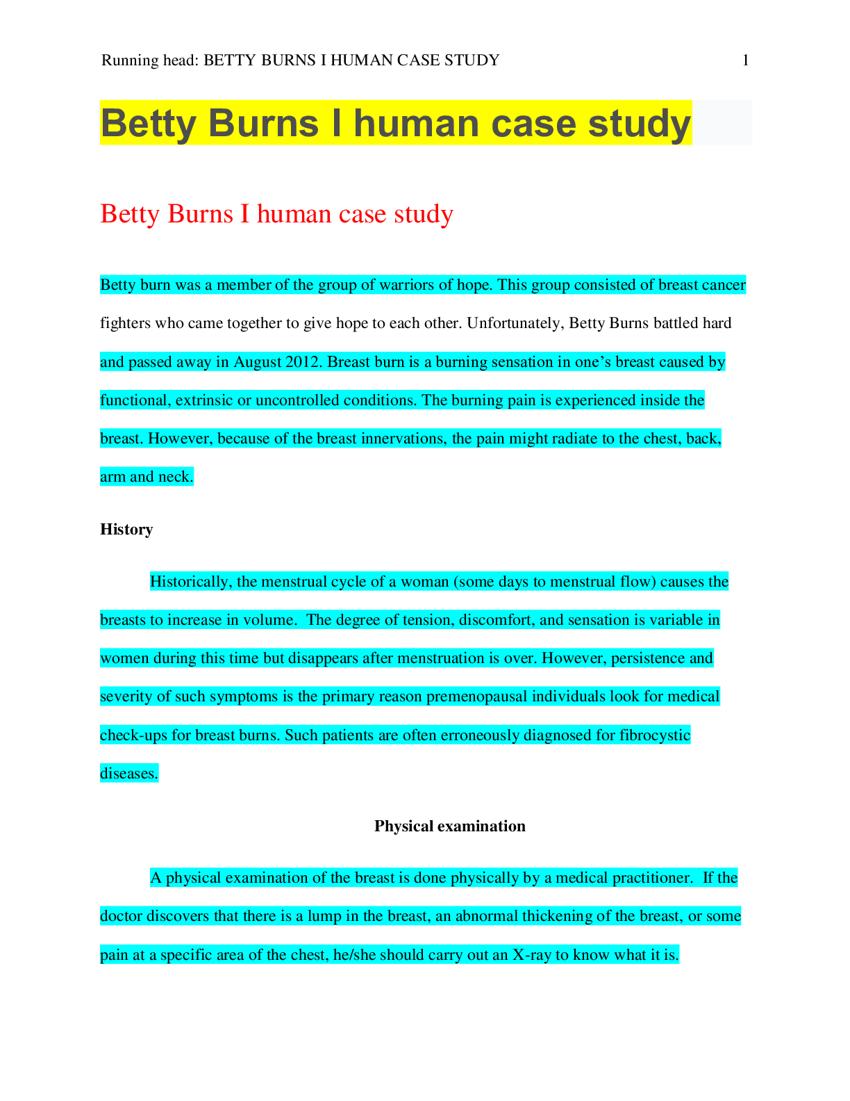
Betty Burns I human case study Name of student Institution This study source was downloaded by 100000830772748 from CourseHero.com on 07-05-2022 04:14:16 GMT -05:00 https://www.coursehero.com/file... /37378541/Betty-Burns-I-human-case-studyediteddocx/ BETTY BURNS I HUMAN CASE STUDY 2 Betty burn was a member of the group of warriors of hope. This group consisted of breast cancer fighters who came together to give hope to each other. Unfortunately, Betty Burns battled hard and passed away in August 2012. Breast burn is a burning sensation in one’s breast caused by functional, extrinsic or uncontrolled conditions. The burning pain is experienced inside the breast. However, because of the breast innervations, the pain might radiate to the chest, back, arm and neck. History Historically, the menstrual cycle of a woman (some days to menstrual flow) causes the breasts to increase in volume. The degree of tension, discomfort, and sensation is variable in women during this time but disappears after menstruation is over. However, persistence and severity of such symptoms is the primary reason premenopausal individuals look for medical check-ups for breast burns. Such patients are often erroneously diagnosed for fibrocystic diseases. Physical examination A physical examination of the breast is done physically by a medical practitioner. If the doctor discovers that there is a lump in the breast, an abnormal thickening of the breast, or some pain at a specific area of the chest, he/she should carry out an X-ray to know what it is. Differential diagnosis In the differential diagnosis, several considerations should be diagnosed. Circumscribed breast wounds such as fibroadenoma and in cysts can be diagnosed. This indicates that a fibrocystic disease might be emerging. It is done by a radiologist who must be familiar with the This study source was downloaded by 100000830772748 from CourseHero.com on 07-05-2022 04:14:16 GMT -05:00 https://www.coursehero.com/file/37378541/Betty-Burns-I-human-case-studyediteddocx/ BETTY BURNS I HUMAN CASE STUDY 3 difference between benign and malignant disease. Fibroadenoma disease has benign tumors made of epithelial and stromal components. Young women with complex fibroadenoma tumors are likely to have cancer unlike those without (Shermis, et al 2017). An examination done on fibroadenoma can indicate a palpable mass of the patient. In young and pregnant women, ultrasonography is used whereas mammography suits older and patients that are not expectant. However, patients below 30 years best suit the US because she is not exposed to radiations and also, the possibility of fibroadenoma is high. Ranking of the differential diagnosis Another differential diagnosis is the skin inflammation that might be as a result of abscesses and mass in the breast. Most women that have breast mass are likely to have breast cancer. The masses can affect any breast tissue like the duct, the lobules, and the overlying skin. Fibrocystic is the common breast mass as indicated by routine autopsy. However, inflammatory carcinoma is aggressive and has the worst prognosis (Balleyguier, et al 2015). Test selection In test selection, several tests can be selected to evaluate breast burns. First, the doctor can carry out a clinical exam on the patient's breast. He/she can check for any changes in the chest by observing the lymph nodes, the neck and the arms of the patient. The doctor will listen to the heart and lungs and determine if other conditions in the chest and abdomen are the causes of the pain. If the results do not show any unusual circumstances, no additional tests would be carried out. This study source was downloaded by 100000830772748 from CourseHero.com on 07-05-2022 04:14:16 GMT -05:00 https://www.coursehero.com/file/37378541/Betty-Burns-I-human-case-studyediteddocx/ BETTY BURNS I HUMAN CASE STUDY 4 Breast biopsy and ultrasound tests can be used as well. The tests are used if there is a suspicion for breast lumps and breast thickening. In a biopsy, the doctor takes a sample of the breast tissue for laboratory analysis. In ultrasound, sound waves are used to produce breast image, with a focus on the area of pain. The results are then examined before diagnosis (Lipscomb, et al 2016). [Show More]
Last updated: 1 year ago
Preview 1 out of 5 pages

Reviews( 0 )
Document information
Connected school, study & course
About the document
Uploaded On
Jul 05, 2022
Number of pages
5
Written in
Additional information
This document has been written for:
Uploaded
Jul 05, 2022
Downloads
0
Views
63


