NUR 3125 MODULE 1: CELLULAR, TISSUE FUNCTION AND GENES
Document Content and Description Below
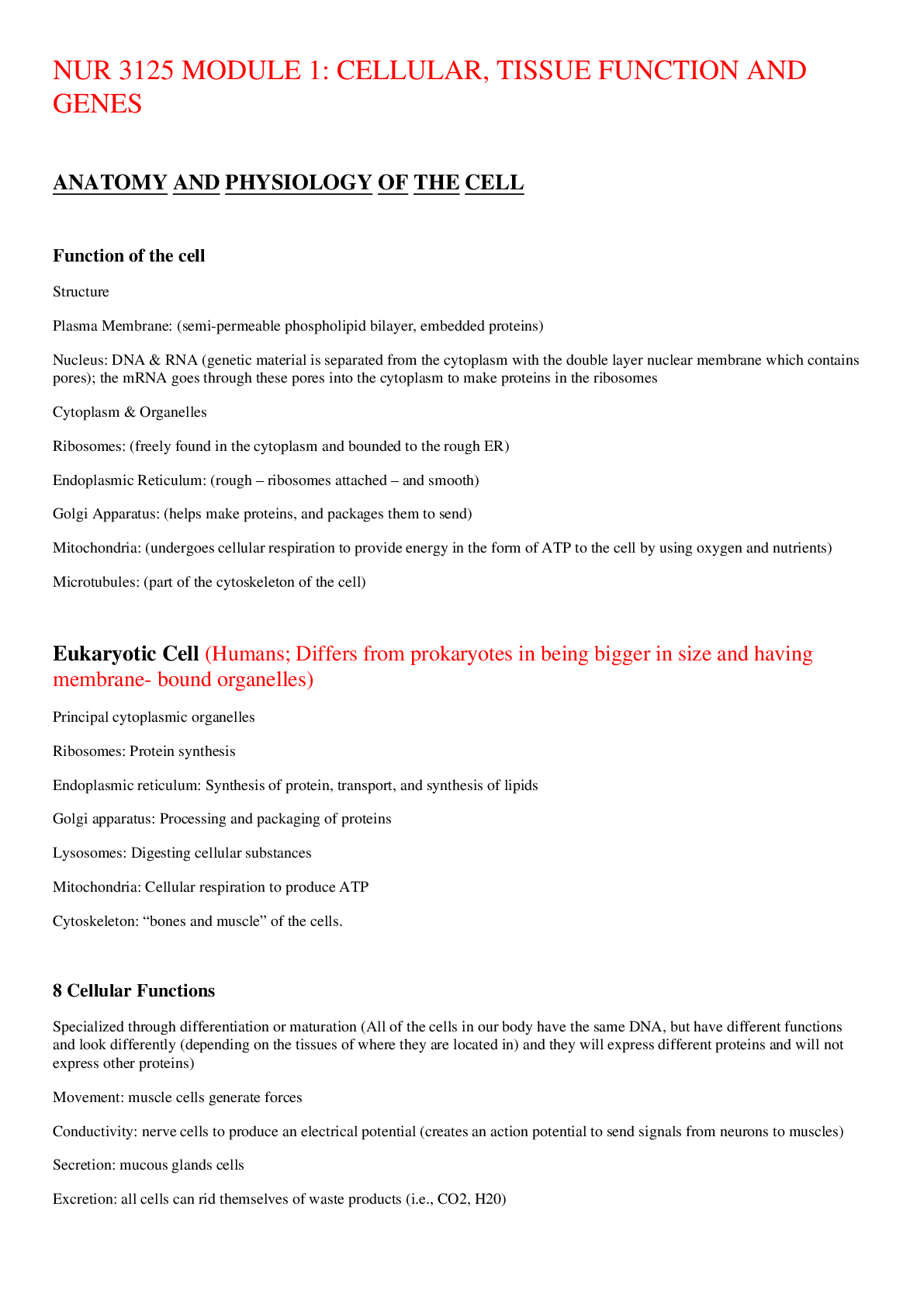
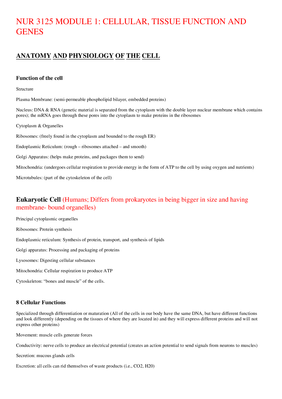
NUR 3125 MODULE 1: CELLULAR, TISSUE FUNCTION AND GENES ANATOMY AND PHYSIOLOGY OF THE CELL Function of the cell • Structure o Plasma Membrane: (semi-permeable phospholipid bilayer, embedded p... roteins) o Nucleus: DNA & RNA (genetic material is separated from the cytoplasm with the double layer nuclear membrane which contains pores); the mRNA goes through these pores into the cytoplasm to make proteins in the ribosomes o Cytoplasm & Organelles ▪ Ribosomes: (freely found in the cytoplasm and bounded to the rough ER) ▪ Endoplasmic Reticulum: (rough – ribosomes attached – and smooth) ▪ Golgi Apparatus: (helps make proteins, and packages them to send) ▪ Mitochondria: (undergoes cellular respiration to provide energy in the form of ATP to the cell by using oxygen and nutrients) ▪ Microtubules: (part of the cytoskeleton of the cell) Eukaryotic Cell (Humans; Differs from prokaryotes in being bigger in size and having membrane- bound organelles) • Principal cytoplasmic organelles o Ribosomes: Protein synthesis o Endoplasmic reticulum: Synthesis of protein, transport, and synthesis of lipids o Golgi apparatus: Processing and packaging of proteins o Lysosomes: Digesting cellular substances o Mitochondria: Cellular respiration to produce ATP o Cytoskeleton: “bones and muscle” of the cells. 8 Cellular Functions • Specialized through differentiation or maturation (All of the cells in our body have the same DNA, but have different functions and look differently (depending on the tissues of where they are located in) and they will express different proteins and will not express other proteins) o Movement: muscle cells generate forces o Conductivity: nerve cells to produce an electrical potential (creates an action potential to send signals from neurons to muscles) o Secretion: mucous glands cells o Excretion: all cells can rid themselves of waste products (i.e., CO2, H20) o Metabolic absorption: all cells can take in and use nutrients o Respiration: all cells absorb oxygen (+ glucose) and transform nutrients into energy (ATP). Cell respiration (by aerobic); Oxidation (reaction in which glucose is oxidized and oxygen is reduced) o Reproduction: most cells can grow tissue or replace cells o Communication: all cells - vital to survive Cell Metabolism • Energy metabolism: Chemical tasks of maintaining cellular functions. o Anabolism: E-using process; build molecules (AA’s - building blocks of proteins) o Catabolism: E-releasing process; break down; converts carbohydrates, fats & proteins into energy needed for cell function. • ATP: major source of cellular energy • Aerobic Metabolism: presence of oxygen • Anaerobic Metabolism: lack of oxygen Membrane Transport Mechanisms • Passive transport (NO ENERGY NEEDED) o Diffusion: Movement of solutes from area of high concentration to area of low concentration (down the concentration gradient). Does not need energy. ▪ Passive Diffusion: Substance diffuses across the plasma membrane (i.e., oxygen, alcohol, CO2, and more). No protein needed for transportation. Diffusion stops when there is an equal concentration of molecules in area ▪ Facilitated Diffusion: A membrane protein facilitates diffusion (i.e., glucose, since it is such a large molecule) • Osmosis: The movement of water down a concentration gradient (NO ENERGY NEEDED) o Occurs across a semipermeable membrane from a region of higher water concentration to a region of lower concentration. (SCENARIO: There are solutes in a container of water molecules which are divided by a semi-permeable membrane. There are more solutes on one side of the membrane making the [water] on that side low versus the lesser number of solutes on the other side, making the [water] on that side high. Via osmosis, the water molecules will go from the [high water] to the [low water] to balance the [water] on both sides. This can/may result in unequal water molecules on either side of the membrane, but it will result in equal [water]. • Active transport: (ATP ENERGY NEEDED) – ATP breaks down into ADP and Phosphate ion o Transportation of a molecule across a plasma membrane against (or up) its concentration gradient ([low] to [high]). Always need energy and a membrane protein. Very important as it regulates the enzyme (Na+/K+ ATPase pump) is in every cell in our body and will help us maintain our Na+/K+ levels (at same time) ▪ Na+ is an ion that is more abundant in the extracellular compartment and the pump will still move more Na+ (OUT) in the cell. ▪ K+ is an ion that is more abundant in the intracellular compartment and the pump will still move more K+ (IN) in the cell. Tissues • Cells of one or more types are organized into tissues, different tissues compose organs, into organ systems, and lastly composing the organism. • Four types of tissues: o Epithelial: Covers most of internal and external body surfaces ▪ Simple squamous epithelium ▪ Transitional epithelium ▪ Stratified squamous epithelium ▪ Simple cuboidal epithelium ▪ Simple columnar epithelium ▪ Stratified columnar epithelium o Connective: Binds different tissues and organs together ▪ Adipose ▪ Cartilage ▪ Bone ▪ Blood o Nerve: Coordinates and controls many body activities ▪ Specialized cells neurons ▪ Glia cells o Muscle: Composed of myocytes ▪ Striated ▪ Cardiac ▪ Smooth Cellular Adaptation [Physiologic (adaptive) vs. Pathologic] • Atrophy – decrease in cellular size/disuse atrophy (i.e., thymus gland, ovaries, gonads) • Hypertrophy –an increase in cell size due to mechanical stimuli. The number of cells remain the same. (i.e., myocyte enlargement in cardiac hypertrophy, left ventricle hypertrophy) • Hyperplasia – an increase in the number of cells. The size of cells remains the same. (i.e., hepatocytes after removal part of the liver) The liver can regenerate cells, but NOT the kidneys. • Metaplasia – the replacement of one mature cell by another. (i.e., the columnar ciliated epithelial cells of the airway will be replaced by stratified squamous epithelial cells and in doing so, the airway lost its original protective mechanism. However, this can be reversed if smoking is removed.) • Dysplasia – (low or high grade) the abnormal changes in the size, shape and organization of mature cell. Not a true adaptive change. It does not indicate cancer, but it may or may not progress to cancer. Like 20% of the time it will progress to cancer (i.e., the epithelial tissue of the cervix can occur if someone has HPV (human papillomavirus). HPV can lead to cervical dysplasia or cervical cancer. Tumor Classification and Nomenclature (tumor = the uncontrollable growth of cells) • Benign tumors (do not metastasize into other parts of the body, they stay in place) o Named according to the tissues from which they arise and include the suffix “- oma” (lipoma, meningioma) and may progress to malignant tumor o We usually want to remove benign tumors (even though they do not metastasize) because eventually they CAN convert into malignant tumors and then we have a problem. Also, depending on where the benign tumors are located, we MUST remove them. (i.e., located in the brain) • Malignant tumors (does metastasize into other parts of the body) o Rapid growth rates with microscopic alterations. (Metastasis = spread far beyond the tissue of origin) o Named according to the tissues from which they arise: Carcinoma (Epithelial); Adenocarcinoma (From ductal or glandular); Sarcoma (Mesenchymal); Lymphoma (Lymphatic); Leukemia (Blood-forming cells) • Carcinoma in situ (CIS) (At first, the cells are still in place, but have the ability to move and they can begin to metastasize as time goes on) o Early-stage cancer o These are pre-invasive epithelial malignant tumors of glandular or epithelial origin that have not broken through the basement membrane or invaded the surrounding stroma (yet) Cell Injury & Death-Causes • Mutations • Physical Agents o Contusions, Lacerations, Fractures, Incised wound, Stab wound, Puncture wound • Radiation Injury • Chemical Injury o Over the counter and prescribed drugs o Leading cause of child poisoning • Nutritional Imbalances (low or high intake of foods) • Hypoxic Injury: (low O2) single most common cause of cellular injury. Multiple causes. Most common cause is ischemia. (i.e., myocardial infraction; the heart has some vessels “culinary arteries” that are going to give O2 to the cells of the heart. For whatever reason, if the culinary arteries get blocked, the area of the heart that is near these arteries will stop receiving oxygenated blood and will go through ischemia (reduced oxygenated blood flow). If this lasts longer than 20 minutes, then cardiac cells will die (necrosis) and heart will not be able to contract. • Free Radical Injury: Oxidative stress due to excessive reactive oxygen species (ROS) Cell Death • Apoptosis: (Programmed cell death/self-destruction, eliminates aged/injured cells) o Normal physiologic process: ▪ Destruction of cells during embryonic process (apoptosis of the cells in the mother that occur during pregnancy when baby is born) ▪ Endometrial cells during menses (apoptosis of cells during the time of menstruation) ▪ Webbed fingers (too little apoptosis = cells don’t allow digits to separate ▪ Breast tissue regression after breastfeeding (apoptosis of breast tissue) o Pathologic (disease) process: ▪ Can be pathologic if too much or too little (dysregulated apoptosis) • Too much apoptosis can be caused by neurological disorders such as Alzheimer’s, ALS, Parkinson’s Disease, etc. • Too little apoptosis can be caused by neurological disorders such as carcinogenesis (survival of abnormal cells), autoimmune disorders, etc. • Necrosis: (Cell death in an organ/tissue that is still alive. Sum of cellular changes after local cell death.) (The death of a cell that should not be dying. It is dying from the lack of nutrients or O2, like in myocardial infarction when there is a lack of O2 supply) o Always pathological/pathophysiological (kidney, heart, adrenal glands) o Involves signs and symptoms of inflammation from organism o Rapid loss of plasma membrane, swelling of organelles. (An explosion of the cell) o Enzymatic digestion of cell components o Coagulation necrosis (heart; ischemia or infarction), Liquefaction necrosis (brain), Caseous necrosis (lungs/tuberculosis disease), Fatty necrosis (abdominal organs, breasts) o Gangrene ▪ Mass of tissue undergoing necrosis-Dry or Wet ▪ Gas gangrene-infection of tissue by anaerobic bacteria (Clostridium) GENES AND GENETIC DISEASES Definitions • Chromosomes: chromatin condensed (23 pairs in humans) • Chromatin: DNA with proteins • Diploid: cells that have 23 pairs of chromosomes • Haploid: cells that have 23 chromosomes • Genes: basic units of inheritance in the chromosomes (a piece of DNA that is going to code for a protein) • Homozygous: Loci on a pair of chromosomes have identical alleles • Heterozygous: Loci on a pair of chromosomes have different alleles • Allele: a specific form of a gene, differing from other alleles by one or a few bases only and occupying the same gene locus as other alleles of the gene; responsible for the variations in which a given trait can be expressed • Gene: a portion of DNA that determines a certain trait; responsible for the expression of traits. • DNA: Deoxyribonucleic acid. Genetic material. Double helix model made of nucleotides. o DNA RNA proteins • Mutation: alteration of genetic material (DNA) • Genotype: the genetic constitution of an individual organism • Phenotype: observable characteristics of an individual resulting from the interaction of its genotype with the environment • Dominant: the observable allele seen when two alleles are together (represented by a capital letter) • Recessive: the allele whose effects are hidden is (represented by a lowercase letter) • Autosomes: the first 22 out of the 23 pairs of chromosomes in males and females o 22 pairs = (44 chromosomes called autosomes) o 1 pair = (2 chromosomes called sex chromosomes) • Sex Chromosomes: the remaining pair of chromosomes (the 23rd pair) o Females = (XX) o Males = (XY) What do Chromosomes look like? How many chromosomes are inherited? This is an example of a male since the 23rd chromosome is Y Genetic Disorders • Single-Gene o Autosomal Dominant (NO CARRIERS; affected = DD & Dd; unaffected = dd) o Autosomal Recessive (CARRIERS = Dd; affected = dd; unaffected = DD) o X-Linked recessive (more common than x- linked dominant) • Multifactorial • Chromosomal Disorders Autosomal Dominant Inheritance • Male/female offspring affected equally • Only one of the parents is usually affected • If one of the parents is heterozygous affected, the children have a 50% chance of being affected • If both parents are heterozygous affected the children have a 75 % chance of being affected • Example: Marfan syndrome Autosomal Dominant Disorders Marfan Syndrome • Inherited disease of connective tissue • Primary causes ocular, skeletal and cardiovascular abnormalities • Affects men/women equally, 1 in 20,000 people • Diagnosis based on physical characteristics and family history Marfan’s Clinical Manifestations • Connective tissue disease • Retinal detachment, myopia • Skeletal: Joint hypermobility (circus contortionist), spinal deformities, “pigeon” chest, long thin body • Heart: Mitral valve prolapse • Vascular: Aortic valve disease, weakness of aorta leading to dissection Autosomal Recessive Inheritance • Male/female offspring affected equally • If both parents are unaffected but are carriers for the trait, each offspring has a 1/4 chance of being affected (25%) • If both parents are affected, all of their children will be affected • If one parent is affected and the other is not a carrier, all of their offspring will be unaffected but will be carriers • If one parent is affected and the other is a carrier, each of the offspring will have a 1/2 chance of being affected • Example: Cystic fibrosis (multi-system disease, but mostly affects the lungs; thick mucus in the respiratory and GI system; airway blockage; tubes from pancreas to duodenum can be blocked as well. Used to have a shortened lifespan of 20 years, but now lifespan increased. Nowadays, they can get lung transplants, but the lungs will get damaged again since this is an autosomal disease, and it is in the genes) Autosomal Recessive Disorder – Cystic Fibrosis • Chloride Transport decreased • Mutation in Gene CFTR (Cystic Fibrosis Transmembrane Regulator) on chromosome 7 • Organs affected by cystic fibrosis: o Sinuses: sinusitis (infections coming from thick mucus blockage) o Lungs: Thick, sticky mucus buildup and bacterial infection o Skin: sweat glands produce salty sweat o Liver: blocked biliary ducts o Pancreas: blocked pancreatic ducts o Intestines: cannot fully absorb nutrients o Reproductive organs: male and female complications • Test: Sweat test shows elevated chloride • Treatment: Preventative therapy and lung transplant Autosomal Recessive Disorder – Phenylketonuria (PKU) • Once a child is born, the hospital checks if the baby has PKU • Defect in amino acid metabolism o The inability of the body to convert the essential amino acid phenylalanine to tyrosine (a precursor to melanin) ▪ The accumulation of phenylalanine leads to mental retardation. The elevated phenylalanine AA’s are thought to reduce IQ levels. • Phenotype: fair skin and hair due to decreased melanin (since phenylalanine is not being converted to tyrosine) • Eczema is higher in PKU patients. • Treatment: Diet restriction of phenylalanine (DO NOT eat foods with phenylalanine; some foods are meat, chicken, beef) • Not recommended to breast feed if infant has PKU! Autosomal recessive disorder – Tay-Sachs Disease • Autosomal recessive genetic disorder • Accumulation of glycolipids in the brain neurons & retina due to failure of lysosome (enzyme) function • Progressive destruction of neurons in brain, spinal cord, autonomic nervous system leading to mental retardation and motor problems • Blindness, seizures, death occurs by 2 to 5 years • Phenotype: cherry red spit on the retina • Predominately in Eastern Jews population, 1: 30. • No treatment but genetic screening available Sex-linked Inheritance • Caused by genes located on sex chromosomes (Female XX, male XY) • X-linked or Y-linked • Most X-linked disorders are recessive • X-linked recessive disease: Usually males more affected because females have another X chromosome to counteract the abnormal gene. Affected males cannot transmit the affected gene to sons but they can to all daughters. • Sons of female carriers have 50% risk of being affected • Males cannot be carriers; they are either affected or unaffected Recessive X-linked (MAINLY affects males) • Example: Color blindness (seen MAINLY in males since they only have 1 X chromosome; hemophilia is another case that is MOSTLY seen in males, but they both can appear in females) Chromosomal Disorders • Two Causes: o Alterations in the structure of one or more chromosomes with rearrangement/deletion of chromosome part o Radiation/chemical exposure o Viral infections • Abnormal number of chromosomes (when meiosis does not happen correctly) o Failure of chromosomes to separate during oogenesis/spermatogenesis Down’s Syndrome/Trisomy 21 (affects both males and females) (SEE LAST PAGE OF NOTES) • Chromosomes 21 has 3 copies instead of 2 (errors in translocation OR meiosis – most common) • Most common chromosomal disorder (caused by abnormal meiosis division, not generally genetically inherited; most trisomy 21 people do not have parents with it) • Risk increases with maternal age-increased risk for exposure to environmental factors • If the women’s eggs are frozen (i.e., at age 25) then the risk of trisomy 21 is very low o The issue is in the age of the egg (not just in the age of the mother) Clinical Manifestations – Down’s Syndrome • Phenotype o Protruding tongue o Flat nasal bridge • Mental retardation • Heart problems • Advice for pregnant mom: o Small ears o Single palmar (simian) crease o < 35 years old = Triple screen. Check for alpha fetoprotein o > 35 years old = Amniocentesis, chorionic villi sampling Turner Syndrome (affects females) • Partial or total inactivation of X chromosome (a change in the number of chromosomes) • Has 45 chromosomes - 22 pairs of autosomes and 1 sex chromosome - usually the X chromosome is there and there is (missing the second sex chromosome) • Present in 1 in 2,500 live births • Often results in spontaneous abortions • Diagnosed through genetic testing • Clinical Manifestations: o Depend on degree of inactivation/deletion of X o Short stature and webbing of neck o Lack of secondary sex characteristics ▪ Underdeveloped breast o Absent ovaries, amenorrhea (doesn’t get period), sterile o Coarctation of aorta and bicuspid aortic valve; large left ventricle o Normal intelligence; may have difficulty driving, nonverbal problem solving, math [Show More]
Last updated: 1 year ago
Preview 1 out of 11 pages
Buy this document to get the full access instantly
Instant Download Access after purchase
Add to cartInstant download
We Accept:

Reviews( 0 )
$13.00
Document information
Connected school, study & course
About the document
Uploaded On
Apr 04, 2023
Number of pages
11
Written in
Additional information
This document has been written for:
Uploaded
Apr 04, 2023
Downloads
0
Views
29

