Lipoproteins Quiz Questions with Correct Solutions (GRADED A)
Document Content and Description Below
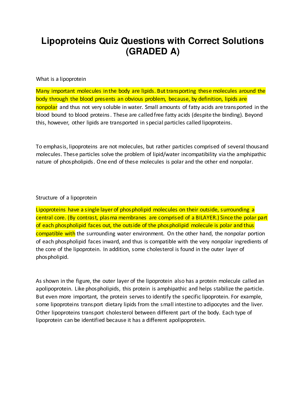
What is a lipoprotein Many important molecules in the body are lipids. But transporting these molecules around the body through the blood presents an obvious problem, because, by definition, lipids a... re nonpolar and thus not very soluble in water. Small amounts of fatty acids are transported in the blood bound to blood proteins. These are called free fatty acids (despite the binding). Beyond this, however, other lipids are transported in special particles called lipoproteins. To emphasis, lipoproteins are not molecules, but rather particles comprised of several thousand molecules. These particles solve the problem of lipid/water incompatibility via the amphipathic nature of phospholipids. One end of these molecules is polar and the other end nonpolar. Structure of a lipoprotein Lipoproteins have a single layer of phospholipid molecules on their outside, surrounding a central core. (By contrast, plasma membranes are comprised of a BILAYER.) Since the polar part of each phospholipid faces out, the outside of the phospholipid molecule is polar and thus compatible with the surrounding water environment. On the other hand, the nonpolar portion of each phospholipid faces inward, and thus is compatible with the very nonpolar ingredients of the core of the lipoprotein. In addition, some cholesterol is found in the outer layer of phospholipid. As shown in the figure, the outer layer of the lipoprotein also has a protein molecule called an apolipoprotein. Like phospholipids, this protein is amphipathic and helps stabilize the particle. But even more important, the protein serves to identify the specific lipoprotein. For example, some lipoproteins transport dietary lipids from the small intestine to adipocytes and the liver. Other lipoproteins transport cholesterol between different part of the body. Each type of lipoprotein can be identified because it has a different apolipoprotein. What does the core of the lipoprotein contain? Most other lipids are transported in the blood as part of lipoproteins, complex particles whose structure includes: core consisting of a droplet of triacylglycerols and/or cholesteryl esters What does the surface of a lipoprotein contain? a surface monolayer of phospholipid, cholesterol, & specific proteins (apolipoproteins), e.g., B-100. How do some lipoproteins differ in the body? How are they classified? Lipoproteins differ in the: 1. ratio of protein to lipids 2. in the particular apoproteins & lipids that they contain. They are classified based on their density Types of Lipoproteins Chylomicron (largest; lowest in density due to high lipid/protein ratio; highest % weight triacylglycerols) VLDL (very low density lipoprotein; 2nd highest in triacylglycerols as % of weight) IDL (intermediate density lipoprotein) LDL (low density lipoprotein, highest in cholesteryl esters as % of weight) HDL (high density lipoprotein; highest in density due to high protein/lipid ratio) In order of increasing mobility, which correlates to density, and decreasing molecular weight, the lipoproteins are: 1. Chylomicrons 2. Very Low Density Lipids (VLDL) 3. Low Density Lipids (LDL) 4. High Density Lipids (HDL) Chylomicrons Metabolism Synthesized in the small intestine (chylomicrons contain ApoB-48). Contain a high content of triacylglycerols (80%-90%). Released into the plasma, and travel through the bloodstream. 1. Get ApoC-II and ApoE from HDL 2. ApoC-II activates lipoprotein lipase (LPL) 3. Degradation of triacylglycerols (TG) by LIPOPROTEIN LIPASE to break down into Glycerol and Fatty Acids. This step allows free fatty acids to go into tissues (e.g. adipose) and glycerol to go to liver. 4. They become much smaller, with only the remnants remaining. 5. ApoC-II is returned to HDL and the rest that remains it taken up by the liver through ApoE docking onto it, facilitating absorption VLDL Metabolism Metabolism of VLDL looks very similar to that of a chylomicron with two major differences—the source and the size of the lipoprotein. VLDL comes from the liver and is smaller than chylomicrons, which are synthesized in the small intestine. Also, it has more cholesterol esters and less TG than chylomicrons, which accounts for some of the size difference. The steps of VLDL metabolism are as follows: 1. VLDL is formed in the liver with ApoB-100, and travels through the bloodstream 2. It gets ApoC-II and ApoE from HDL 3. Apo C-II activates lipoprotein lipase (LPL) 4. Degradation of TG by LPL to break down into Fatty Acids and Glycerol. This step allows free fatty acids to go into tissues (e.g. adipose) and glycerol to go to liver. 5. Recycling step: returning Apo C-II and ApoE to HDL. In the process of VLDL giving off FAs and Glycerol, it becomes IDL, and then LDL after giving off Apo C-II and ApoE to HDL 6. Finally it is taken up through binding of ApoB-100 as LDL HDL metabolism HDL is mainly formed in the liver with ApoA-I (Nascent HDL), but it can also come from the small intestine (ApoA-1 is in BOTH) HDL has a flat structure, like an empty bag, for collecting cholesterol from peripheral tissues. Has a high protein content, and is the most dense lipoprotein (and smallest) Its job is to then collect free cholesterol from peripheral tissues, forming cholesterol esters, and become larger and rounder The final step is to be taken up by the liver so that the cholesterol esters can undergo further processing What is the overarching function of lipoproteins? The overarching function of lipoproteins is to maintain the homeostasis of lipids in the body - to get it out to peripheral tissues when needed and to remove it when it is not needed All lipoproteins are in the bloodstream, which can be considered the highway, and each carries similar things in different ratios to and from specific tissues In bloodstream there is exchange of lipoproteins to accommodate needs of different tissues, what coordinates the whole process? The Liver coordinates the whole process, making components readily available for when tissues need fats and serves as a dumping ground for any extra cholesterol that is not needed (taken back by HDL) and can also take up the remnants of the chylomicron Mobilization of fatty acids and TGs from adipose stored for different uses and delivered to the appropriate tissues via chylomicrons & VLDL. 1. Lipids (TG & FA) are transported to tissues via Chylomicrons (from intestine) and VLDL (from liver) 2. Lipids are also transported from adipose to muscle and liver via FFA/Albumin 3. Cholesterol is transported to tissues by LDL and back to the liver by HDL (whatever wasn't needed) For everything to function correctly, a specific protein is needed for each function. Those listed in red are the ones Dr. Zhang points out as the most important, which we'll be dealing with: ApoB-100 vs. ApoB-48 Both proteins are encoded by the same gene, but the RNA editing process allows tissue specificity by deamination, changing C to U so the intestinal cells gets a stop codon from the change to U, creating a nonsense STOP codon allowing translation of only 48% of the mRNA. B100: 100% mRNA translated found in the liver and found in VLDL and LDL B48: 48% mRNA translated found in chylomicrons made my intestinal cells. Where is lipoprotein lipase found? LPL is an extracellular enzyme that is anchored by heparin sulfate to the capillary walls of most tissues, but predominantly those of adipose tissue, cardiac, and skeletal muscle. Adult liver does not have this enzyme LPL is activated by apoC2 on circulating lipoprotein particles, hydrolyzes the TAG contained in these particles to yield fatty acids and glycerol. Lipoprotein Lipase (LPL) and ApoC-II Interaction 1. Adipocyte LPL is induced by Insulin and then synthesized in the Rough ER 2. LPL matures in the Golgi, becoming dimerized, and is then secreted into the interstitial space 3. It is translocated to the lumen of a capillary where it waits for either Chylomicron or VLDL to bring ApoC-II 4. ApoC-II activates LPL so it can hydrolyze triacylclycerol from the lipoprotein, extracting the FA to be able to bring it to tissues, glycerol goes to liver. Chylomicron and ApoC-II, ApoE 1. Chylomicrons are made by intestinal epithelial cells and derived from dietary lipids (exogenous). The particle released by intestinal mucosal cell is called a "nascent" chylomicron because it is functionally incomplete only has ApoB48. When it reaches the plasma, the particle rapidly modified, receiving apolipoproteins E and C. The source of these proteins is circulating HDL. 2. ApoC-II and ApoE are added from HDL 3. ApoC-II interacts with LPL as described above to bring FA to tissues 4. Then as it becomes smaller, it becomes chylomicron remnants (ratio of TG:Chol of ~1:1) 5. ApoE assists chylomicron remnant docking at liver cells to be reabsorbed Abetalipoproteinemia is a lack of ApoB-48 and ApoB-100 and results in deficiency in making chylomicrons and VLDL, disabling absorption of fat-soluble vitamins Type I Hyperlipoproteinemia can be a deficiency of LPL or ApoC-II 1. LPL deficiency would result in more lipoproteins (chylomicrons & VLDL) in the bloodstream because LPL is not available to extract Glycerol and free fatty acids and release ApoC-II back to HDL 2. ApoC-II deficiency has the same result because there is nothing to activate LPL, so indirectly there is effectively reduced LDL functioning Type II Hyperlipoproteinemia is a deficiency in ApoE, which would cause inability of chylomicron remnants to be taken up by the liver, resulting in increased numbers of chylomicrons in the blood VLDL and ApoC-II 1. VLDL is made by liver cells and derived from liver-synthesized FA (endogenous), plus any FA from chylomicron remnants, and packaged as triacylgylcerol. 2. Nascent VLDL has ApoB100, and ApoCII and ApoE are added on from HDL 2. Density increases as size decreases transitioning from VLDL to IDL to LDL (ratio of TG:Chol of ~1:1 in LDL) 3. IDL is taken up by liver and peripheral tissues via ApoB100 Lipoproteinemias that affect chylomicrons will also affect VLDL, such as: 1.Abetalipoproteinemia 2. Type I Hyperlipoproteinemia 3. Type III Hyperlipoproteinemia Lipoproteinemias that affect chylomicrons will also affect VLDL, such as Abetalipoproteinemia, Type I Hyperlipoproteinemia, and Type III Hyperlipoproteinemia Type IV hyperlipoproteinemia excess of VLDL ApoB100-mediated LDL Endocytosis and Hyperlipidemia 1. LDL Receptors are cell membrane proteins, meaning synthesized in RER, then mature proteins are secreted out from Golgi 2. Mature LDL Receptor interacts with plasma LDL particle, causing an indentation of the cell, and eventually pinching off inside the cell so that LDL is inside a vesicle in the cell 3. Next, protons are pumped in to increase acidity, making an Endosome, which eventually will separate the contents of the vesicle, freeing the LDL from the receptors. 4. The vesicle containing receptors can go back to the surface of the cell recycling them, while the rest of the contents of the vesicle is taken up by a lysosome which will lyse, diffusing its contents, which can be protein, FA, cholesterol ester, and cholesterol. Focusing on free cholesterol: 3 major effects of the three different end points of free cholesterol Effect 1) Decrease in LDL receptor synthesis (downregulation assures there is enough cholesterol in the cell but not as much to overwhelm the cell) Effect 2) Decrease HMG CoA reductase to reduce uptake & synthesis of cholesterol Effect 3) Increase ACAT (acyl CoA cholesterol acyltransferase) to store free cholesterol for later use in the form of cholesterol ester droplets, which is more hydrophobic and good for long term storage High Density Lipids (HDL) Synthesized by the liver and the intestine in its nascent form and secreted into the blood Notable characteristics of HDL are that it is the most dense of all lipoproteins, consisting of mainly protein with some phospholipid and cholesterol with little lipid. Its main function is to transfer ApoC-II and ApoE onto Chylomicrons and VLDL. It also participates in moving cholesterol from tissues back to liver in a kind of "reverse" cholesterol transport, recycling of what is not currently needed. On a larger scale, HDL has many roles in the body, including: 1. Mediation of reverse cholesterol transport 2. Exhibits antioxidant activity via inhibition of LDL oxidation 3. Has anti-inflammatory effects in vasculature by inhibiting oxidized LDL-induced upregulaion of cell adhesion molecules 4. Inhibition of platelet activation 5. Enhance endothelial vasodilation 6. Upregulation of NOS, mediating anti-apoptosis Overview of "Reverse" cholesterol transport "Reverse" cholesterol transport takes lipids from peripheral tissues back to the liver. 1. ApoA1 is synthesized by liver, processed through ABCA-1 (complicated, and not very well understood mechanisms) to join nascent HDL -ABCA1 is an ATP Binding Cassette protei -ABCA1 uses energy from ATP hydrolysis to transport material, specifically, transporting cholesterols from peripheral tissues to HDL 2. ApoA-I activates PCAT 3. PCAT (same as ACAT with a different substrate) reaction converts Cholesterol to C-ester, which is non-polar and moves to the middle of the HDL, causing it to become ball-shaped 4. Exchanges of various phospholipids, TG, and CE take place -Exchange can occur with the help of plasma proteins: cholesterol ester transfer protein (CETP) and phospholipid transfer protein (PLTP) -They transfer cholesterol esters from HDL to VLDL in exchange for TG and phospholipids 5. Finally, HDL either drives past Liver depositing cholesterol via hepatic lipase, or binding a receptor on the liver to become incorporated 6. Receptors include the ApoE receptor, and more commonly, the scavenger receptor SRB-1 Fish-eye disease PCAT deficiency (not emphasized) Tangier disease mutation of ABCA1 causing inability to eliminate cholesterols. Signs and symptoms include yellow-orange, enlarged tonsils, very low levels of HDL (reason for not being able to eliminate cholesterols), an enlarged liver and spleen Gain-of-function and Loss-of-function mutations of CETP and PLTP can result in what? Gain-of-function and Loss-of-function mutations of CETP and PLTP result in decreased ability to facilitate phospholipid exchange Overview 1. Chylomicrons are formed after eating a fat-containing meal (digestion yields monoacylgylcerols and long-chain fatty acids which are converted into triacylglycerol and packaged into the chylomicrons) 2. Chylomicrons are released from small intestine and in the bloodstream they receive ApoC-II and ApoE from HDL (originally from liver) 3. Chylomicron then goes to peripheral tissue where LPL will remove its triacylglycerols for the tissues to use 4. Then it returns to the liver where ApoE will help it dock 5. VLDL is made in the liver from cholesterol that the liver makes itself. Otherwise it follows the same path as chylomicrons 6. LDL is synthesized and stimulated by Insulin 7. If RNA editing is defective and ApoB-100 is transcribed in both intestine and liver, then chylomicrons could be deficient because there is no ApoB-48, or more problematic, would be that toxins normally metabolized by liver after chylomicron docking was disrupted and instead went straight to peripheral tissues, that would be a problem - this is just Dr. Zhang's hypothesis and something he wanted us to think about What are the three ways liver can obtain cholesterol? 1. From recylcled chylomicron remnants 2. Endogenously, using de novo synthesis of cholesterol 3. Exogenously, from cholesterol synthesis in extrahepatic tissues, by making HDL ApoA1 and taking up HDL-containing colesterol There are 3 routes by which cholesterol can leave the liver: 1. Secretion on VLDL 2. Secretion of free cholesterol into bile 3. Conversion to bile acid/salts Cholesterol is endogenously made according to the amount lacking from the diet, where does it occur? What is the process enhanced by? Occurs in the cytoplasm Process is enhanced by Insulin What is the key precursor and key enzyme of endogenous synthesis of cholesterol? Acetyl CoA is the major presursor and HMG-CoA Reductase is the key enzyme, and also the regulatory step as the product (Cholesterol) inhibits this enzyme. Comparing Synthesis of Ketone Bodies vs. Cholesterol Location is different: Ketone Bodies synthesized in Mitochondria vs. Cholesterol synthesized in cytoplasm Hormone regulator is different: Ketone Body synthesis is induced by glucagon vs. Cholesterol synthesis is induced by insulin Key enzyme is different: Ketone body synthesis requires HMG-CoA Synthase at its regulatory step, not HMG-CoA Reductase Precursor (Acetyl CoA) is the same Regulation of HMG CoA Reductase can be hormonal, negative feedback from product (cholesterol), or energy dependent 1. Hormonal: Insulin an Thyroxine increase transcription/expression, and glucagon and glucocorticoids decrease transcription/expression. Feeding conditions lead to regulation of cholesterol - the more you intake, the less that's synthesized. 2. Increased Cholesterol inhibits Transcription factor SCREBP (sterol-response element binding protein), which stops transcription. 3. Proteolytic degradation - the enzyme can sense the sterol level, so this is in a way the opposite of #2, where the product causes inhibition, and here where its degredation causes regulation 4. Covalent modification by AMP-kinase & phosphatase is controlled by energy levels such that increased ATP allows AMP-Kinase to phosphorylate HMG CoA reductase, rendering it inactive Steatosis Steatosis is when fatty liver is more than 5-10% of dry weight. Fat is synthesized in liver, but not supposed to be stored there. Fat sources, Insulin Resistance causes Steatosis Steatosis causes Increased FA mobilization: More lipids available (increased blood TG), Increased FA synthesis (increased CHO intake). Insulin deficiency or resistance, obesity or Alcoholism Decreased outflow/secretion VLDL synthesis slowed and Inhibited FA oxidation (NADH is increased in alcoholism) lcoholism and Fatty Liver 1. NADPH & Acetyl CoA = key products, which mimics fasting conditions and enhances gluconeogenesis 2. BUT, you cannot finish gluconeogenisis because the excess NADPH blocks reaction with pyruvate or any intermediate of TCA cycle, turning pyruvate into lactate and favors oxaloacetate to malate, favoring substrates gluconeogenesis, and leading to acidosis 3. Glucagon mobilizes fat storage (simultaneously with protein mobilization), translocating FA from adipose to liver and prepares for ketogenesis. NADPH & ADH levels increase, and TCA Cycle cannot go forward so there is back-up of Acetyl CoA (it cannot be gotten rid of in a timely manner) and the FA accumulated from adipose is left in liver 4. Eventually VLDL fail to get sent out 5. Not only is the size of the liver increased, but also the amount of contents and fat 6. The problem with Alcohol is that its metabolism is not regulated, you just process it as it comes in—the more you drink the more you digest Fructose Fructose metabolism is also less regulated than glucose metabolism, like alcohol, and can increase long term storage of FAs, TGs, & accumulation of fat in visceral areas -Increased calories -If used for energy, it works the same as glucose, but if not, it is only stored as fat Overoxidation/oxidative stress can cause lipotoxicity can cause liver disfunction Atherosclerosis and Cardiovascular Disease Atherosclerotic plaque formation is a reversible process and is age-related. As plaque capsule erodes, creating an early plaque fissure, it may heal or it may grow until it completely occludes the vessel lumen. A rupture of a plaque capsule causes formation of a thrombus, which results in an intraluminal occlusion, completely blocking off the vessel. Lipoprotein oxidation leads to atherosclerosis 1. Oxidized LDL causes endothelial injury which calls monocytes to that location and adhere to the damaged cells 2. They are pulled into subendothelium and transformed into macrophages that consume excess oxidized lipoproteins, becoming foam cells 3. These larger foam cells accumulate and release cytokines and growth factors which cause migration of smooth muscle cells from the tunica media into the tunica intima 4. They proliferate, and take up more lipids potentially making more foam cells 5. This increases the "staying time" between the vessel and the tissues, impairing normal lipoprotein activity 6. The more LDL, the more permanent this condition is 7. The oxidation is the problem, so if antioxidants can be increased, it can reduce the percentage of LDL that is oxidized, reducing the amount of foam cells 8. Oxidative stress enhances this process leading to atherosclerosis Coronary Artery Disease "equivalents" which equate to a 10 year risk of coronary artery disease >20% are: 1. Diabetes mellitus 2. Aortic aneurysm 3. Peripheral vascular disease 4. Symptomatic carotid artery disease 5. Transient ischemic attack 6. Stroke Metabolic Syndrome is a group of risk factors linked to overweight and obesity. It is defined by having at least 3 of the following clinical signs: 1. Abdominal adiposity (accumulation of fat in visceral area) 2. Hypertriglyceridemia (accumulation of fat in the bloodstream) 3. Low HDL (& high LDL) 4. Hypertension 5. Fasting hyperglycemia CVD Risk Factors - all of the above diseases or symptoms can be impacted by the following risk factors: Positive risk factors (increasing risk of disease) 1. Age: Males ≥ 45 years and Females ≥ 55 year or premature menopause 2. Family history of premature CHD 3. Smoking 4. Hypertension 5. HDL-cholesterol < 35 mg/dL 6. Diabetes 7. Obesity Negative risk factor (decreasing risk of disease) 1. HDL-cholesterol ≥ 60 mg/dL Ideal LDL-Cholesterol Levels are based on risk category according to the sum of positive and negative factors from the above list. The higher the risk, the lower the optimal LDL goal should be. Low Risk means score of 0-1: LDL goal < 160 mg/dL Moderate Risk means score of 2-3: LDL <130 mg/dL High Risk score of 4 or more: LDL ≤ 100 mg/dL Physical/Environmental Causes of Hyperlipidemia 1. Smoking 2. Hypertriglyceridemia, due to high fat diet, diabetes, obesity, etc. 3. Hypertension, due to stress levels, salt intake, meds, etc. 4. Homocysteine levels —a separate risk indicator. Due to decreased vitamin B12, vitamin B6, and/or folate there is increased Homocysteine. Genetic Hyperlipidemias as discussed earlier, know that they will cause elevated levels of chylomicrons, VLDL, LDL, etc. but don't need to know more detail than that Treatment of high cholesterol 1. statins 2. bile acid sequestrants 3. Fibric acid/Fibrates 4. ezetimibe 5. Omega 3 6. Lifestyle changes Statins inhibit HMG CoA Reductase in order to decrease cholesterol synthesis and increase LDL receptors and LDL clearance Side effects include liver dysfunction but this is rare This is the BEST treatment available Bile acid sequestrants prevent bile acid (from liver, gallbladder, to small intestine and recycled back after digestion) and cholesterol to be recycled in gut, lowering LDL cholesterol Side effects include bloating, constipation, and TG increase Fibric acid/Fibrates the synthetic ligand of PPAR-α (peroxisome proliferator-activated receptor), increases transcription of genes that degrade lipids and increase LPL expression, lowering VLDL Side effects: nausea, skin rash, gallstones, myopathy if combined w/statins Niacin/ Nicotinic acid reduces B-containing LPs, (mechanism unknown) 1. This results in increased HDL by blocking uptake by liver, decreased mobilization of TG, and decrease VLDL and LDL synthesis 2. It is useful in treating Type IIb hyperlipoproteinemia (both VLDL and LDL are elevated) Side effects: flushing, nausea, glucose intolerance, gout Ezetimibe inhibits intestinal absorption of cholesterol (the second component of Vytorin) This reduces LDL, TG, and apoB Side effects: well-tolerated, contraindicated for patients with liver disease Omega-3 PUFA inhibit hepatic TG synthesis and increase chylomicron TG clearance via LPL Works at controlling TG levels and raises HDL Side effects: dyspepsia, nausea, may increase bleeding time, can increase LDL with increasing TG (cause unknown) This method attempts to balance the ratio between Omega-6 and Omenga-3 Lifestyle changes with or without drugs Diet should be adjusted via: More fruits, vegetables, nuts, whole grains, and soy. Less Monounsaturated oils, red meat, and animal fat Control weight and stress levels Get regular exercise (30 min+/day) Quit smoking [Show More]
Last updated: 8 months ago
Preview 1 out of 21 pages
Buy this document to get the full access instantly
Instant Download Access after purchase
Add to cartInstant download
We Accept:

Also available in bundle (1)

CCRP AACVPR TESTS COMPILATION PACKAGE DEAL (100% Correct Solutions)
CCRP AACVPR TESTS COMPILATION PACKAGE DEAL (100% Correct Solutions)
By Goodluck Academia 8 months ago
$20.5
13
Reviews( 0 )
$10.50
Document information
Connected school, study & course
About the document
Uploaded On
Oct 03, 2023
Number of pages
21
Written in

Additional information
This document has been written for:
Uploaded
Oct 03, 2023
Downloads
0
Views
66
.png)

.png)
.png)