*NURSING > EXAM > Lindsey Jones 2015 RRT Clin Sim Study Guide Exam 107 Questions with Verified Answers.100% CORRECT (All)
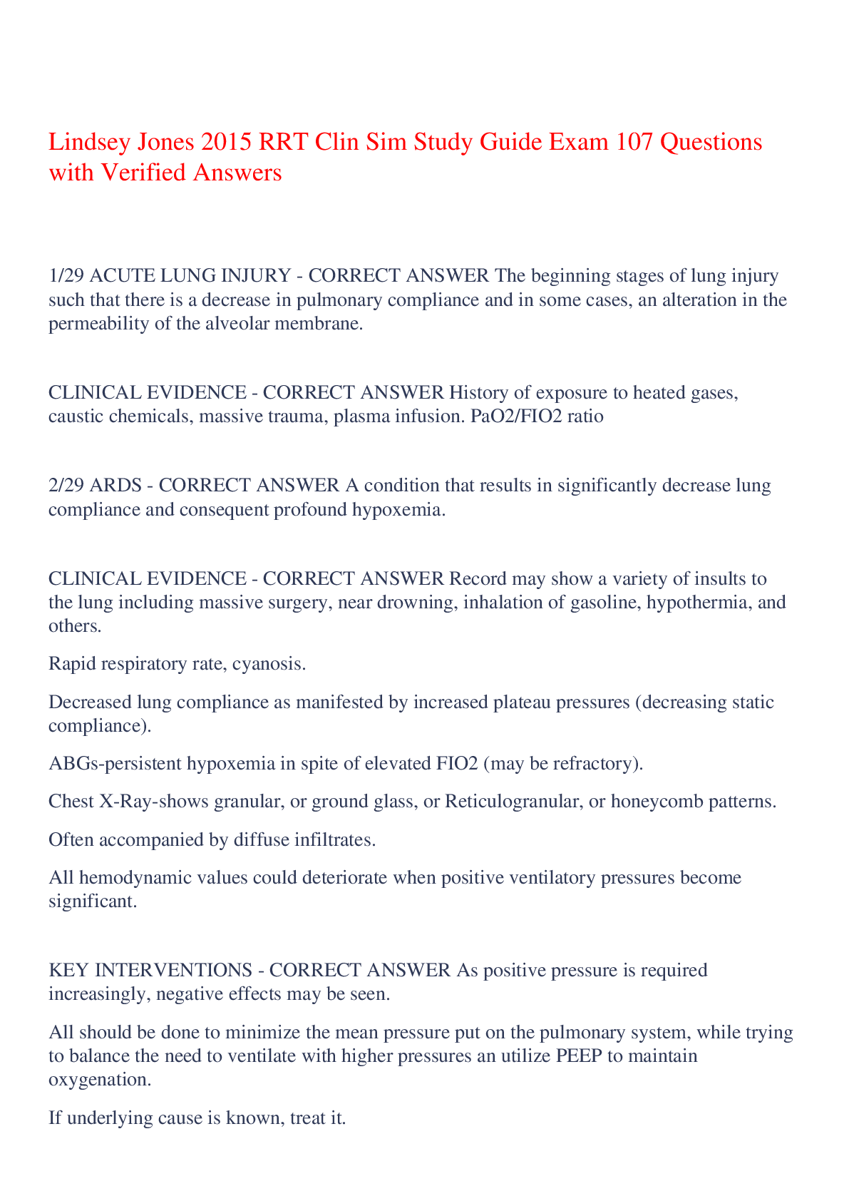
Lindsey Jones 2015 RRT Clin Sim Study Guide Exam 107 Questions with Verified Answers.100% CORRECT
Document Content and Description Below
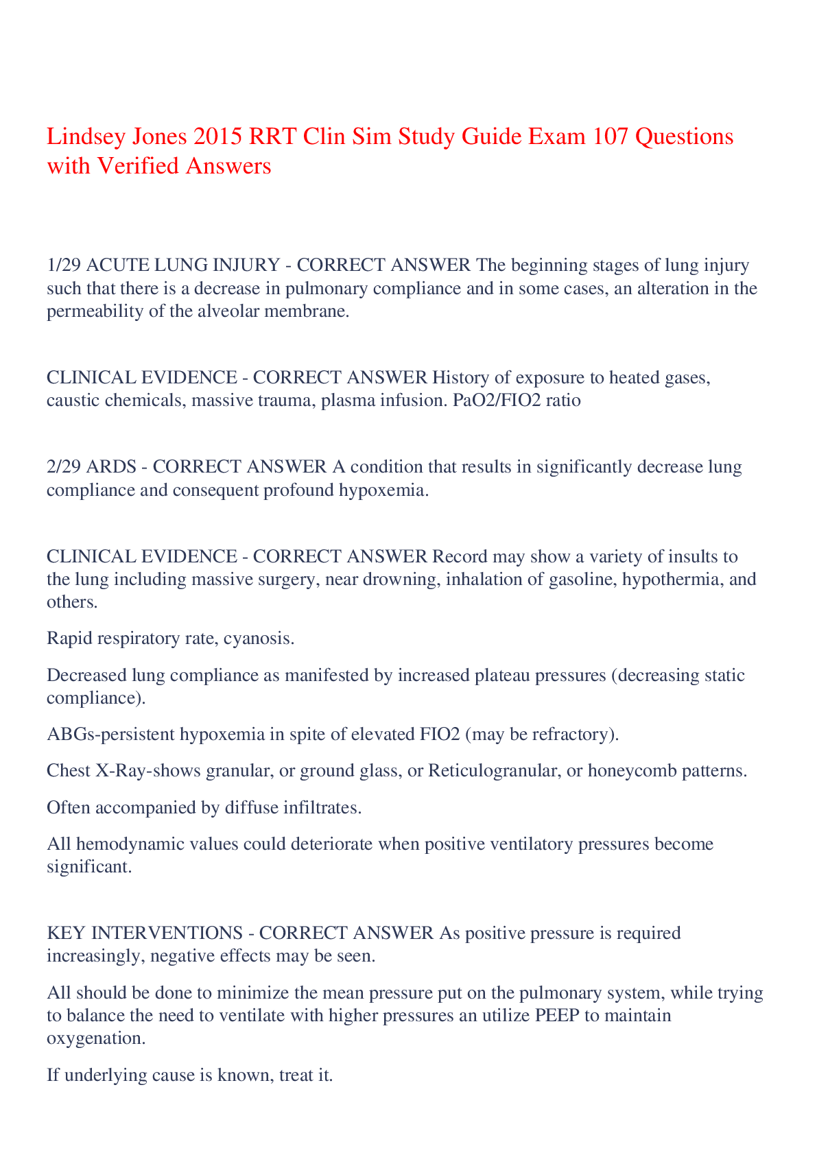
Lindsey Jones 2015 RRT Clin Sim Study Guide Exam 107 Questions with Verified Answers 1/29 ACUTE LUNG INJURY - CORRECT ANSWER The beginning stages of lung injury such that there is a decrease in p... ulmonary compliance and in some cases, an alteration in the permeability of the alveolar membrane. CLINICAL EVIDENCE - CORRECT ANSWER History of exposure to heated gases, caustic chemicals, massive trauma, plasma infusion. PaO2/FIO2 ratio <300 PF ratio <200 = ARDS 2/29 ARDS - CORRECT ANSWER A condition that results in significantly decrease lung compliance and consequent profound hypoxemia. CLINICAL EVIDENCE - CORRECT ANSWER Record may show a variety of insults to the lung including massive surgery, near drowning, inhalation of gasoline, hypothermia, and others. Rapid respiratory rate, cyanosis. Decreased lung compliance as manifested by increased plateau pressures (decreasing static compliance). ABGs-persistent hypoxemia in spite of elevated FIO2 (may be refractory). Chest X-Ray-shows granular, or ground glass, or Reticulogranular, or honeycomb patterns. Often accompanied by diffuse infiltrates. All hemodynamic values could deteriorate when positive ventilatory pressures become significant. KEY INTERVENTIONS - CORRECT ANSWER As positive pressure is required increasingly, negative effects may be seen. All should be done to minimize the mean pressure put on the pulmonary system, while trying to balance the need to ventilate with higher pressures an utilize PEEP to maintain oxygenation. If underlying cause is known, treat it. After emergency situation is past, keep FIO2 at 0.6 or below and use PEEP. Keep increasing PEEP until an obvious degradation in hemodynamic values (especially cardiac output) is witnessed. As ventilatory pressure get higher, OK to consider alternate methods of ventilation including pressure control, high frequency, etc. Keep VT at or below 6 ml/kg Utilize the ARDS.net protocols (in this book) EXAM CHALLENGE - CORRECT ANSWER ARDS can be a very disquieting case. Usually persistent increases in PEEP are needed. Do not be afraid to increase PEEP significantly. Also, most often, cardiac output or some other hemodynamic value will fall indicating a need to decrease PEEP in spite of profound hypoxemia. When doing so always return to the previous acceptable setting and then increase FIO2 as needed. 3/29 EMPHYSEMA - CORRECT ANSWER Abnormal condition of the alveoli resulting in destruction and loss of elasticity. CLINICAL EVIDENCE - CORRECT ANSWER Barrel Chest Accessory muscle use Digital clubbing of the nail beds Significant history of smoking & or occupational exposure to smoke or other pulmonary irritants. Chest X Ray- increased AP Diameter, flattened diaphragms, hyperlucency, diminished pulmonary markings. CBC- polycythemia, increased WBC due to possible infection. ABGs-compensated respiratory acidosis (high PaCO3, normal pH), moderate to severe hypoxemia. PFT-Flows are decreased especially middle sized airways (FEF 25-75%) and FEV 1. Wheezing and diminished breath sounds. KEY INTERVENTIONS - CORRECT ANSWER Oxygen Therapy-low FIO2 (0.24 to 0.28) or 1 to 2 lpm nasal cannula. Oxygen conserving devices such as liquid oxygen or trans-tracheal oxygen. Home care education on devices and equipment cleaning. Aids to help quit smoking such as nicotine replacement therapy. Bronchodilation medication via MDI or aerosol nebulizers (given with room air). Corticosteroids. Use ET tubes with subglottic suction ports to prevent VAP. May remove from mechanical ventilation early and place on NIV immediately. EXAM CHALLENGE - CORRECT ANSWER Therapists may be tempted to utilize high FIO2 because of the severity of the hypoxemia. They may also be tested with an emergency, the only time it is appropriate to use 100% oxygen on a COPD patient. 4/29 CHRONIC BRONCHITIS - CORRECT ANSWER Condition where the patient has a productive cough 25% of the year for at least 2 consecutive years. CLINICAL EVIDENCE - CORRECT ANSWER Productive cough, purulent sputum production Exposure to pulmonary irritants, like history of smoking Frequent infections Dyspnea Chest X-ray - could be normal, or may show hyperlucency, diminished pulmonary markings. CBC- possibly increased WBC due to possible infection. ABGs_ could be normal or very slight respiratory acidosis and hypoxemia PFT- flows are decreased especially middle sized airways (FEF 25-75%) and FEV1 KEY INTERVENTIONS - CORRECT ANSWER Anything that promotes good pulmonary hygiene such as chest physiotherapy, hydration therapy when sputum is thick. Fluid therapy if dehydrated. Oxygen therapy for hypoxemia Aerosolized bronchodilator therapy Antibiotic Tetracycline may be preferable EXAM CHALLENGE - CORRECT ANSWER The most distinguishing characteristic is that the cough is productive and has been so for a good portion of the year. 5/29 PULMONARY EDEMA / CHF - CORRECT ANSWER Significant reduction in cardiac output. Involvement of fluid penetrating the alveolar capillary membrane into the lungs. CLINICAL EVIDENCE - CORRECT ANSWER History of CFH or pulmonary hypertension Tachypnea, tachycardia, anxiety Cold, clammy, diaphoretic Pink frothy secretions (may be described as marked congestion) Edema of fluids (especially pedal edema) Pitting edema (+2, +3) ABGs - ventilatory failure with moderate to severe hypoxemia Chest X-ray - Butterfly pattern, fluffy infiltrates Increased hemodynamic pressure (PCWP, PAP, CVP) KEY INTERVENTIONS - CORRECT ANSWER Treat as an emergency! 100% oxygen Administer diuretic medication furosemide (Lasix) Cardiac inotropic stimulating drugs such as digoxin, digitalis if increased PCWP and PAP Be prepared to treat ventilatory failure with mechanical ventilation EXAM CHALLENGE - CORRECT ANSWER This case may feel complicated because it involves the heart and hemodynamic values. It is usually easily identified by pink frothy secretions and a butterfly pattern on the chest X-ray. You may need to make the distinction between pulmonary edema caused by cardiac problems and that which is caused by alveolar capillary membrane problems (ARDS). If it is cardiac, then you must treat the heart. 6/29 HEART SURGERY - CORRECT ANSWER Any kind of surgery on the heart. CLINICAL EVIDENCE - CORRECT ANSWER Do well rounded assessment prior to surgery including vital signs, family history of cardiac illness. Preoperative assessments of breath sounds Baseline data including basic spirometry of all types including FEV1/FVC% and pre and post bronchodilator studies ABGs - preoperative for baseline KEY INTERVENTIONS - CORRECT ANSWER Always assess ventilatory volumes and be prepared to mechanically ventilate Incentive spirometry every hour after surgery for lung expansion and alveolar ventilation. If unable (unconscious) use simple ventilatory assisting devices such as IPPB or CPAP with mask. Be on the alert for cardiac arrest- perform CPR without reservation or consideration of the heart surgery. EXAM CHALLENGE - CORRECT ANSWER This case is not too complicated. You may feel hesitant to do CPR on someone fresh out of surgery. Just do it. 7/29 MYOCARDIAL INFARCTION / ARRHYTHMIA - CORRECT ANSWER Ischemia of the hart causing muscle damage and potential failure. CLINICAL EVIDENCE - CORRECT ANSWER History of chest pain, radiating pain down the left arm Family history of disease Diaphoretic, nausea, tachycardia Cold, diaphoretic and clammy to the touch Dyspnea ABGs - hypoxemia ECG (EKG) - pronounced Q waves and S_T segment elevation Just prior to the MI, may see flipped T waves Cardiac enzymes including CPK, LDH, SGOT are elevated KEY INTERVENTIONS - CORRECT ANSWER Emergency - 100% oxygen. Oxygen at adult therapeutic level (40 to 60 %) upon suspicion of first presentation of signs and/or symptoms Treat arrhythmias Bradycardia with Atropine PVCs with Lidocaine or oxygen Pulseless ventricular tachycardia with defibrillation and chest compressions Ventricular Fibrillation with defibrillation Note: For Ventricular fibrillation, defibrillate at 360 joules - repeat as needed. Do not exceed 360 joules. Note: For Atrial fibrillation of flutter, do synchronized cardioversion - start at 50 joules. EXAM CHALLENGE - CORRECT ANSWER Will likely need to treat arrhythmias with appropriate medication and/or defibrillation. 8/29 ABDOMINAL SURGERY - CORRECT ANSWER Surgery of the abdominal area for various reasons. CLINICAL EVIDENCE - CORRECT ANSWER All general visual assessments All general bedside assessments including all vitals Ventilatory volumes (VC, VT, FEV1) compared to pre-surgery baselines KEY INTERVENTIONS - CORRECT ANSWER Establishing baselines in pulmonary function testing flows and volumes. Start patient on incentive spirometry (SMI therapy) prior to surgery, every hour after surgery Initial SMI therapy goals should be 1/2 the pre-surgical baseline value. If unable to achieve 1/2 the pre-surgical volume, then lower the goal to just above what the patient can accomplish. If patient cannot do SMI because of sedation, then administer IPPB to keep alveoli inflated Use positive pressure (IPPB) if needed after surgery if patient is unconscious. EXAM CHALLENGE - CORRECT ANSWER Abdominal surgery is usually a very general, non-complicated case involving preventative care and follow up. 9/29 LARYNGECTOMY - CORRECT ANSWER Surgery done to address or remove cancer of the larynx. CLINICAL EVIDENCE - CORRECT ANSWER Surgical record: Surgery radical (entire larynx) or simple (cord removal) Medical history will show cancer in the upper airway Signs of airway obstruction after surgery. Usually caused by blood within a few hours after the surgery. KEY INTERVENTIONS - CORRECT ANSWER If radical surgery (entire larynx removed) then the tracheostomy become permanent. If not radical then a temporary laryngectomy tube is placed but must be replaced in 3 to 6 weeks. Prevent aspiration! Wait at least a week before oral ingestion of liquid and longer for food. Thorough pulmonary hygiene through suctioning Use cool aerosol or ultrasonic nebulizer to keep secretions thin and hydrated. Once the surgery is done, you can no longer, orally intubate the patient. Even if temporary laryngectomy tube is in place, you must intubate and/or ventilate through that tube! EXAM CHALLENGE - CORRECT ANSWER In this case, you are always looking for post-surgical complications like blood clots in the laryngeal tube. Often, you will have to mechanically ventilate this patient through the laryngectomy tube. 10/29 THORACIC SURGERY - CORRECT ANSWER Can have a variety of complications from thoracic surgery CLINICAL EVIDENCE - CORRECT ANSWER Always monitoring chest tube drainage adequacy Looking for potential complications: Hypovolemic shock, low hemodynamic values including blood pressure Subcutaneous emphysema Elevated ventilatory pressures Chest X-ray- to confirm proper re-inflation of the lung and proper placement of chest tubes KEY INTERVENTIONS - CORRECT ANSWER Anything that promotes expansion of the lungs u=including incentive spirometry, IPPB, and positive pressure mechanical ventilation If a lobectomy or pneumonectomy, ventilatory volumes should be set lower Fluid therapy if volume is a problem (often is). EXAM CHALLENGE - CORRECT ANSWER Your ability to deal with and troubleshoot chest tube maintenance is tested in this simulation. Sometimes this case is combined with chest trauma. 11/29 ASTHMA - CORRECT ANSWER Abnormal constriction of the bronchials resulting in sputum production and narrowed airways. CLINICAL EVIDENCE - CORRECT ANSWER Accessory muscle use Tachycardia Dyspnea Wheezing Congested cough Wet, clammy skin ABGs- possible respiratory acidosis, could be hypoxic Chest X-ray - hyperinflation, scattered infiltrates, flattened diaphragms. In allergic cases, may see elevated eosinophil count which can cause yellow sputum Watch out for "patient speaking in one-word sentences"- indicates and emergency PFT- Decreased flows in FEV1 but diffusion is normal as manifested by DLCO KEY INTERVENTIONS - CORRECT ANSWER Oxygen therapy initially, even in the absence of conclusive data like SPO2 and ABGs Aerosolized bronchodilator therapy Xanthenes medication given IV (Aminophylline, etc.) Promote pulmonary hygiene Corticosteroids such as oral or IV Prednisone If repeated bronchodilator treatment does not work, consider possible diagnosis of Status Asthmaticus Instruct patient on the use of an Asthma Action plan, a plan to self-monitor and record daily results EXAM CHALLENGE - CORRECT ANSWER When doing PFTs, always do a pre and post bronchodilator study. Consider effective if 12% or more improvement is noted. Always start oxygen first when presenting in the ER - part of the National Asthma Guidelines. 12/29 STATUS ASTHMATICUS - CORRECT ANSWER Asthma that will not respond to bronchodilation therapy usually persists more then 24 hours. CLINICAL EVIDENCE - CORRECT ANSWER Historically non-responsive to bronchodilators. Patient will report the need to take many bronchodilator treatments before feeling better. Accessory muscle use and retractions Dyspnea, wheezing, and congested cough Wet, clammy skin, pulses paradoxus ABGs - possible respiratory acidosis when tiring, alkalosis at first due to anxiety, could be hypoxic Chest X-ray - hyperinflation, scattered infiltrates, flattened diaphragms. KEY INTERVENTIONS - CORRECT ANSWER May deteriorate quickly, so if progression is shown, intubate mechanically ventilate before full ventilatory failure. Use subcutaneous epinephrine - 1 mL of 1:1000 strength. May need to give every 20 minutes for up to three consecutive doses- no more than three. Utilize continuous bronchodilator treatments and Heliox if needed. Address all three components of Asthma: Inflammation - corticosteroids are key Bronchoconstriction - bronchodilators Sputum - airway clearance, hydration, thinning of sputum if needed. EXAM CHALLENGE - CORRECT ANSWER Questions on this will challenge you ability to recognize impending ventilatory failure. it is very important that you treat it before full ventilatory failure. There is a frequent need to repeat actions, such as bronchodilator treatments, which may make you uncomfortable. Do not be afraid to administer several bronchodilators in succession. The same is true of the subcutaneous epinephrine. If you give one dose, you will likely have to give another, and possibly another. Continue if symptoms show no signs of relief. 13/29 CHEST TRAUMA - CORRECT ANSWER May consist of any trauma leading to fractured ribs or flail chest CLINICAL EVIDENCE - CORRECT ANSWER Circumstantial history (motor vehicle accident, etc.) Respiratory rate and pattern is fast and shallow due to pain May have obvious trauma (bruising) on chest wall Sharp chest pain, especially at the top of each breath Paradoxical chest movement if ribs are broken in two places (flail chest) Pneumothorax is possible (see signs and symptoms of pneumothorax) Chest x-ray - may reveal broken ribs, usually isolated in same area KEY INTERVENTIONS - CORRECT ANSWER Anything that encourages deep (adequate) breathing in spite of pain such as IPPB, incentive spirometry, coughing. Watch for ventilatory fatigue and eventual ventilatory failure Mechanically support ventilation when it is evident ventliatory failure is impending. If possible do not wait until full ventilatory failure. Treat partial pneumothorax if greater than 20% - i.e. insert chest tubes Treat hemothorax, with chest tubes or thoracentesis Treat tension pneumothorax with a large-bore 14 gauge needle Mechanical ventilation at lower tidal volumes - initial VT of 6-7 mL/kg is acceptable PEEP therapy at 5-10 acceptable EXAM CHALLENGE - CORRECT ANSWER This case is usually easy to recognize. You may be tempted by options that address the broken ribs when, in fact, you simply need to address ventilation. Very commonly, this case will lead to pneumothorax or partial pneumothorax or hemothorax. 14/29 HEAD TRAUMA - CORRECT ANSWER Potential loss of ventilatory drive as a result of drug overdose (usually a narcotic) CLINICAL EVIDENCE - CORRECT ANSWER Sometimes trauma is visual with blood contusions on the head History is trauma related, often automobile accident Look and act sleepy, difficult to arouse respiratory rate and pattern is low and/or shallow and irregular Papillary response to light may be unequal or inadequate If intracranial pressure monitor is in place, may see ICP greater than 20 cm H20 KEY INTERVENTIONS - CORRECT ANSWER Must constrict vessels in the head by keeping PaC02 between 25-30 mm Hg. Adjust FIO2 to maintain high normal levels (PaO2 of 100 mm Hg). Avoid increased ICP by minimizing PEEP usage. Suction only when needed due to elevating peak pressures. Avoid anything that will increase mean arterial pressure (MAP). Sedation is important, but should monitor exhaled volumes and pressures closely Use of drugs such as Mannitol (cerebral diuretic medication) when ICP is at or above 20 cm H20 Use Dilantin and establish an airway if seizure activity is observed EXAM CHALLENGE - CORRECT ANSWER Unique to this situation is the need to monitor ICP readings and avoid anything that increases MAP. You will likely need to suction this patient to keep peak pressures down but the very act of doing so may elevate ICPs. 15/29 NECK / SPINAL INJURY - CORRECT ANSWER Any trauma threatening the physical structure of the neck. Can include neck or spinal surgery. CLINICAL EVIDENCE - CORRECT ANSWER Historical relevance, some sort of accident such as diving an automobile. Visible damage to the neck Altered conscious level Pulse must be palpated brachially or femorally VT, VC, PEFR, and other ventilatory volumes may quickly deteriorate Neck x-ray - will show injury KEY INTERVENTIONS - CORRECT ANSWER Always be prepared to quickly assist and/or promote ventilation. If intubation is required, always use MODIFIED jay thrust. If given the option, always intubate with a bronchoscope so damage can be visualized and care can be taken to avoid inflicting further damage. Alternatively, a blind nasal intubation is acceptable. Spinal injury may require long-term mechanical ventilation. EXAM CHALLENGE - CORRECT ANSWER Your knowledge of special intubation techniques is what is being tested in this type of simulation. 16/29 BURN TRAUMA / CO POISONING - CORRECT ANSWER Results from direct exposure to fire and or smoke. Directly threatens airway and oxygen carrying capacity of the blood. CLINICAL EVIDENCE - CORRECT ANSWER Diagnosis is based largely on history - exposure to fire or smoke. Often occurs in occupational related cases (fire fighter) Visible burns about the body and face Signed nasal and or eyebrow hairs "Cherry-red" color of face with CO poisoning Patient is often confused or unresponsive Stridor, hoarseness Breath sounds - wheezing, rhonchi, rales ABGs - intially decreased PaCO2, normal PaO2, decreased saturation. Latter may develop into respiratory acidosis Chest X-ray - may be clear at first, but later may show pulmonary edema and markedly decreased lung compliance COHb - 20% or more KEY INTERVENTIONS - CORRECT ANSWER Protect airway by establishing an artificial airway immediately. Particularly if there s respiratory distress and there are burns about the face. For CO poisoning - start 100% oxygen immediately - even if only suspect it - do not wait for COHb results. Continue oxygen therapy until COHb level is below 10%.- may use hyperbaric medicine if offered. Practice Reverse isolation (protect the patient from staff) Mechanically ventilate as needed. Avoid use of Anectine (paralyzing agent) as it can cause hyperkalemia in burn trauma patients. EXAM CHALLENGE - CORRECT ANSWER Fairly common case on the test. Remember to focus on the airway and on oxygen carrying capacity of the blood. Remember to employ isolation techniques. Otherwise, provide general respiratory therapy. 17/29 MYASTHENIA GRAVIS - CORRECT ANSWER Neuromuscular abnormality where muscles experience paralysis starting from the head down to the feet including ventilatory muscles. CLINICAL EVIDENCE - CORRECT ANSWER May have a history of Myasthenia Gravis if not the initial onset Droopy facial muscles and eyelids (Ptosis) Patient will describe slowly feeling weakness generally but feels better with rest. Double vision (diplopia) Dysphagia (difficulty swallowing) Shrinking VT, VC, MIP Tensilon Challenge Test - positive for Myasthenic crisis if improvement is noted upon the administration of Tensilon KEY INTERVENTIONS - CORRECT ANSWER If Tensilon improves condition then, anticholinesterase therapy is indicated including: Neostigmine (prostigmine) Mestinon (pyridostigmine) OK to do additional Tensilon challenge test to observe progression. If symptoms improve with Tensilon and then worsen, must reverse the anticholinesterase with Atropine. Always monitor spontaneous ventilatory volumes (VT and VC) as well as MIP. Never treat Myasthenia gravis with Tensilon - only use to diagnose. Be totally prepared to intubate and mechanically ventilate prior to Tensilon challenge since it could take out the respiratory drive. When VC falls off rapidly (especially if below 1.0 L), then intubate and mechanically ventilate. EXAM CHALLENGE - CORRECT ANSWER This can be a very tricky simulation and it is likely that it will show up on the exam. especially important is your use of Tensilon to diagnose it and an understanding of the dangerous effects it could have. Must always be prepared to assume ventilation. VT, VC, and MIP are key in monitoring this patient for degradation in ventilatory status. Intubate prior to full failure if possible. 18/29 GUILLAIN-BARRE' SYNDROME - CORRECT ANSWER An insidious neuromuscular problem involving muscle paralysi. Paralysis begins in the lower extremities and moves upward, including the ventilatory muscles. CLINICAL EVIDENCE - CORRECT ANSWER Medical history or patient complaint of recnt influenza-type sickness. Complaint of sluggish lower extremities Shrinking VT, VC, MIP ABGs- impending or current ventilatory failure. Spinal tap- will show increased protein in the spinal fluid KEY INTERVENTIONS - CORRECT ANSWER Be primarily concerned with loss of ventilation, monitor ventilatory volumes (VC, VT) and MIP. Be patient about intubation and mechanical ventilation. Onset can be slow. Primary treatment will involve mechanical ventilation and letting the syndrome run its course. Prolonged weaning is not necessary (but is OK) once the disease as run its course. Such evidence is simply manifested by the return of VC, VT, MIP. EXAM CHALLENGE - CORRECT ANSWER Like most neuromuscular cases, you will be tested in your ability to recognize deterioration in ventilatory muscles. In this case, onset can be slow, so don't jump-the-gun and mechanically ventilate too early. Only do so as VC falls below 1.0 L. 19/29 SHOCK - CORRECT ANSWER Condition where tissues oxygenation is in jeopardy due to a sudden decrease in blood flow. CLINICAL EVIDENCE - CORRECT ANSWER Historical evidence of an event or massive trauma or hypothermia, etc. General appearance - cold, clammy, dusky, cyanotic Tachycardia, tachypnea, hypotensive Temperature may be below normal Reduction in urine output ABGs - hypoxemia and ventilatory failure Reduction in common hemodynamic values (CVP, PAP, PCWP) and cardiac output. Types of Shock There are different types of shock. Each requires a different treatment. (see Types of Shock in this book). KEY INTERVENTIONS - CORRECT ANSWER Mechanically ventilate with ventilatory failure. Fluids Main treatment involves treating the original problem (that which caused the shock). This can be highly variable. EXAM CHALLENGE - CORRECT ANSWER Shock will test your ability to recognize it and monitor the patient for ventilatory failure. 20/29 PULMONARY EMBOLI - CORRECT ANSWER Situation where the pulmonary artery becomes obstructed and dead-space ventilation results. Sometimes called deadspace disease. CLINICAL EVIDENCE - CORRECT ANSWER History of recent major surgery or trauma (amputations, clotted massive bleeding sightss) Complaint of chest pain and dyspnea Elevated vitals including pulse, respirations, and blood pressure Breath sounds - wheezing and medium rales PEC02 (Capnography) decreasing PEC02 during normal PaCO2 ABGs - persistent hypoxemia in spite of increasing FIO2 V/Q scan will ventilation without adequate perfusion Patient will be described as "OK one minute, but suddenly became short of breath" KEY INTERVENTIONS - CORRECT ANSWER Anticoagulation therapy with Heparin or Coumadin Note: must monitor clotting tests PTT for Heparin PT for Coumadin Clot-busting medication such as streptokinase. May also use a bolus of heparin Installation of retrievable vena cava filter, especially future clot formation is possible. Mechanical ventilation as needed. Emergency level oxygen - 100% Place a retrievable inferior vena cava filter to prevent clots from reaching the lungs, especially if the nature of the problem cannot withstand any blood thinning agents (like a hemorrhagic stroke). EXAM CHALLENGE - CORRECT ANSWER This case primarily involves recognizing the pulmonary emboli and treating it with anticoagulation medications. You will likely have to monitor clotting times., PTT or PT. otherwise, involves general respiratory therapy. 21/29 AIDS - CORRECT ANSWER Disease of the immune system commonly resulting in pneumocystis carinii, a type of pneumonia. CLINICAL EVIDENCE - CORRECT ANSWER Previous history of HIV positive test results Emaciation, unexplained weight-loss, diarrhea, low-grade fevers, night sweats, Commonly homosexual activity or drug usage id admitted Positive HTLV III ELISA test- positive for HIV Bronchoscopy - from lung washings or biopsy may show pneumocystis carinii KEY INTERVENTIONS - CORRECT ANSWER Exercise Universal Precautions Aerosolized Pentamadine - usually done monthly When administrating Pentamadine, use one-way valves and filters. 22/29 PULMONARY HYPERTENSION - CORRECT ANSWER Excessive pressure in the pulmonary vasculature, especially the pulmonary artery CLINICAL EVIDENCE - CORRECT ANSWER Hemodynamic value, pulmonary artery pressure (PAP). Normal is 25/8 mm Hg. Associated with pathologies involving the right heart. (COPD, PFO, Cor Pulmonale). KEY INTERVENTIONS - CORRECT ANSWER Inhaled Nitric Oxide Other pulmonary BP lowering medications (see pulmonary vasodilators in Pharmacology Review). 23/29 DRUG OVERDOSE - CORRECT ANSWER Potential loss of ventilatory drive as a result of drug overdose (usually a narcotic) CLINICAL EVIDENCE - CORRECT ANSWER Historical drug use as told by previous admissions or family or paramedics Sometimes poor self-hygiene, emaciated Respiratory rate and pattern is low and/or shallow ABG- often show pure respiratory acidosis and/or ventilatory failure KEY INTERVENTIONS - CORRECT ANSWER #1 priority in this case is intubation to protect the airway, prevent aspiration of stomach contents, and facilitate manual ventilation. Monitor closely as ventilation can cease in an instant (due to possible suppression of the CNS) If narcotic overdose (usually is) then use narcotic reversing medication such a Narcan (Nalaxon) Support ventilation until drugs are out of system. EXAM CHALLENGE - CORRECT ANSWER The most important part of this pathology is the need for immediate intubation while recognizing that there may not be a need to mechanically ventilate until ventilatory status deteriorates. 24/29 CYSTIC FIBROSIS - CORRECT ANSWER An inherited disorder resulting in the mass production of thick mucus in the lungs. CLINICAL EVIDENCE - CORRECT ANSWER Family history of disease, siblings may have it. Emaciated in appearance and body frame may be small or age Sputum production of thick voluminous purulent secretions Can look like a young COPD patient, barrel-chested Decreased flow rates such as FEV1 Chest X-ray - looks like COPD, hyperinflation, increased A-P diameter, diaphragm flattening Sweat Chloride Test - show sweat chloride > 60 mEq/L KEY INTERVENTIONS - CORRECT ANSWER Primary treatments relates to the need to mobilize and remove secretions. Secretion removal promotion therapies: PEP therapy devices Chest physiotherapy with postural drainage Hydration devices such as heated aerosol or ultrasonic Nebulization Vibration therapy Oxygen as needed Antibiotic therapy when infection is present - often is Medications used commonly include Tobramycin and Pulmozyme (Dornase Alpha) 25/29 METHENOGLOBINEMIA - CORRECT ANSWER Presence of methenoglobinemia in the circulating blood - caused by use of some recreational drugs or use oral anesthetics, such as Benzocaine or Benzene. CLINICAL EVIDENCE - CORRECT ANSWER Central cyanosis in spite of high FIO2 and very elevated PaO2 Fatigue Shortness of breath Headache Dark-brown or dark blue hood (chocolate-colored) KEY INTERVENTIONS - CORRECT ANSWER IV Methylene Blue 26/29 PNEUMONIA - CORRECT ANSWER Collection and/or consolidation of sputum as a result of a bacterial or viral agent entering the lung on inhalation. CLINICAL EVIDENCE - CORRECT ANSWER Fever, dyspnea, chills, cyanosis, rhonchi and rales Increased WBC if bacterial, decreased WBC if viral Scattered infiltrates on chest x-ray KEY INTERVENTIONS - CORRECT ANSWER Oxygen therapy first Suctioning and other bronchial hygiene efforts Antibiotics Penicillin for gram positive organisms Gentamycin, or other 'mycin' antibiotics for gram negative organisms 27/29 PLEURAL EFFUSION - CORRECT ANSWER Development of excess fluid in the pleural space causing some amount of lung space shrinkage or collapse. CLINICAL EVIDENCE - CORRECT ANSWER Sharp chest pains in the area Mediastinal shift away from the effusion Obliteration of costophrenic angles on x-ray (lateral decubitus) Fluid may shift when patient is in different positions - proof of pleural effusion KEY INTERVENTIONS - CORRECT ANSWER Thoracentesis to remove fluid if small Chest tubes in the pleural space if lung is more than 20% collapsed. 28/29 PULMONARY TUBERCULOSIS - CORRECT ANSWER Pulmonary tissue destructive disease as a result of inhalation of the tubercly bacilli CLINICAL EVIDENCE - CORRECT ANSWER Night sweats Hemoptysis (frank or non-frank blood) Expectoration of lung tissue during coughing Formation of cavitations in the lung on x-ray KEY INTERVENTIONS - CORRECT ANSWER Isoniazid (INH) and other medications (Rifampin, Ethambutol, Streptomycin) Strict Respiratory Isolation Minimize coughing 29/29 DIABETES - CORRECT ANSWER Condition related to failure of the renel system resulting in the inability to dispose of CO2. Respiratory result is often respiratory ketoacidoses CLINICAL EVIDENCE - CORRECT ANSWER History of diabetes Lethargy, confusion, unresponsiveness Respiratory rate and pattern- significant in depth and rate with an irregular rhythm (Kussmaul's) Pedal Edema ABGs- Profound metabolic acidosis Urine output is markedly decreased (less than 20 ml per hour) Blood glucose - > 160 mg (Normal 80-120 mg) KEY INTERVENTIONS - CORRECT ANSWER Must watch for ventilatory failure from prolonged ventilatory effort and fatigue. Administer elctrolytes (K+, Na+, HCO3-< Cl-) as needed. Provide fluid as needed. Correct ketoacidosis. EXAM CHALLENGE - CORRECT ANSWER May be tempted by profoundly acidotic pH. Only determine respiratory failure through the CO2, or a sudden decrease in ventilatory volumes and breathing rate. [Show More]
Last updated: 7 months ago
Preview 1 out of 21 pages
Buy this document to get the full access instantly
Instant Download Access after purchase
Add to cartInstant download
We Accept:

Reviews( 0 )
$9.00
Document information
Connected school, study & course
About the document
Uploaded On
Nov 23, 2023
Number of pages
21
Written in
Additional information
This document has been written for:
Uploaded
Nov 23, 2023
Downloads
0
Views
20

