*NURSING > HESI MED SURG > Med-Surg NSG 331 (NSG331) (Med-Surg NSG 331 (NSG331)) NSG 331 Med-Surg Final Exam Review _ A+ guide (All)
Med-Surg NSG 331 (NSG331) (Med-Surg NSG 331 (NSG331)) NSG 331 Med-Surg Final Exam Review _ A+ guide Latest Spring 2020/2021
Document Content and Description Below
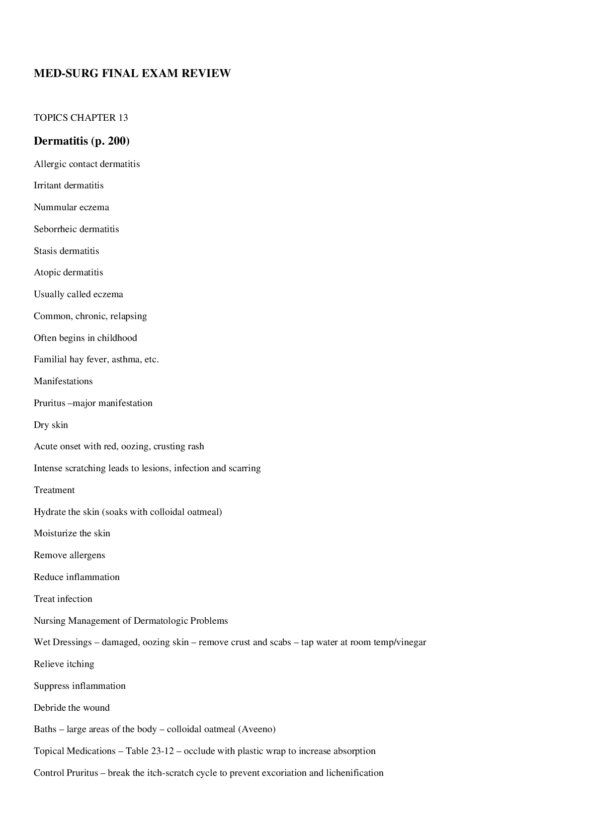
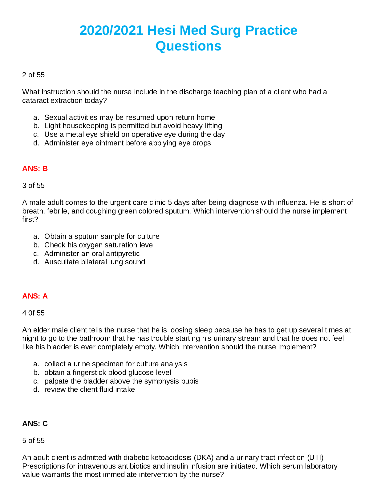
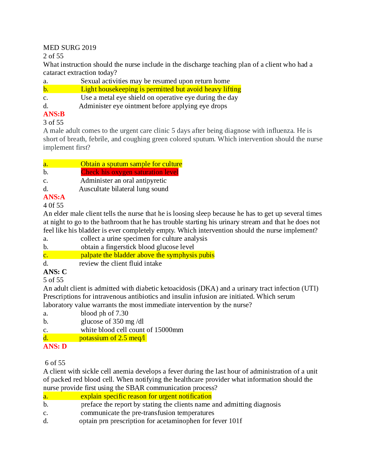
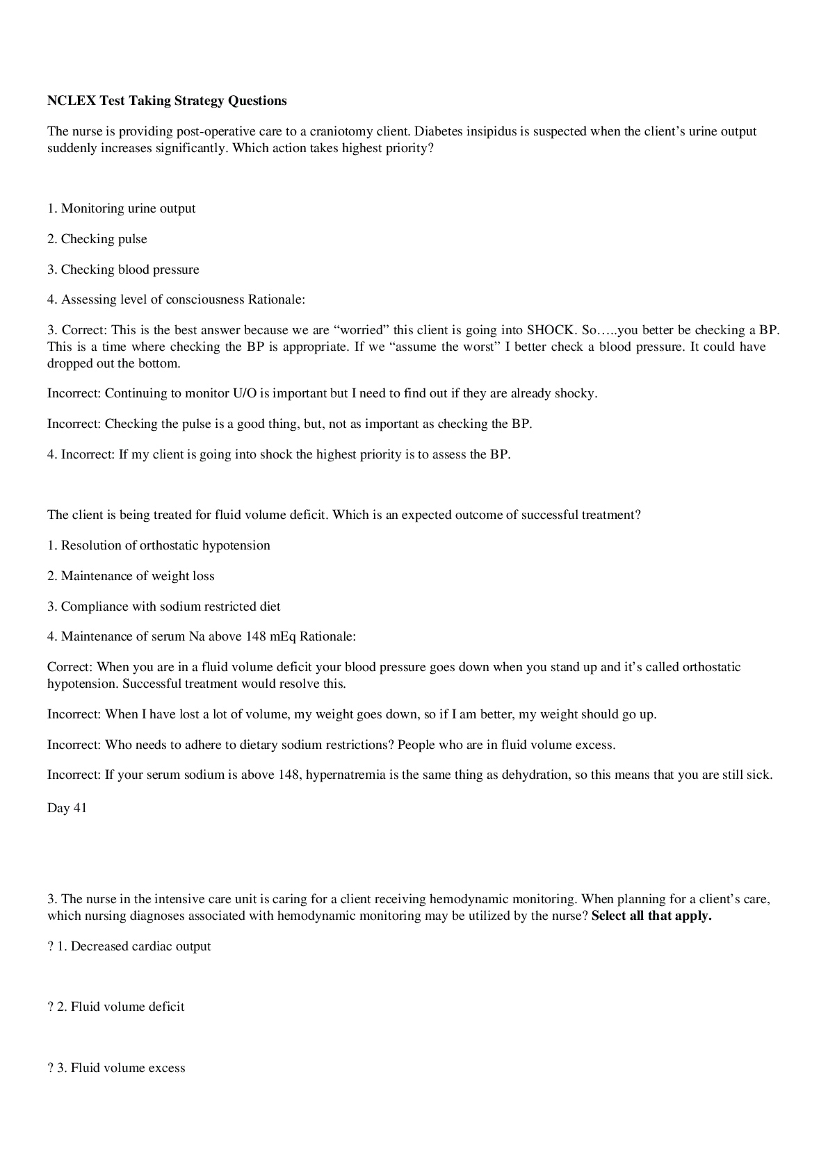
Exam (elaborations) Med-Surg NSG 331 (NSG331) (Med-Surg NSG 331 (NSG331)) NSG 331 Med-Surg Final Exam Review _ A+ guide Latest Spring 2020/2021 MED-SURG FINAL EXAM REVIEW TOPICS CHAPTER 13 Dermatitis ... (p. 200) 1. Allergic contact dermatitis 2. Irritant dermatitis 3. Nummular eczema 4. Seborrheic dermatitis 5. Stasis dermatitis 6. Atopic dermatitis • Usually called eczema • Common, chronic, relapsing • Often begins in childhood • Familial hay fever, asthma, etc. • Manifestations Pruritus –major manifestation Dry skin Acute onset with red, oozing, crusting rash Intense scratching leads to lesions, infection and scarring • Treatment Hydrate the skin (soaks with colloidal oatmeal) Moisturize the skin Remove allergens Reduce inflammation Treat infection 7. Nursing Management of Dermatologic Problems • Wet Dressings – damaged, oozing skin – remove crust and scabs – tap water at room temp/vinegar Relieve itching Suppress inflammation Debride the wound • Baths – large areas of the body – colloidal oatmeal (Aveeno) • Topical Medications – Table 23-12 – occlude with plastic wrap to increase absorption • Control Pruritus – break the itch-scratch cycle to prevent excoriation and lichenification Lichenification – thickening of the epidermis with exaggerated markings resembling a washboard o Caused by chronic itching / rubbing of the skin • Prevention of Spread – gloves and adamant hand washing • Prevention of Secondary Infections – particularly to existing skin lesions • Specific skin care – educate patient on skin care after dermatologic procedures and hygiene Wounds kept moist and covered heal more rapidly, leave scab/crust undisturbed CHAPTER 16 Dehydration Fluid Volume Deficit (FVD): Hypovolemia • Causes: decr intake, vomiting, fever, diarrhea, NG loss, hemorrhage, 3rd spacing, incr insensible loss, diab insipid • S&S: dry, pale cold clammy skin, wt loss, decreased turgor and cap refill, tachycardia, postural hypotension, high HCT level, low U/O, low grade temp, altered mental status, seizures, coma, restlessness, drowsiness • Treatment: underlying cause, oral/IV fluids (0.9% NS), blood (if d/t hemorrhage), rest, nutrition • Nursing measures: VS, I&O, postural hypotension (safety) Hypothalamic-pituitary regulation o Body fluid deficit / Increases in plasma osmolarity activates osmoreceptors in hypothalamus Activates thirst and release of ADH from posterior pituitary to retain water (distal tubules) Thirst mechanism major defense against dehydration - Elderly have reduced thirst mechanism o Dec BP, nausea, pain, hypoglycemia, hypoxemia all stimulate ADH release; postop stress response, receiving analgesics/anesthesia = ADH release and decreased osmolality o Dry mouth will cause a person to drink even when there is no body water deficit Renal Regulation o Kidneys regulate water balance by adjusting urinary volume and excretion of electrolytes o Avg adult = kidneys reabsorb 99% of filtrate = 1.5L urine per day Kidney issues = less ability to regulate = edema, etc. Adrenal cortical Regulation o Release of hormones to regulate water and electrolytes Glucocorticoids – cortisol – anti-inflammatory = increase serum glucose levels Mineralocorticoids – aldosterone =enhance Na retention/K+ excretion (dec Na = RAAS activation Hormones regulate amt of water is retained Cardiac Regulation o Natriuretic peptide (antagonist to RAAS) – cardiomyocytes: response to incr atrial pressure & incr Na Decrease blood volume = atrial NP (ANP) and b-type NP (BNP) – renal tubules – excrete Na/H2O Activate to decrease volume – elevated = CHF – inhibit aldosterone, renin, ADH, angiotensin II Gastrointestinal Regulation o Oral intake accounts for most water (D/V = losing fluid/electrolytes) o Secretes approximately 8000mL of digestive fluid that is reabsorbed (small amt eliminated in feces) D/V – prevents reabsorption – fluid and electrolyte loss Insensible water loss = Sweating Sensible water loss = Excessive sweating – exercise, fever, excessive environmental heat Hypo/Hypernatremia (p. 279) Hypo/hyperK+ (p. 281) Magnesium Imbalance (p. 286) Fluid Volume Excess/Overload (p. 276) Acid/Base Imbalances (p. 288) CHAPTER 21 Bacterial Conjunctivitis (p. 371) 1. Pinkeye most common infection 2. Occurs in any age group 3. Manifestations: • Discomfort • Pruritus • Redness • Mucopurulent drainage 4. Typically occurs in one eye THEN spreads 5. MEDS—besifloxacin 6. Teach—handwashing & avoid contact with infected person Cataracts (p. 373-376) 1. Def: opacity within the lens 2. Etiology & Pathophysiology • Most: age related = senile cataracts • Blunt or penetrating Trauma • Congenital factors – i.e. maternal rubella • Radiation or ultraviolet exposure - UV • Steroids – systemic or long-term corticosteroids • Ocular inflammation • Diabetes Mellitus – develops cataracts at a younger age 3. Clinical Manifestations • Decrease in vision - visual decline is gradual, rate of cataract development varies from patient to patient. • Abnormal color perception • Totally opaque lens creates the appearance of a white pupil. • Glare - result of light scatter d/t lens opacities - may be significantly worse at night when pupil dilates. 4. Diagnostic Studies - Diagnosis is based on decreased visual acuity or other complaints of visual dysfunction. • History and physical • Visual acuity measurement • Ophthalmoscopy (Direct and Indirect) – observe opacity • Appearance of white pupil • Slit lamp microscopic exam – observe opacity • Glare testing – potential acuity testing in selected patients • Keratometry / A-scan ultrasound – if Sx is planned • Visual field perimetry to determine cause of visual loss 5. Interprofessional Care • Nonsurgical Visual aids Changing eyewear prescription Reading glasses Magnifiers Increased lighting • Surgical Preop Dilating drops & nonsteroidal anti-inflammatory eye drop Mydriatic or cycloplegic drugs Intraop Phacoemulsification—most common cataract surgery Extracapsular cataract extraction—for advanced cataracts IOL lens placed at end of procedure Postop Antibiotic drops to prevent infection & corticosteroid drops Avoid bending, stooping, coughing, lifting 6. Nursing Management A/f surgery, pt experiences little or no pain NOTIFY—intense pain bc indicates hemorrhage, infection, or increased IOP NOTIFY—increased or purulent drainage, redness, deceased visual acuity Glaucoma (p. 379-352) 1. Def: increased IOP and consequences of elevated pressure, optic nerve atrophy, and peripheral visual loss 2. Etiology & Pathophysiology • Primary Open-Angle Glaucoma Most common type Outflow of aqueous humor is decreased Drainage channels become clogged Damage to optic nerve can result • Primary Angle-Closure Glaucoma Lens bulging forward as a result of the aging process Pupil dilation 3. Clinical Manifestations • POAG Develops slowly w/o Sx Eventually tunnel vision • PACG Sudden, excruciating pain in or around the eye N/V Seeing colored halos around lights, blurred vision and ocular redness 4. Diagnostic Studies • Elevated IOP (normal 10-21 mm) • POAG—slit lamp microscopy reveals normal angle • PACG—slit lamp microscopy reveals narrow or flat angle, edematous cornea, fixed or dilated pupil, ciliary injection 5. Interprofessional Care • Keep IOP low to prevent optic nerve damage • Chronic Open-Angle Glaucoma Drugs, initial treatment Argon Laser Trabeculoplasty—noninvasive option when drugs fail • Acute Angle-Closure Glaucoma Ocular emergency Miotics, oral/IV hyperosmotic agents, isosorbide, mannitorl solution 6. Nursing Management • Preventable • Comprehensive ophthalmic exam q2-4y for ppl 40-64, 1-2y for ppl > 65 • All β-adrenergic blocking glaucoma agents are contraindicated in the patient with bradycardia, heart block greater than first-degree heart block, cardiogenic shock, and overt cardiac failure. • AVOID anticholinergic drugs: Benadryl, Tagament, Prozac, Paxil, Oxybutynin, Topamax, Bactrim, etc. Sensorineural and Conductive Hearing Loss (p. 387-388) 1. Sensorineural Hearing Loss • Presbycusis Hearing loss due to aging Causes o Aging process o Injury to hair cells of organ of Corti o Atrophy to lymph-producing cells, and blood vessels in wall of cochlea interrupting nutrients Changes o External Ear: incr. cerumen; incr. hair growth; loss of elasticity o Middle Ear: atrophy of tympanic membrane o Inner Ear: hair cell degeneration; neuron degeneration; calcification of ossicles; vestibular apparatus changes. o Brain: decline in ability to filter sounds Assessment Findings o External Ear: impacted ear canal; visible hair; collapsed ear canal. o Middle Ear: Conductive hearing loss o Inner Ear: diminished sensitivity to high-pitched sounds; impaired speech reception; alteration is balance and body orientation. o Brain: sensitive to loud noises, inability to hear in loud environments • Sudden Hearing Loss – sudden, unexpected loss of hearing (usually in one ear) – medical emergency • Congenital • Noise-induced • Benign and Malignant Tumors • Meniere’s Disease • Vaccines – MMR – Rubella first 8 weeks of pregnancy – avoid pregnancy 3 months after vaccines, test for Abs • Ototoxic substances 2. Sensorineural vs. Conductive Hearing Loss • Sensorineural Impaired function of inner ear or vestibulocochlear nerve (CN VIII) Causes: hereditary, trauma, infection, immune disease, DM Sounds are muffled – audiogram = loss of 2000-4000 dB Hearing aid may only make sounds louder, not clearer • Conductive Outer or middle ear conditions prevent transmission of sound via air to inner ear. Causes: otitis media, mastoiditis, impaction, foreign bodies, otosclerosis Presence of bone-air gap Person speaks softly Find cause and treat. Hearing aid Meniere’s Disease (p. 386) 1. Usual onset 30-60 years old 2. Every 5 of 100 persons 3. Cause is unknown • Trauma • Recent viral infection • Allergies • Genetic predisposition 4. Aural: feeling of fullness in the ear 5. S&S: sudden attacks of vertigo, tinnitus, hearing loss, nausea, vomiting 6. After attack: vertigo 2-4 hrs, dizziness, unsteadiness, gait changes, depression, moody, VS WNL, hearing loss 7. Disorder that affects both vestibular and auditory function 8. Caused by excess endolymph in the vestibular and semicircular canals 9. Remission and relapses without apparent causes 10. Clinical Manifestations • Episodic incapacitating vertigo • Tinnitis or a roaring sound • Fluctuating progressive sensorineural hearing loss • Feeling pressure or fullness in the ear (aural) • Attacks increase in frequency and symptoms 11. Diagnosis • History and physical • Diagnostic testing Audiometric tests – speech discrimination, tone decay Vestibular tests – caloric test, positional test – Romberg Electronystagmography Neurologic exam Glycerol test 12. Collaborative Care • Acute Attacks Drug therapy o Antihistamine o Anticholinergics o Benzodiazepines o Antiemetics o Antivertigo drugs Bedrest • Non-acute management Diuretics Antihistamines Calcium channel blockers Antivertigo drugs Benzodiazepines Low sodium diet, restrict caffeine, alcohol, nicotine, foods with MSG 13. Nursing Management • Safety • Potential hearing loss • Fluid and electrolyte balance • Minimize vertigo • Minimize modifiable stressors • Avoid flickering lights/watching TV • Sudden head movement 14. Surgical Intervention • Indications Frequent attacks that incapacitate Quality of life is affected Possible loss of job • Options Endolymphatic shunt Vestibular nerve resection Labyrinthectomy CHAPTER 23 Skin Cancer (p. 410-413) 1. Nonmelanoma Skin Cancers • Actinic Keratosis – AKA solar keratosis Premalignant skin lesions – most common of all pre-cancerous skin lesions Hyperkeratotic papules and plaques on sun exposed areas Incidence: nearly all older white population Lesion: irregularly shaped, flat, slightly erythematous papule with indistinct borders and overlying hard keratotic scale or horn Treatment: cryosurgery, surgical removal, topicals, dermabrasion, etc. o Aggressive d/t to inability to distinguish from squamous cell carcinoma • Basal Cell Carcinoma Locally invasive malignancy arising from epidermal basal cells Most common, least deadly – middle aged to older adults o Occurs most likely at the site of a previous trauma – scar, thermal burns, injuries Erythematous, pearly, sharply defined, may become nodular and ulcerative Treatment, surgical excision, chemosurgery, electrosurgery, cryosurgery o Hard to distinguish from Melanoma – tissue biopsy needed to confirm Dx Rarely metastasize – if left untreated can lead to massive tissue destruction • Squamous Cell Carcinoma Malignant tumor of squamous cells of epidermis = keratinizing epidermal cells: sun exposed skin o At the base of an actinic keratosis or another lesion Invasive, can be highly aggressive and metastasize - Less common than Basal Cell Carcinoma o Can lead to death if not treated early and/or correctly Pipe, cigar, cigarettes – contribute to formation of SCC on mouth and lips Immunosuppression leads to a dramatic increase in the incidence of SCC o Renal transplant Pts – 253-fold increase in the risk for SCC Treatment: surgical excision, radiation, chemotherapy 2. Malignant Melanoma • Tumor arising in melanocytes • Cutaneous melanoma: MM that arises from the skin • Most deadly skin cancer • Ability to metastasize to any organ including brain and heart • Risk factors: Chronic UV exposure – damages the DNA and causes mutations in genetic code Fair skin and eyes Genetic factors – 5-10% incidence if family member has it Immunosuppression and dysplastic nevi (moles) – 25% of cases – continue to produce melanin • Prognosis determinants – tumor thickness at time of diagnosis Breslow measurement – depth of tumor in mm Clark level – depth of invasion of tumor • Nearly 100% curable if excised at stage 0 (in situ) 3. ABCDE’s of Melanoma • A. Asymmetry • B. Border irregularity • C. Color • D. Diameter • E. Evolving 4. Atypical / Dysplastic Nevus • Appearance between common nevi(mole) and melanoma • Increased risk for melanoma • Often > 5mm across, irreg. border, variegated color, freq. multiple, after puberty, on back but may be on scalp or buttocks • Excisional biopsy for suspicious lesions Psoriasis (p. 417) 1. Background • Chronic noninfectious autoimmune inflammatory disease • Occurs worldwide • Equal in both genders • 15-50 years (big range) – usually develops before age 40 • Vulgaris is Latin for “common” • Genetic system disease with immunologic basis • Frequently manifests in the skin and joints 2. Clinical Manifestations • Red raised patches/white layer • May or may not itch • Symmetrical • Distribution: scalp, elbows, knees, genital and sacral regions • May become systemic • Course unpredictable 3. Treatment • Suppress T-Cell Activation (topical steroids,intralesion injections,retinoids,keratolytic agents) • Promote Desquamation (UVL with topicals) • Systemic Treatments (Methotrexate and Cyclosporine) 4. Nursing Measures • Self-concept issues • Teaching about medications • Risk for infections • Control causative factors CHAPTER 26 Head and Neck Cancer (p. 491-496) 1. Incidence: • 2-3% of all malignancies • Men>women • May involve Nasal cavity Para-nasal sinuses Nasopharynx Oropharynx Larynx Oral cavity Salivary glands • Most people have advance disease at time of diagnosis 2. Risk Factors: • Cigarette smoking (85% of cancers) • Alcohol • Occupational exposure to asbestos, wood dust, mustard gas, petroleum products • Chronic laryngitis • Voice abuse • Genetics • HPV infection • Poor oral hygiene 3. Manifestations • Early – vary with location of the tumor Oral cavity – white (leukoplakia)/red (erythroplakia) patch in mouth, ulcer that does not heal, change in the fit of dentures Lump in throat, change in quality of voice Laryngeal - Hoarseness that lasts for more than 2 weeks Sore throat (unilateral), otalgia (ear pain), swelling or lumps in the neck 4. Interprofessional Care • Diagnostic Assessment Hx & Physical exam Indirect pharyngoscopy and laryngoscopy Endoscopy Biopsy Chest x-ray Barium swallow CT / MRI / PET scan • Management Surgery o Vocal cord stripping – removal of outer layer of tissue on vocal cords (early stage) – does not change speech o Laser surgery – inserted to vaporize / remove tumor o Cordectomy – part/all vocal cords are removed (changes speech – hoarse voice(partial); loss of voice (full removal) o Partial or total laryngectomy – removal (full or partial) of larynx o Pharyngectomy – part/all of throat is removed o Lymph node removal with neck dissection o Tracheostomy – stoma / alternate pathway o Reconstructive procedures Radiation therapy Chemotherapy Targeted therapy Physical therapy Occupational therapy Speech therapy Laryngectomy 1. Manifestations • Hoarseness • Pain in throat • Dysphagia • Neck masses 2. Diagnosis • Visual exam of larynx • Biopsy • CT/MRI • Chest x-ray • Barium swallow study 3. Treatment • Early: partial laryngectomy, chemo, radiation, temp. trach., soft voice • Advanced cancers: total laryngectomy, radical neck, permanent trach. Stoma. No voice, unable to smell, decr. taste 4. Nursing management for laryngectomy • Watch for complications: Airway obstruction Hemorrhage – monitor VS Carotid artery rupture Fistula formation • Elevate HOB – decreases edema and reduces pressure on esophagus NO FLAT BEDS • Flex neck forward • Trach/stoma care • Wound assessment/care • NG feedings – d/t location of surgery and complications of chemo and radiation 5. Nursing diagnosis for laryngectomy • Risk for Aspiration • Ineffective Airway Clearance • Risk for Impaired Gas Exchange • Impaired Nutrition: Less than Body Requirements • Risk for Infection 6. Artificial Larynx • Discharge teaching Stoma care Self tube feedings Fluids Humidification Suction prn No swimming Shower with guard Carry ID Cover stoma when outside Continue speech therapy No smoking CHAPTER 27 Pneumonia (p. 500-506) 1. Etiology • Most likely to occur when defense become incompetence or overwhelmed by the virulence or quantity of infectious agents • Organisms that cause pneumonia reach the lung by three ways: Aspirations of normal flora from the nasopharynx or oropharynx. Many organisms that cause pneumonia are normal inhabitants of the pharynx in healthy adults Inhalation of microbes present in the air. Examples include Mycoplasma pneumonia and fungal pneumonias Hematogenous spread from a primary infection elsewhere in the body. Examples are streptococci and staphylococcus aureus from infective endocarditis. 2. Pathophysiology of Pneumonia • Slightly different depending on organisms, but they all cause inflammatory response • Consolidation occurs when the normally air-filled alveoli become filled with fluid and debris • Mucus production increases 3. Clinical Manifestations • Cough, fever, chills, dyspnea, tachypnea, and pleuritic chest pain • Cough may or may not be productive • Sputum: green, yellow, or bloody • Older adult may not have classic symptoms: Confusion or stupor Hypothermia rather than fever Nonspecific manifestations: diaphoresis, anorexia, fatigue, myalgias, and HA. Fine or coarse crackles If consolidation occurs: o Bronchial breath sounds o Egophony (a change in the sounds of the voice) o Increases fremitus Patient with pleural effusion may exhibit dullness to percussion over the affected areas 4. Classifications of Pneumonia • Causative Agents Bacteria Viruses Mycoplasma organisms Fungi Parasites Some chemicals • Clinical Classification Community-acquired pneumonia (CAP) o Acute infection of the lung occurring in patients who have not been hospitalized or resided in a long-term care facility within 14 days of the onset of symptoms o Treatment: At home or hospitalization depending on severity Empiric antibiotic therapy – the initiation of treatment before definitive diagnosis or causative agent is confirmed. Should be started as soon as CAP is suspected Hospital-acquired pneumonia (HAP) also known as nosocomial pneumonia o Ex. Ventilator-associated pneumonia (VAP) – a type of HAP, refers to pneumonia that occurs more than 48 hours after endotracheal intubation o Treatment Initiated based on risk factors, early verses late onset, and probable organism Antibiotic therapy is adjusted after sputum culture results are back if needed HAP and VAP are associated with longer hospital stays, increased associated costs, sicker patients, and increased risk of morbidity and mortality o Major problems in treatment is multi-drug resistance organisms Ex. Primary culprits include methicillin-resistant staphylococcus aureus and gram-negative bacilli • Other Types Aspiration Pneumonia o Conditions that increase risk Decreased LOC (decreases gag and cough reflexes) Difficulty swallowing Insertion of a NG tube with or without feeding o Typically more than one organism is identified on sputum culture, including aerobes and anaerobes o Usually a bacterial infection o Aspiration of gastric acid content causes chemical (noninfectious) pneumonitis, which may not require antibiotic therapy but secondary bacterial infections can occur 48 to 72 hours later. Necrotizing Pneumonia o Rare complication of bacterial lung infection o Characterized by liquefaction and sometimes cavitation of lung tissue o Causative organisms include: staphylococcus, klebsiella, and streptococcus o Lung abscesses typically occur o S&S: immediate respiratory insufficiency and/or failure, leukopenia, and bleeding in airways o Treatment: long term antibiotic therapy and possible surgery Opportunistic Pneumonia o Inflammation and infection of the lower respiratory tract in immunocompromised patients o At risk for bacterial and viral pneumonia o The person may also develop an infection from micro-organisms that do not normally cause disease, such as pneumocystis jiroveci and cytomegalovirus P. jiroveci rarely occurs in the healthy individual but is the most common from of pneumonia in people with HIV Slow and subtle onset with symptoms of fever, tachycardia, dyspnea, nonproductive cough and hypoxemia Chest xray shows diffuse bilateral infiltrates Treatment consist of Bactrim, Septra either IV or orally depending on severity CMV, a herpes virus, can cause viral pneumonia Most are asymptomatic or mild, but severe can occur in people with impaired immune response. Most common life threatening infectious complications after hematopoietic stem cell transplant Treatment: anti-viral medications and high dose immunoglobulin 5. Types of Pneumonia • Pneumocystis jiroveci pneumonia (PJP) 6. Complications of Pneumonia • Atelectasis • Pleurisy • Pleural effusion • Bacteremia • Pneumothorax • Meningitis • Acute Respiratory Failure • Sepsis/Septic Shock • Lung abscess – not a common complication 7. Diagnostic Studies • Chest x-ray • Sputum specimen for culture and gram stain • Blood cultures are done for the severely ill patient • ABGs • C-reactive proteins (CRP) and pro-calcitonin are being explored as possible ways to help physicians distinguish between pneumonia from cardiac and respiratory failure 8. Interprofessional Care for Pneumonia • Pneumococcal vaccine • Prompt treatment with antibiotics is essential • Supportive care • No definitive treatment for majority of viral pneumonias • Antivirals for influenza pneumonia • Drug Therapy • Nutrition 9. Nursing Assessment • Subjective Data Past health history: lung cancer, COPD, diabetes, malnutrition, chronic debilitating disease Use of antibiotics, corticosteroids, chemotherapy, or immunosuppressants Recent abdominal or thoracic surgery Recent intubation Tube feedings Smoking Alcoholism Respiratory infections Nutritional intake Activity Dyspnea Cough Pain • Objective Data Vital Signs Oxygen saturation Fever Restlessness or lethargy Splinting affected area Tachypnea Asymmetric chest movements Use of accessory muscles Crackles Friction rub Dullness on percussion Increased tactile fremitus Sputum amount and color Tachycardia Changes in mental status Nursing Management • Nursing diagnosis Impaired gas exchange Ineffective breathing pattern Acute pain (chest) Activity intolerance • Outcomes Clear breath sounds Normal breathing patterns No signs of hypoxia Normal chest x-ray Normal WBC count Absence of complications related to pneumonia 10. Nursing Implementation • Health Promotion • Prevent pneumonia in at risk patients • Acute Care • Acute Intervention Tuberculosis (p. 506-511) 1. Background • Infectious disease caused by Mycobacterium tuberculosis • Lungs most commonly infected o but any organ can be infected • 1/3 of world’s population has TB • Leading cause of death in patients with HIV/AIDs • Prevalence is decreasing in the United States 2. Risk Factors for TB • Homeless • Residents of inner-city neighborhoods • Foreign-born persons • Living or working in institutions (includes health care workers) • IV injecting drug users • Poverty, poor access to health care • Immunosuppression 3. Multi-drug resistant tuberculosis (MDR-TB) • Resistance to 2 of the most potent first-line anti-TB drugs • Extensively drug-resistant TB (XDR-TB) resistant to any fluoroquinolone plus any injectable antibiotic • Several causes for resistance occur Incorrect prescribing Lack of case management Nonadherence 4. Etiology and Pathophysiology • Spread via airborne particles • Can be suspended in air for minutes to hours • Transmission requires close, frequent, or prolonged exposure • NOT spread by touching, sharing food utensils, kissing, or other physical contact • Factors that influence the likelihood of transmission number of organisms expelled into the air concentration of organisms length of time of exposure immune system of the exposed person • Once inhaled, particles lodge in bronchioles and alveoli • Local inflammatory reaction occurs Ghon lesion or focus – represents a calcified TB granuloma (the hallmark of a primary TB infection) Infection walled off and further spread stopped The formation of a granuloma is a defensive mechanism aimed at walling off the infection and preventing further spread • Only 5% to 10% will develop active TB • Aerophilic (oxygen-loving) – causes affinity for lungs • Infection can spread via lymphatics and grow in other organs as well Cerebral cortex Spine Epiphyses of the bone Adrenal glands 5. Classification • Classes - TABLE 27-8 0 = No TB exposure 1 = Exposure, no infection 2 = Latent TB, no disease 3 = TB, clinically active 4 = TB, not clinically active 5 = TB suspected • Primary infection When bacteria are inhaled and initiate an inflammatory reaction most people’s immune system will keep them from actually developing the disease • Latent TB infection (LTBI) Infected but no active disease positive skin test but are asymptomatic cannot transmit to others but can development active TB immunosuppression, DM, poor nutrition, aging, pregnancy, stress, and chronic disease can precipitated the reactivation of LTBI • Active TB disease Primary TB - if it develops within the first two years Reactivation TB (post-primary) - TB disease occurring 2 years after the initial infection if the disease is laryngeal or pulmonary, the patient is considered infectious and can transmit the disease to others. 6. Clinical Manifestations • LTBI – asymptomatic • Pulmonary TB Takes 2-3 weeks to develop symptoms Initial dry cough that becomes productive Constitutional symptoms (fatigue, malaise, anorexia, weight loss, low-grade fever, night sweats) Dyspnea and hemoptysis late symptoms • Can also present more acutely High fever Chills, generalized flu-like symptoms Pleuritic pain Productive cough Crackles and/or adventitious breath sounds • Extrapulmonary TB manifestations dependent on organs infected ex. renal TB can cause dysuria and hematuria ex. bone and joint TB may cause severe pain ex. TB meningitis causes HA, vomiting, and lymphadenopathy • Immunosuppressed people and older adults are less likely to have fever and other signs of an infection Carefully investigate respiratory problems in HIV patients o Rule out opportunistic diseases A change in cognitive function may be the only initial sign of TB in an older person 7. Complications • Appropriately treated pulmonary TB heals without complications, except for scarring and residual cavitation within the lung • Miliary TB Large numbers of organisms spread via the bloodstream to distant organs Fatal if untreated Manifestations progress slowly and vary depending on which organs are infected Fever, cough, and lymphadenopathy occur Can include hepatomegaly and splenomegaly • Pleural TB - specific type of extrapulmonary TB Chest pain, fever, cough, and a unilateral pleural effusion are common Pleural effusion o Bacteria in pleural space cause inflammation. o Pleural exudates of protein-rich fluid Empyema o Large numbers of tubercular organisms in pleural space Diagnosis is confirmed by AFB cultures and pleural biopsy • TB pneumonia Large amounts of bacilli discharged from granulomas into lung or lymph nodes Manifests as bacterial pneumonia • Other organ development Spinal destruction Bacterial meningitis - affects central nervous system Peritonitis 8. Diagnostic Studies • Tuberculin skin test (TST) AKA: Mantoux test Uses purified protein derivative (PPD) injected intradermally Assess for induration in 48 – 72 hours Presence of induration (not redness) at injection site indicates development of antibodies secondary to exposure to TB • Tuberculin skin test (TST) Positive if ≥15 mm induration in low-risk individuals Response ↓ in immunocompromised patients o Reactions ≥5 mm considered positive two step skin test is used to prevent misinterpretation o recommended for health care workers and for individuals who have a decreased response to allergens • Interferon-γ gamma release assays (IGRAs) Blood tests that detects T-cells in response to Mycobacterium tuberculosis Includes QuantiFERON-TB and T-SPOT.TB tests Rapid results - few hours Several advantages over TST but more expensive o one patient visit o not subject to reader bias o have no booster phenomenon o are not affected by priot bacillus Calmette-Guerin (BCG) vaccination • Chest x-ray Cannot make diagnosis solely on x-ray o because other diseases, such as sarcoidosis, can mimic the appearance of TB May appear normal in a patient with TB Upper lobe infiltrates, cavitary infiltrates, lymph node involvement, and pleural and/or pericardial effusion suggest TB • Bacteriologic studies Required for diagnosis Consecutive sputum samples obtained on 3 different days Stained sputum smears examined for AFB Culture results can take up to 8 weeks Can also examine samples from other suspected TB sites o gastric washings o CSF o fluid from effusion or abscess 9. Interprofessional Care • Hospitalization not necessary for most patients many people can continue to work and maintain their lifestyles with few changes • Infectious for first 2 weeks after starting treatment if sputum + restrict visitors and avoid travel on public transportation and trips to public places good handwashing and oral hygeine • Drug therapy used to prevent or treat active disease • Need to monitor compliance adherence is critical 10. Drug Therapy - TABLE 27-11 & 27-12 • Active disease Treatment is aggressive Two phases of treatment o Initial (8 weeks) o Continuation (18 weeks) Four-drug regimen o Isoniazid - avoid alcohol DRUG ALERT: Isoniazid • ETOH may increase hepatotoxicity of the drug • Instruct the patient to avoid drinking ETOH during treatment • Monitor for Sx of hepatitis b/f and while taking drug o Rifampin (Rifadin) o Pyrazinamide - contraindicated in liver disease, pregnancy o Ethambutol Patients should be taught about side effects and when to seek medical attention Liver function should be monitored Alternatives are available for those who develop a toxic reaction to primary drugs o rifabutin and rifapentine (Priftin) • Directly observed therapy (DOT) Noncompliance is major factor in multidrug resistance and treatment failures Requires watching patient swallow drugs Preferred strategy to ensure adherence May be administered by public health nurses at clinic site expensive but essential if patient is non-compliant • Latent TB infection - TABLE 27-13 Usually treated with Isoniazid for 6 to 9 months HIV patients should take Isoniazid for 9 months Alternative 3-month regimen of Isoniazid and rifapentine OR 4 months of rifampin If patient is resistant to isoniazid then a 4 month therapy with rifampin may be indicated because of severe liver injury and deaths, the CDC does not recommend the combination of rifampin and pyrazinamide for treatment of LTBI • Vaccine Bacille-Calmette-Guérin (BCG) vaccine to prevent TB is currently in use in many parts of world live, attenuated strain of mycobacterium bovis In United States, not recommended except for very select individuals Can result in positive PPD reaction 11. Nursing Assessment • History chronic illness, TB, or any immunosuppressive medications social and occupational history to determine risk factors • Physical symptoms Productive cough Night sweats Afternoon temperature elevation Weight loss Pleuritic chest pain Crackles over apices of lungs • Sputum collection 12. Nursing Diagnosis • Ineffective breathing pattern • Ineffective airway clearance • Risk for infection • Noncompliance • Ineffective health management 13. Nursing Goals • Comply with therapeutic regimen • Have no recurrence of disease • Have normal pulmonary function • Take appropriate measures to prevent spread of disease 14. Nursing Implementation • Health Promotion Ultimate goal in the United States is eradication Selective screening programs in high-risk groups to detect TB Treatment of LTBI reduces the number of carriers Follow-up positive TST results with chest x-ray Reportable disease to health department for identification and assessment of contacts and risk to community Address social determinants of TB o reduce HIV, poverty, overcrowded living conditions, malnutrition, smoking, drug & alcohol abuse improve access to health care • Acute Care Airborne isolation o Single-occupancy room with 6-12 airflow exchanges/hour o Health care workers wear high-efficiency particulate air (HEPA) masks Immediate medical workup including: o chest x-ray o sputum smear and culture Appropriate drug therapy Educate patient o cover mouth with tissue when sneezing, coughing, etc. o tissue should be thrown in paper bag or flushed down toilet o hand washing especially after handling tissue with sputum o If patient leaves negative pressure room, a mask should be worn to prevent exposure to others identify and screen people that have come in contact with the patient diagnosed with TB • Ambulatory Care sputum specimen for AFB smear should be obtained monthly at minimum until 2 consecutives are negative educate to minimize exposure to close contacts o homes should be well ventilated o while still infectious the patient should sleep alone spend time outdoors minimize time in congregate settings or on public transportation educate patient about how important it is to be compliant to drug therapy notifying the health department is required. educate patient about relapse manifestations o because 5% relapse smoking cessation is important 15. Evaluation • Expected Outcomes Resolution of disease Normal pulmonary function Absence of any complications No further transmission of TB Lung Cancer (p. 513-519) 1. Etiology • Smoking • High levels of pollution • Radiation • Asbestos • Radiation, coal dust, uranium, chromium 2. Pathophysiology • Mutated epithelial cells • Paraneoplastic syndrome—hormones, cytokines, enzymes, antibodies that destroy healthy cells 3. Clinical Manifestations • Nonspecific, appear late • Persistent cough • Blood-tinged sputum • Dyspnea or wheezing • Late manifestations: Anorexia Fatigue Weight loss N/V Pericardial effusion Cardiac tamponade Dysrhythmias 4. Diagnostic Studies • Chest x-ray, initial • CT scanning for eval lung mass, location & extent of mass • BIOBSPY for definitive diagnosis 5. Interprofessional Care • Surgical therapy Surgical resectioning in NSCLC stages I to IIIA w/o mediastinal involvement Pneumonectomy—removal of one entire lung Lobectomy—removal of one or more lobes • Radiation therapy For NSCLC and SCLC Curative, palliative, or adjuvant • Chemotherapy Primary treatment for SCLC • Immunotherapy 6. Nursing Management • Smoking cessation Cor Pulmonale (p. 533) 1. Pulmonary hypertension may progress and lead to hypertrophy of the right ventricle of the heart (Cor Pulmonale). The right ventricle dilates and may eventually lead to right-sided heart failure 2. Pathophysiology of Cor Pulmonale • Sustained pulmonary hypertension • Right ventricular hypertrophy 3. Manifestations • Dyspnea, possible lung crackles • Distended neck veins • Hepatomegaly with right upper quadrant tenderness • Peripheral edema • Weight gain 4. Diagnostics • Chest x-ray • Right-sided cardiac catheterization • Echocardiogram • BNP levels CHAPTER 28 COPD (p. 557-576) 1. Description • airflow limitation not fully reversible • Usually progressive • Abnormal inflammatory response of lungs, primarily caused by cigarette smoking and other noxious particles or gases • Definitions previously included chronic bronchitis and emphysema • Chronic bronchitis is an independent disease and may precede or follow the development of airflow limitation • Described as the presence of cough and sputum production for at least 3 months in each of 2 consecutive years • Emphysema is a pathologic term that explains only one of several structural abnormalities in COPD ● Defined as the destruction of alveoli 2. Significance • Third leading cause of death in United States • Kills more than 133,000 Americans each year • COPD should be considered in any person who is over 40 with a smoking history of 10 or more pack-years 3. Etiology • Risk factors Cigarette smoking ● Major factor ● Several different effects on the respiratory tract ○ Hyperplasia of cells Goblet cells Reduces airway diameter Increases the difficulty in clearing secretions ○ Reduces ciliary activity ○ Abnormal dilation of the distal air space with destruction of alveolar walls ○ Chronic enhanced inflammation of various parts of lung with structural changes and repair ○ Causes oxidative stress and imbalance between proteases that break down connective tissue in the lung and antiproteases that protect the lungs Occupational chemicals and dust ● Dust, vapors, irritants, or fumes in the workplace, symptoms of lung impairment consistent with COPD can develop. Air pollution ● High levels of urban air pollution are harmful to people with existing lung disease ● Coal and other biomass fuels that are used for indoor heating and cooking Severe recurring respiratory infections ● Especially in childhood ● HIV infections have an accelerated development of COPD ● TB is also a risk factor α1-antitrypsin deficiency ● An autosomal recessive disorder that may affect the lungs or liver. ● Genetic factor for COPD 4. Pathophysiology • Defining features Not fully reversible airflow limitations during forced exhalation due to Loss of elastic recoil Airflow obstruction due to mucus hypersecretion, mucosal edema, and bronchospasm Primary process is inflammation o Inhalation of noxious particles and gases o Mediators released cause damage to lung tissue o Airways inflamed o Parenchyma destroyed ● Inability to expire air is the main characteristic ○ Primary site for airway limitation is in the smaller airways ○ Air is progressively trapped and residual air is greatly increased ○ Decreased elastic recoil along with increased residual air make passive expiration of air difficult ○ Chest hyper-expands and becomes barrel shaped ● Gas exchange abnormalities result in hypoxemia and hypercapnia as the disease worsens ○ Bullae and blebs are eventually formed ○ V/Q mismatch and hypoxemia result ● Pulmonary vascular changes resulting in mild to moderate pulmonary hypertension may occur late in the course of COPD ● Excess mucus production resulting in chronic productive cough, is a feature of individuals with predominant chronic bronchitis ○ Not all COPD patients have excess mucus production ● Pulmonary hypertension may progress and lead to hypertrophy of the right ventricle of the heart (Cor Pulmonale). The right ventricle dilates and may eventually lead to right-sided heart failure ● COPD is a systemic disease because of chronic inflammation ○ CV disease ○ Cachexia (muscle wasting) ○ Osteoporosis ○ DM ○ Metabolic syndrome 5. Clinical Manifestations • Develops slowly • Diagnosis is considered with • Chronic cough or sputum production • Dyspnea • Exposure to risk factors • Dyspnea usually prompts medical attention Occurs with exertion in early stages Present at rest with advanced disease • Causes chest breathing Use of accessory and intercostal muscles Inefficient breathing •May experience wheezing and chest tightness • Laryngeal wheezes or on auscultation • polycythemia and cyanosis Hypoxemia Increased production of red blood cells Bluish-red color of skin Hemoglobin concentrations may reach 20 g/dL (200 g/L) or more • Characteristically underweight with anorexia • Chronic fatigue • Paroxysmal coughing may be so severe that patient faints or fractures ribs • Physical examination findings Prolonged expiratory phase Wheezes Decreased breath sounds ↑ Anterior-posterior diameter (barrel chest) Tripod position Pursed lip breathing 6. Diagnostic Studies • History and physical exam • Diagnosis confirmed by spirometry • FEV1/FVC ratio <70% • Increased residual volume Chest x-ray - not diagnostic but may show a flat diaphragm 6-minute walk test - if the patient oxygen saturation is less than 88% at rest they qualify for supplemental oxygen COPD Assessment Test (CAT) Clinical COPD Questionnaire (CCQ) ABGs Echocardiogram or multigated acquisition (MUGA) (cardiac blood pool) scan Evaluates right and left sided ventricular function Sputum for culture and sensitivity That do not respond to antibiotic therapy while hospitalized ABG typical findings in later stages Low PaO2 ↑ PaCO2 ↓ pH ↑ Bicarbonate level found in late stages of COPD 7. Classification of COPD 8. COPD Complications •Exacerbations of COPD ● Manifestations ○ •Signaled by change in usual ■ •Dyspnea ■ •Cough ■ •Sputum ○ •Associated with poorer outcomes ○ •Primary causes ■ oBacterial and viral infections ○ •Signs of severity ■ oUse of accessory muscles ■ oCentral cyanosis ● Treatment ○ oShort-acting bronchodilators ○ oOral systemic corticosteroids ○ oAntibiotics ○ oSupplemental oxygen therapy •Pulmonary Hypertension •Acute respiratory failure ● Causes ○ oExacerbations ○ oDiscontinuing bronchodilator or corticosteroid medication ○ oOveruse of sedatives, benzodiazepines, and opioids ○ oSurgery or severe, painful illness involving chest or abdomen •Depression/anxiety ● •COPD patients experience many losses ● •If patient becomes anxious because of dyspnea, teach pursed lip breathing 9. Interprofessional Care • Stable COPD Treated as outpatients Hospitalized for complications o Acute exacerbations o Acute respiratory failure Evaluate for environmental or occupational irritants Influenza virus vaccine Pneumococcal vaccine - smokers age 19 or older and all COPD patients Smoking cessation - most important intervention Drug therapy - improve exercise capacity, overall health, and reduces exacerbations o Bronchodilators Relax smooth muscle in the airway Improve ventilation of the lungs ↓ Dyspnea and ↑ FEV1 Inhaled route is preferred. o Commonly used bronchodilators β2-Adrenergic agonists Anticholinergics Methylxanthines In COPD patients with FEV1 < 60% o Inhaled long-acting bronchodilators (LABA) o Inhaled corticosteroids (ICS) In patients with severe COPD and chronic bronchitis o rofumilast (Daliresp) Antibiotic therapy o Azithromycin (Zithromax) Phosphodiesterase inhibitor o §Roflumilast (Daliresp) Combivent Respimat (ipratropium and albuterol) • O2 therapy is used to Keep O2 saturation > 90% during rest, sleep, and exertion, or PaO2 > 60 mm Hg • Long-term O2 therapy improves Survival Exercise capacity Cognitive performance Sleep in hypoxemic patients • O2 delivery systems are high- or low-flow Low-flow is most common - dependent on patient’s respiratory pattern Low-flow is mixed with room air, and delivery is less precise than high-flow High-flow fixed concentration - independent of the patient’s respiratory pattern o Venturi mask ● Humidification Used because O2 has a drying effect on the mucosa Supplied by nebulizers, vapotherm, and bubble-through humidifiers ● Methods of oxygen delivery Low flow High flow ● Complications of oxygen therapy Combustion o No smoking or flames around oxygen o No smoking sign on door CO2 narcosis o COPD patients with hypercapnia develop a tolerance for high CO2 levels o So this could reduce the drive to breath o Pulse ox and ABGs are used as a guide to determine what FIO2 level is sufficient o Venturi mask may be used o Assess mental status and vitals before starting and shortly after administering • O2 toxicity May result from prolonged exposure of high oxygen delivery Monitor ABGs frequently All levels above 60% and oxygen used for longer than 24 hours should be considered potentially toxic Levels of 40% and below used for short periods of time may be regarded as relatively nontoxic • Absorption atelectasis • Infection Heated nebulizers present the greatest risk Constant use of humidifiers promote bacteria growth • Long-term O2 therapy (LTOT) at home improves (more than 15 hrs/day) Prognosis Mental status Exercise intolerance • Chronic O2 therapy at home reduces Hematocrit Pulmonary hypertension • Chronic O2 therapy at home Periodic re-evaluations are necessary to determine duration of use • Respiratory and physical therapy - • High-frequency chest wall oscillation The Vest •Breathing retraining oDecreases dyspnea, improves oxygenation, and slows respiratory rate §Pursed lip breathing (PLB) ● Prevent bronchiolar collapse and air trapping ● Prolonged exhalation ● Slows respiratory rate and easier to learn §Diaphragmatic (abdominal) breathing ● Achieve maximum inhalation ● Slow respiratory rate •Effective coughing ● “Huffing cough” - is an effective forced expiratory technique that you can easily teach the patient. TABLE 28-23 •Chest physiotherapy - primarily used for patients with excessive bronchial secretions who have difficulty clearing them §Percussion - performed in the appropriate postural drainage positions with the hands in a cuplike position, with the finger and thumbs closed §Vibration - accomplished by tensing the hand and arm muscles repeatedly and pressing mildly with the flat of the hand on the affected area while the patient slowly exhales a deep breath. §Postural drainage ● Drain secretions towards a larger airway ● Aerosolized bronchodilators and hydration are usually administered before ● Trendelenburg should not be used on patients with chest trauma, hemoptysis, heart disease, PE, or head injury and other conditions where the patient is not stable • Airway clearance devices Acapella o Vibrates lungs to shake free mucous plugs o Improves clearance of secretions o Faster and more tolerable than CPT • Nutritional therapy Weight loss is a symptoms of poor prognosis oMalnutrition in COPD patients is multifactorial §Increased inflammatory mediators §Increased metabolic rate §Lack of appetite To decrease dyspnea and conserve energy Rest at least 30 minutes before eating Avoid exercise for 1 hour before and after eating Use bronchodilator Supplemental O2 may be helpful High-calorie, high-protein diet is recommended Eat five to six small meals to avoid bloating and early satiety • Surgical therapy •Lung volume reduction surgery (LVRS) Remove diseased lung to enhance performance of remaining healthy lung tissue Results in decreased airway obstruction and increased room for remaining normal alveoli to expand ● Allows diaphragm to return to normal shape, allowing patient to breathing efficiently Bronchoscopic lung volume reduction surgery One-way valves are placed in the airways leading to the diseased parts of the lung. Collapses a certain segment of the lung Similar result as LVRS Bullectomy Bullae are large air sacs that form from destroyed alveoli One or more large bullae are removed to improve lung function Lung transplantation Single lung—Most common because of donor shortages Prolongs life Improves functional capacity Enhances quality of life Gerontologic considerations •Reduced lean body mass and decreased respiratory muscle strength may increase dyspnea and lower exercise tolerance •This causes Poorer ADL status Higher incidence of acute exacerbations ● SMOKING CESSATION IS A KEY INTERVENTION •COPD complicated by co-morbidities Cardiovascular disease Serious infections Osteoporosis Psychologic problems Impaired cognition Lung cancer ● Some medications used to treat common disorders in older adults, such as HTN, can worsen COPD symptoms ○ Nonspecific beta blockers should be avoided ○ ACE- inhibitors may cause a dry cough or worsen a present one ● Usually have impaired QOL ○ As the nurse, focus on what can improve their QOL ○ Psychological and emotional support becomes imperative to help them achieve successful outcomes ● End stage COPD ○ Get palliative care and/or hospice Planning •Goals Prevention of disease progression Ability to perform ADLs Relief from symptoms No complications related to COPD Knowledge and ability to implement long-term regimen Overall improved quality of life Implementation ● Health promotions ○ Smoking cessations ○ Early diagnosis and treatment of respiratory infections and exacerbations of COPD ○ Know family history and seek pulmonologist to doing early screenings •Activity considerations •Exercise training leads to energy conservation §In upper extremities, it may improve muscle function and reduce dyspnea Modify ADLs to conserve energy §Hair care, shaving, showering (sit down while performing) §O2 during activities of hygiene Walk 15 to 20 minutes a day at least 3 times a week with gradual increases §Adequate rest should be allowed §Exercise-induced dyspnea should return to baseline within 5 minutes after exercise ● Pulmonary rehabilitation ○ Exercise training, smoking cessations, nutritional counseling, and educations ○ Smoking cessation is critical to patient’s success ○ Works best if the best starts during the moderate stage of COPD ○ Relieves dyspnea, fatigue, improves emotional function, and enhances the sense of control that individuals have over their COPD COPD end of life considerations • Symptoms can be managed, but COPD cannot be cured • End-of-life issues and advanced directives are important topics for discussion • Palliative care, end-of-life and hospice care are important in advanced COPD Evaluation • Expected Outcomes Return to baseline respiratory function Demonstrate an effective rate, rhythm, and depth of respirations Experience clear breath sounds Maintain clear airway by effective coughing PaCO2 and PaO2 return to levels normal for patient Awareness of need to seek medical attention Behaviors minimizing risk of infection No infection Maintenance of normal body weight Normal serum protein levels Feeling of being rested Improvement in sleep pattern CHAPTER 32 Hypertension (p. 684-698) • Incidence o Affects 1 in 3 adults in USA o One of most modifiable risk factor o High priority health concerns identified in Healthy People 2020 o 83% of people>20 were aware they had hypertension • Risk Factors o Nonmodifiable Family history Age Gender ethnicity o Modifiable Tobacco/alcohol use Diabetes Elevated serum lipids Excess dietary sodium Obesity Sedentary lifestyle Stress Regulation of Blood Pressure • Sympathetic Nervous System (SNS) • Baroreceptors • Vascular Endothelium – regulates vaso-dilating/constricting substances – nitric oxide (NO), prostacyclin (vasodilators) and endothelin (ET) (vasoconstrictor) • Renal System – RAAS, prostaglandins, natriuretic peptides • Endocrine System – epinephrine – incr HR and contractility Hypertension • Pathophysiology of PRIMARY • Persistently elevated systemic vascular resistance (SVR) • Abnormalities in any mechanism involved in maintenance of normal BP • Genetic links • Water and sodium retention • Stress and increased SNS activity – anger, fear, and pain – physiologic responses to stress • Altered renin-angiotensin-aldosterone system (RAAS) • Insulin resistance and hyperinsulinemia – incr. insulin levels stimulate SNS activity – impair NO mediated vasodilation; vascular hypertrophy; increased renal sodium reabsorption • Endothelial dysfunction – prolonged vasoconstriction response – high levels of endothelin • Eighth report of the Joint National Committee on Prevention, Detection, and Treatment of High BP • Goal: Reduce blood pressure • Either you have HTN or you don’t • If you are over 60 and do not have chronic kidney disease or diabetes, your goal is <150/90 mmHg • For everyone else, goal is 140/90 mm Hg • Three first line choices o Thiazide diuretics o Calcium channel antagonists o ACE inhibitors or ARBS • Recording Postural BP • Etiology • Primary o Also called essential or idiopathic hypertension o Elevated BP without an identified cause o 90% to 95% of all cases o Exact cause unknown but several contributing factors • Secondary o Elevated BP with a specific cause o 5% to 10% of adult cases o Clinical findings relate to underlying cause o Treatment aimed at removing or treating cause o Causes • Acute Stress • Vascular Disorders • Endocrine Disorders • Neurologic Disorders • Medications • Problems with Pregnancy • Renal Disorders • Severe Anemia • Tyramine-Containing Foods Gerontologic Considerations • Risk for cardiovascular disease (CVD) increases with age • CVD leading cause of death in adults > 65 years of age • Cardiovascular changes result of aging, disease, environmental factors, and lifetime behaviors • Increased collagen, decreased elastin • Decreased response to stress • Heart valves become thick and stiff • Number of pacemaker cells decrease • Decreased number and function of β-adrenergic receptors • Blood vessels thicken and less elastic • Increase in SBP and decrease or no change in DBP • Incompetent venous valves • Orthostatic hypotension • Postprandial hypotension • BP goal for people > 60 is < 150/90 • Preferred antihypertensive drugs • Thiazide diuretic • Calcium channel blockers • ACE inhibitors or ARBs • Caution use of NSAIDS CHAPTER 33 Atherosclerosis (p. 702-703) Peripheral Artery Disease – thickening of artery walls, resulting in narrow arteries of upper/lower extremities • Etiology o Primary Cause: atherosclerosis -thickening of intima (inner wall) and media (middle wall) of artery o Other causes: Embolism Thrombosis Trauma Vasospasm Edema o Risk Factors: similar to CAD Diabetes Smoking (most important) Hypertension CKD Obesity Hyperlipidemia Phlebitis Surgery Autoimmune disease Increasing age, sedentary lifestyle, stress • Manifestations of Arterial Insufficiency o Exertional pain o Nocturnal pain o Critical Limb Ischemia - Ischemic rest pain lasting more than 2 weeks, arterial leg ulcers, gangrene of leg o Claudication manifestations – ischemic muscle pain caused by exercise, resolves w/in 10 minutes or less o Foot, calf, thigh, or buttock pain – paresthesia as well; neuropathy o Pain worse with exertion o Pain relieved with several minutes rest o Pain relieved with dependent position o Occlusion Sites • Assessments and Findings with PAD o Decreased skin temperature o Shiny skin o Skin hairless over lower extremity o Dystrophic toenails o Slow wound healing in legs, muscle atrophy in extremities o Bilateral leg diminished pulses throughout • Clinical Findings with PAD o Dependent rubor when leg dependent o Skin pallor when leg elevated o Distal extremity color change with position o Impotence • Medical Management of PAD o Promote Arterial Flow o Reduce Risk o Smoking Cessation o Control of Comorbid Diseases o Exercise o Prevention of Injury • Diagnosis o ABI using doppler ultrasound – ankle-brachial index = ankle SBP/higher of left and right SBP o Doppler - segmental blood pressures – thigh, below the knee, ankle (Pt is supine) o Transcutaneous oximetry o Treadmill examination o Arteriography • Nursing Care o Assessment o Teaching Foot Care Risk reduction - Lower BMI to below 25, smoking cessation, Hgb A1C Walking program / exercise therapy – 30-45 minutes at least 3 days a week, minimum of 3 mos. o Pain management o Foot care • Nursing Management of Surgical Patient with Arterial Bypass • Preop o Obtain baseline vital signs o Document character of peripheral pulses, comparing one side to the other o Careful cardiac and pulmonary evaluation o Pt and family teaching • Postop o Assess pulses, color, swelling of extremity o Assess for bleeding o Assess for infection o Be alert for compartment syndrome • Surgical Management of PAD • Angioplasty – Percutaneous transluminal angioplasty – balloon tipped catheter inserted and inflated • Atherectomy – removal of the obstructive plaque • Stent Placement – expandable metallic devices – balloon to open up artery • Arterial Bypass – re-route the artery around the blockage Major Sites of Peripheral Atherosclerotic Occlusive Disease: Acute Arterial Ischemia • Sudden interruption in the arterial blood supply to a tissue, organ, or extremity that, if left untreated, can result in tissue death • Etiology and Pathophysiology o Embolization from the heart d/t heart condition such as: Infective endocarditis, mitral valve disease, atrial fibrillation, cardiomyopathies, prosthetic heart valves o Traumatic injury to extremity • Clinical Manifestations: 6 P’s o Pain o Pallor o Pulselessness o Paresthesia o Paralysis o Poikilothermia CHAPTER 37 Abdominal Aneurysm (p. 810-814) • Incidence: men>women, Caucasians, increases with age, multiple locations • Etiology: involve aortic arch and thoracic (1/4) and/or abdominal aorta (3/4 of aneurisms) • Most abdominal aortic aneurisms = below the renal arteries • Growth rates increased in tobacco users • Causes: degenerative, congenital, mechanical, inflammatory or infectious • Risk factors: age, male, HTN, CAD, family history, tobacco use, High cholesterol, PAD, CAD, hx CVA, obesity • Location • Thoracic Aorta Aneurysm (TAA) • Ascending aorta/aortic arch • Abdominal aortic aneurysms (AAA) • Symptoms • Often asymptomatic • Most common manifestation • Deep diffuse chest pain • Pain may extend to interscapular area • Angina • Transient ischemic attacks • Coughing and shortness of breath • Hoarseness and/or dysphagia • Jugular distention • Often asymptomatic • May mimic pain associated with abdominal or back disorders • May cause back pain, epigastric discomfort, altered bowel elimination, intermittent claudication • May spontaneously embolize cause “blue toe syndrome” – patchy mottling of feet and toes in presence of pedal pulses • True aneurysm: wall of artery forms the aneurysm, with at least one vessel layer still intact • Fusiform aneurysm: circumferential and relatively uniform in shape • Saccular aneurysm: pouch-like with a narrow neck connecting the bulge to one side of arterial wall • False aneurysm/pseudoaneurysm: not an aneurysm; disruption of all arterial wall layers with bleeding that is contained by surrounding anatomical structures • Result of trauma, infxn, peripheral artery bypass graft surgery, arterial leakage after cannulae removal True vs. False Aneurysm Aortic Aneurysm: • Complications • Rupture—serious complication (most common in tobacco users) o Rupture into retroperitoneal space Bleeding may be tamponaded by surrounding structures, thus preventing exsanguination/death. Severe back pain May/may not have back/flank ecchymosis (Grey Turner’s sign) o Rupture into thoracic or abdominal cavity Massive hemorrhage - DEATH Most do not survive long enough to get to the hospital • If they do = hypovolemic shock, tachy, hypotension, pale clammy skin, decr. Urine output, altered LOC, abdominal tenderness • Diagnostic Studies o Echocardiography – assess fxn of aortic valve o Ultrasonography – screening and monitor size of aneurysm o CT scan – MOST ACCURATE t [Show More]
Last updated: 1 year ago
Preview 1 out of 113 pages
Reviews( 0 )
Document information
Connected school, study & course
About the document
Uploaded On
Mar 11, 2021
Number of pages
113
Written in
Additional information
This document has been written for:
Uploaded
Mar 11, 2021
Downloads
0
Views
60

 Test Bank.png)

.png)

 Correct Study Guide, Download to Score A.png)

