Pathophysiology > STUDY GUIDE > NURS 242 Pathophysiology Final Test 2020 (Complete Guide for Exam Preparation, Download to Secure HI (All)
NURS 242 Pathophysiology Final Test 2020 (Complete Guide for Exam Preparation, Download to Secure HIGHSCORE)
Document Content and Description Below
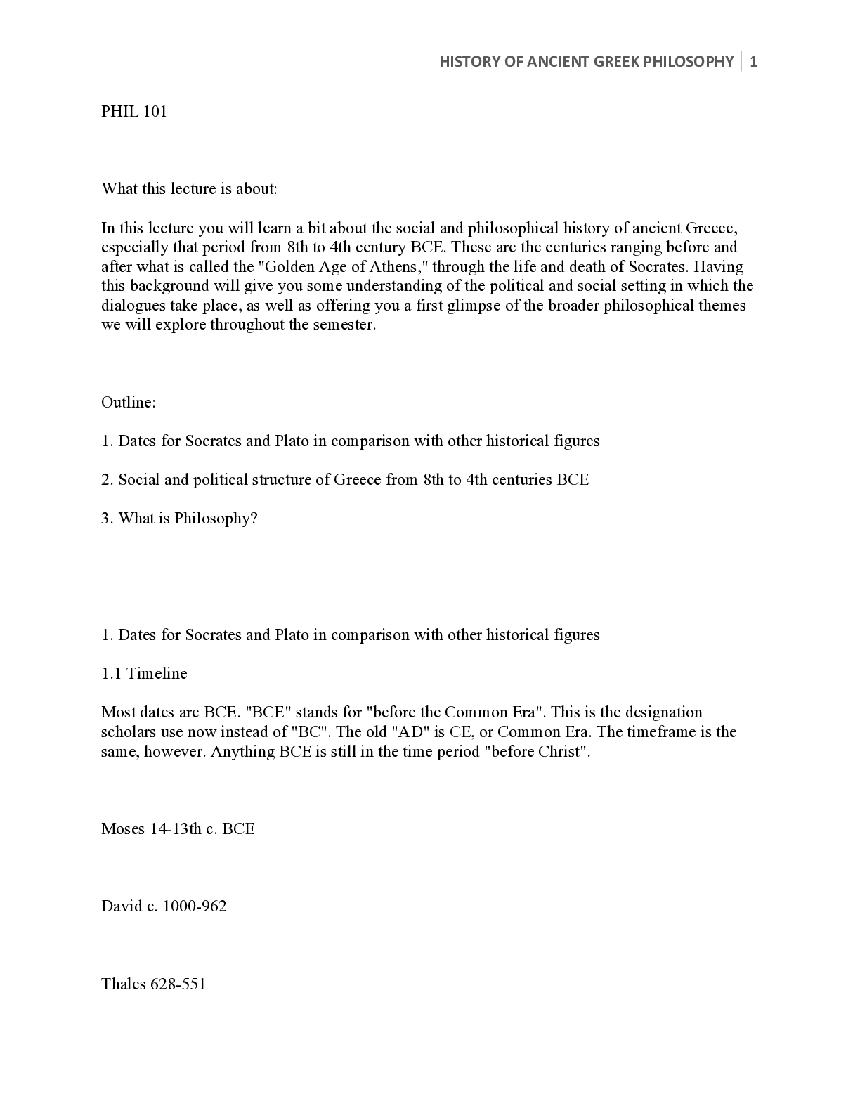
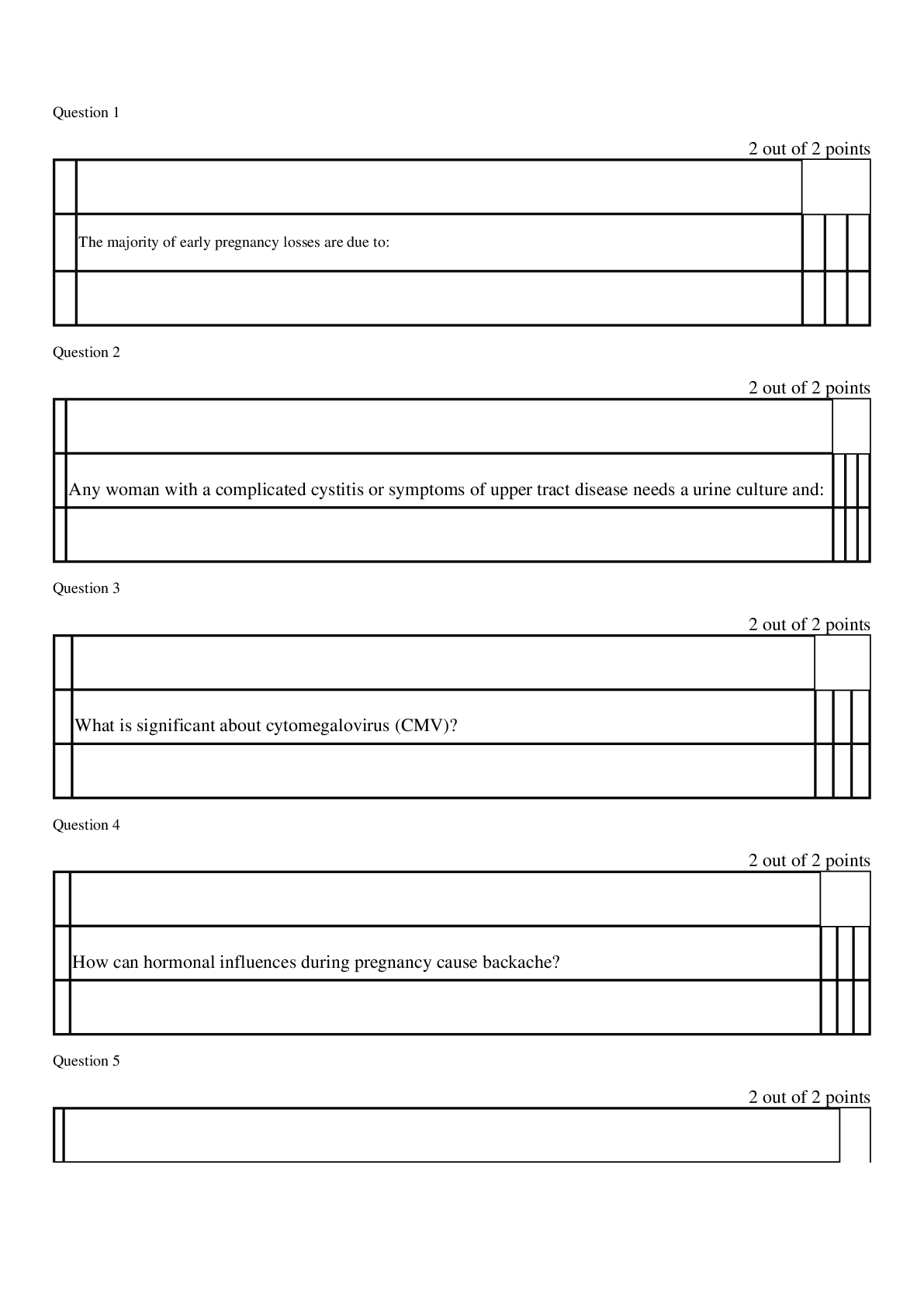
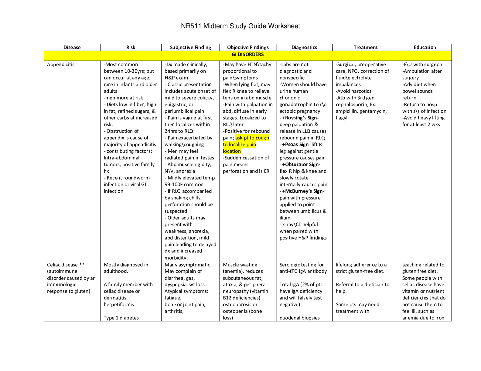
Chapter 25 Describe the pathophysiology of glomerulonephritis Glomerulonephritis represents a group of kidney diseases that result from inflammation and injury of the glomerulus. It may occur as a ... primary condition in which the glomerular abnormality is the only disease present, or as a secondary condition in which the glomerular abnormality results from another disease, such as diabetes mellitus or SLE. Most cases of primary and many cases of secondary glomerular disease probably have an immune origin. Glomerular disorders alter the permeability of the glomerular capillary membrane to plasma proteins and blood cells to produce either a nephritic or nephrotic syndrome. The nephritic syndromes, which evoke an inflammatory response in the glomeruli and a decrease in glomerular permeability, are characterized by hematuria with red cell casts in the urine, diminished GFR, azotemia, oliguria, and hypertension. The nephrotic syndromes, which increase glomerular capillary membrane permeability, are characterized by massive proteinuria, hypoalbuminemia, generalized edema, lipiduria, and hyperlipidemia. ■ Chronic glomerulonephritis represents the chronic phase of a number of specific types of glomerulonephritis. Compare and contrast nephrotic syndrome and nephritic syndrome. Glomerular disorders alter the permeability of the glomerular capillary membrane to plasma proteins and blood cells to produce either a nephritic or nephrotic syndrome. The nephritic syndromes, which evoke an inflammatory response in the glomeruli and a decrease in glomerular permeability, are characterized by hematuria with red cell casts in the urine, diminished GFR, azotemia, oliguria, and hypertension. The nephrotic syndromes, which increase glomerular capillary membrane permeability, are characterized by massive proteinuria, hypoalbuminemia, generalized edema, lipiduria, and hyperlipidemia Chapter 26 Compare and contrast acute kidney injury and chronic kidney disease Acute kidney injury is abrupt in onset and often is reversible if recognized early and treated appropriately. In contrast, chronic kidney disease is the end result of irreparable damage to the kidneys. It develops slowly, usually over the course of a number of years and often requires dialysis therapy or transplantation. Acute kidney injury (AKI) is an abrupt reduction in kidney function, as evidenced by an elevation in serum creatinine, reduction in urine output, the need for dialysis, or a combination of these factors. ■Acute kidney injury can result from decreased blood flow to the kidney (prerenal injury), from conditions that interfere with the elimination of urine from the kidney (postrenal injury), or from disorders that disrupt the structures in the kidney (intra-renal injury). Chronic kidney disease (CKD) is a pathophysiologic process that results in the loss of nephrons and a decline in renal function as determined by a measured or estimated decrease in the GFR that has persisted for more than 3 months. Chronic kidney disease can result from diabetes, hypertension, glomerulonephritis, systemic lupus erythematosus Polycystic kidney disease. Chronic kidney disease (CKD), which represents a progressive decline in kidney function due to the permanent loss of nephrons, can result from diabetes, hypertension, glomerulonephritis Other kidney diseases. Regardless of the cause, the consequences of nephron destruction in CKD disrupt the filtration, reabsorption, and endocrine functions of the kidneys. ■The glomerular filtration rate (GFR) is best measure of kidney function. Chronic disease is defined as either diagnosed kidney damage or a GFR of less than 60 mL/min/1.73 m2 for 3 months or more, and kidney failure as a GFR of less than 15 mL/min/1.73 m2, usually accompanied by most of the signs and symptoms of uremia or a need to start renal replacement therapy. ■The manifestations of CKD alterations in fluid, electrolyte, and acid–base balance; anemia and coagulopathies; cardiovascular complications; disorders of calcium and phosphate metabolism and skeletal disorders; and impaired elimination of drugs that are excreted by the kidney as well as nitrogenous wastes signs and symptoms of the uremic state, such as o neuromuscular disorders o gastrointestinal disturbances o immune disorders o sexual dysfunction o Discomforting skin changes ■2 Types of Treatment for CKD Conservative management and renal replacement therapy. Conservative management treatment is to prevent or retard deterioration in remaining renal function and assist the body in compensating for the existing impairment. Renal replacement therapy (dialysis or kidney transplantation) is indicated when advanced uremia and serious electrolyte problems are present. Causes of CKD in infants and children Congenital malformations (e.g., renal dysplasia and obstructive uropathy) inherited disorders (e.g., polycystic kidney disease) Acquired diseases (e.g., glomerulonephritis), and metabolic syndromes (e.g., hyperoxaluria). Problems associated with CKD in children Growth impairment Delay in sexual maturation More extensive bone abnormalities than in adults. Although all forms of renal replacement therapy can be safely and reliably used in children, CCPD, NIPD, and transplantation optimize growth and development. ■Normal aging is associated with a decline in the GFR, which makes elderly persons more susceptible to the detrimental effects of nephrotoxic drugs and other conditions that compromise renal function. Describe the pathophysiology of prerenal, intrinsic, and postrenal PRERENAL: The causes of AKI commonly are categorized as prerenal, intra-renal, and postrenal1–7 (Fig. 26-1). Collectively, prerenal and intra-renal causes account for 80% to 95% of AKI cases.3 Causes of kidney injury within these categories are summarized in Chart 26-1. Prerenal kidney injury Most common form of AKI characterized by a decrease in renal blood flow. Reversible if the cause of the decreased renal blood flow can be identified and corrected before kidney damage occurs. Normal kidneys receive 22% of the cardiac output. Large blood supply is required to remove metabolic wastes and regulate body fluids and electrolytes. Normal kidney can tolerate relatively large reductions in blood flow before renal damage occurs. Pathophysiology of the Prerenal Kidney Injury As renal blood flow falls, the glomerular filtration rate (GFR) decreases sodium and other substances that are filtered by the glomeruli is reduced, and the blood flow needed for the energy dependent mechanisms that reabsorb these substances is reduced (see Chapter 24). As the GFR and urine output approach zero, oxygen consumption by the kidney approximates that required to keep renal tubular cells alive. When blood flow falls below this level, which is about 25% of normal, ischemic changes occur. Due to high metabolic rate, the tubular epithelial cells are most vulnerable to ischemic injury. Improperly treated, prolonged renal hypoperfusion can lead to ischemic tubular necrosis with significant morbidity and mortality. Causes of prerenal injury Profound depletion of vascular volume (e.g., hemorrhage, loss of extracellular fluid volume) Impaired perfusion due to heart failure and cardiogenic shock, and decreased vascular filling because of increased vascular capacity (e.g., anaphylaxis or sepsis). Elderly persons are at high risk because of their predisposition to hypovolemia and their high prevalence of renal vascular disorders. Some vasoactive mediators, drugs, and diagnostic agents stimulate intense intra-renal vasoconstriction and can induce glomerular hypoperfusion and prerenal injury. endotoxins, radiocontrast agents such as those used for cardiac catheterization cyclosporine (an immunosuppressant drug that is used to prevent transplant rejection) amphotericin B (an antifungal agent), Epinephrine, and high doses of dopamine. These agents also cause acute tubular necrosis (to be discussed). Drugs that impair renal adaptive mechanisms and can convert compensated renal hypoperfusion into prerenal injury. For example, angiotensin II is constricts the efferent arterioles of the kidney as a means of preserving the GFR in situations of arterial hypotension or volume depletion. The angiotensin converting enzyme (ACE) inhibitors and angiotensin receptor blockers (ARBs) reduce the effects of angiotensin II on renal blood flow. Reduce intraglomerular pressure and may have a renal protective effect in persons with hypertension or type 2 diabetes. When ACE or ARB’s combined with diuretics may cause prerenal injury in persons with decreased blood flow due to large-vessel or small-vessel kidney disease. Nonsteroidal antiinflammatory drugs Postrenal (obstruction of urine outflow from the kidney) Intrinsic (damage to structures within the kidney) FIGURE 26-1. Types of acute kidney injury. CHART 26-1 Causes of Acute Kidney Injury INTRINSIC or Intra renal Injury Cause of stress incontinence is intrinsic urethral deficiency, which may result from congenital sphincter weakness, as occurs with meningomyelocele. It may be acquired as a result of trauma, irradiation, or sacral cord lesions. Stress incontinence in men may result from trauma or surgery to the bladder outlet, as occurs with prostatectomy. Neurologic dysfunction, as occurs with impaired sympathetic innervation of the bladder neck, impaired pelvic nerve innervation to the intrinsic sphincter, or impaired pudendal nerve innervation to the external sphincter, may also be a contributing factor. Intrinsic (damage to structures within the kidney) Intra-renal Injury Intra-renal injury results from conditions that damage structures within the kidney. The major causes of intra-renal injury are ischemia associated with prerenal injury, injury to the tubular structures of the nephron, and intra-tubular obstruction. Acute glomerulonephritis and acute pyelonephritis also are intra-renal causes of AKI. However, injury to the tubular structures of the nephron (acute tubular necrosis) is the most common cause and often is ischemic or toxic in origin. POSTRENAL Injury Postrenal Bilateral ureteral obstruction Bladder outlet obstruction Postrenal injury results from Obstruction of urine outflow from the kidneys. Obstruction can occur in the ureter (i.e., calculi and strictures), bladder (i.e., tumors or neurogenic bladder), or urethra (i.e., prostatic hyperplasia). Prostatic hyperplasia is the most common underlying problem. Because both ureters must be occluded to produce renal injury, obstruction of bladder out flow rarely causes AKI unless one of the kidneys already is damaged or a person has only one kidney. Treatment Treating the underlying cause of obstruction so that urine flow can be reestablished before permanent nephron damage occurs. Acute kidney injury can result from decreased blood flow to the kidney (prerenal injury), from conditions that interfere with the elimination of urine from the kidney (postrenal injury), or from disorders that disrupt the structures in the kidney (intra-renal injury). Chapter 28 Identify all secretions of the gastrointestinal tract. Include the source and function of each. Salivary Secretions –regulated by ANS o Saliva is secreted by the salivary glands. parotid, submaxillary, sublingual, and buccal glands o Three functions Protection and lubrication. Rich in mucus, which protects the oral mucosa and coats the food Protective antimicrobial action. Cleans the mouth and contains the enzyme lysozyme, which has an antibacterial action Contains ptyalin and amylase, which initiate the digestion of dietary starches Gastric Secretions o Pyloric glands secrete gastrin and mucus for protection of the pyloric mucosa o Oxyntic glands contain three types of cells Mucous neck cells, which secrete mainly mucus; Peptic (chief cells), which secrete large quantities of pepsinogen; Parietal (oxyntic cells), which secrete HCl and intrinsic factor, which is needed for vitamin B12 absorption o Three substances stimulate HCl secretion by the parietal cells: acetylcholine, gastrin, and histamine. Pepsinogen that is secreted by the peptic cells is rapidly converted to pepsin (a protein-digesting enzyme) when exposed to the low pH of the gastric juices o Prostaglandin E2, which inhibits acid secretion and stimulates mucus production. It is an important factor in the maintenance of the gastric mucosal barrier. Intestinal Secretions Small intestine, most of the digestion and absorption of food takes place, secretes digestive juices and receives secretions from the liver and pancreas o Brunner glands, is concentrated at the site where the contents from the stomach and secretions from the liver and pancreas enter the duodenum. Secrete large amounts of alkaline mucus that protect the duodenum from the acid content in the gastric chyme and from the action of the digestive enzymes. o Mucus; isotonic alkaline fluid (pH 6.5 to 7.5) secreted by specialized cells in the crypts of Lieberkühn; surface enzymes that aid absorption o mucins secreted by goblet cells, lubricate the mucosal surface and protect it from mechanical injury from solid food particles The large intestine secretes mainly mucus and bicarbonate. Mucus not only protects the mucosa of the colon, but also facilitates compaction of the feces. Bicarbonate adheres to the mucus and acts as a buffer, protecting the mucosa from acid by-products of bacterial metabolism within the feces. ANS activity strongly influences mucus production in the bowel Major Gastrointestinal Hormones • Cholecystokinin CCK – Stimulates contraction of the gall bladder – Stimulates secretion of pancreatic enzymes – Slows gastric emptying • Secretin – Stimulates secretion of bicarbonate-containing solution by the pancreas and liver • Gastrin – Stimulates secretion of gastric aid and pepsinogen – Increases gastric blood flow – Stimulates gastric smooth muscle contraction – Stimulates growth of the gastric, small intestine, and colon mucosa Identify all layers of the GI tract. It consists of four distinct layers: the inner mucosal layer; the underlying submucosal layer, the muscularis externa, and the outer serosal layer 1. The mucosa, or inner mucosal layer a. Made up of an epithelium lining; an underlying loose connective tissue, called the lamina propriae; and the muscularis mucosa b. Composed of smooth muscle cells that can contract and change the shape and surface area of the mucosal layer c. Production of the mucus that protects and lubricates the inner lining of the GI tract lumen, secretion of the digestive enzymes and substances that break food down, absorption of the breakdown products of digestion, and maintenance of a barrier to prevent the entry of noxious substances and pathogenic organisms d. Lymphatics within the mucosa serve as the body’s first line of immune defense. e. Because of the regenerative capabilities of the mucosal layer, injury to this layer heals rapidly without leaving scar tissue 2. The Submucosa, submucosal layer, or second layer a. Consists of dense connective tissue, aggregates of adipose tissue, and occasional glands b. Large blood vessels that send branches to the mucosa, muscular externa, and adventitia c. Lymphatic vessels, as well as nerves that control motility and the secretory activity of glands in the mucosal layer. 3. Muscularis Extema a. Consists of two concentric and relatively thick layers of smooth muscle: i. An inner layer made up of circularly arranged smooth muscle cells ii. An outer layer of longitudinally arranged smooth muscle iii. Between the two muscle layers is a connective tissue layer that contains nerves that control smooth muscle movement, as well as blood and lymphatic vessels b. Contraction of the smooth muscle in this layer mixes and churns the GI contents and facilitates its movement along the GI tract. 4. Serosa and Adventitia a. The outermost layer of the GI tract b. Serosa is a serous membrane consisting of a layer of simple squamous epithelium, called the mesothelium, and a small amount of underlying connective tissue. c. Adventitia consisting only of connective tissue Discuss the function of the GI system Digestion involves the dismantling of foods into their constituent parts, a process that requires hydrolysis, enzyme cleavage, and fat emulsification. Hydrolysis is the breakdown of a compound that involves a chemical reaction with water. Enzyme cleavage requires the use of enzymes to cut substances into smaller components. Emulsification involves the breakdown of large globules of dietary fat into smaller particles. Absorption is the process of moving nutrients and other materials from the external environment of the GI tract into the internal environment. Absorption is accomplished by active transport and diffusion. Absorption of carbohydrates, proteins and fats Carbohydrates Carbohydrates must be broken down into monosaccharides 单 , or single sugars, before they can be absorbed from the small intestine. Digestion of starch begins in the mouth with the action of amylase. The brush border enzymes convert the disaccharides into monosaccharides that can be absorbed [Show More]
Last updated: 1 year ago
Preview 1 out of 45 pages
.png)
Buy this document to get the full access instantly
Instant Download Access after purchase
Add to cartInstant download
We Accept:

Reviews( 0 )
$14.00
Document information
Connected school, study & course
About the document
Uploaded On
Mar 12, 2021
Number of pages
45
Written in

Additional information
This document has been written for:
Uploaded
Mar 12, 2021
Downloads
0
Views
47


 LATEST VERIFIED GUIDE FOR EXAM PREPARATION, Graded A.png)
 LATEST VERIFIED GUIDE FOR EXAM PREPARATION, Graded A.png)
 LATEST VERIFIED GUIDE FOR EXAM PREPARATION, Graded A.png)
 LATEST VERIFIED GUIDE FOR EXAM PREPARATION, Graded A.png)
 LATEST VERIFIED GUIDE FOR EXAM PREPARATION, Graded A.png)
 LATEST VERIFIED GUIDE FOR EXAM PREPARATION, Graded A.png)
 LATEST VERIFIED GUIDE FOR EXAM PREPARATION, Graded A.png)