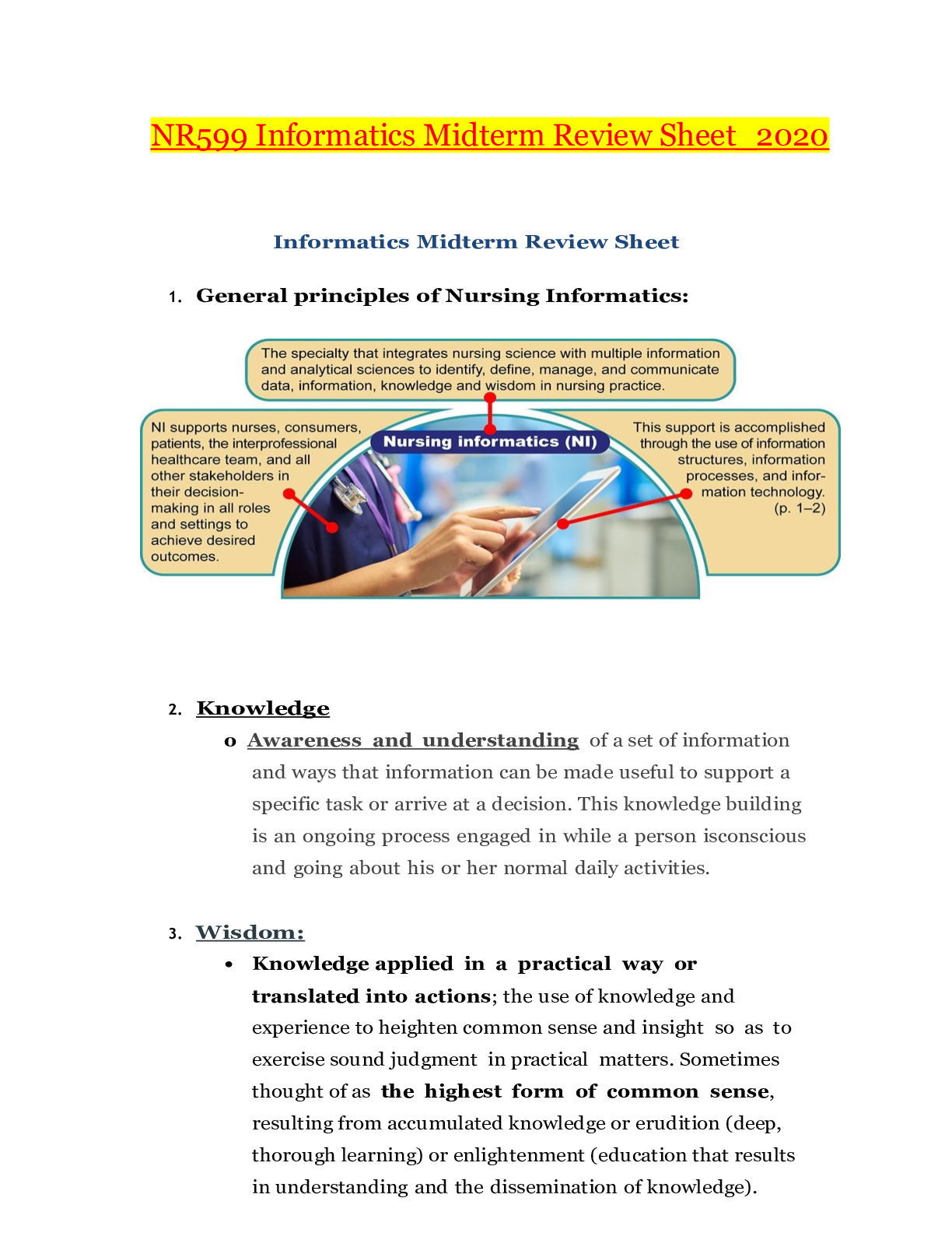
Pathophysiology > Study Notes > Harsh Mohan Textbook of Pathology 6th Edition | Textbook of Pathology 6th Edition_LATEST (All)
Harsh Mohan Textbook of Pathology 6th Edition | Textbook of Pathology 6th Edition_LATEST
Document Content and Description Below
TEXTBOOK OF PATHOLOGY EXTBOOKOF ATHOLOGYNodular lesions in diabetic kidney Aspergillosis lung The photographs on the cover of the textbook depict images of common diseases: Pap smear invasive ... carcinoma cervix Squamous cell carcinoma aerodigestive tract Cavitary tuberculosis lung Chronic ischaemic heart disease Blood smear acute myeloid leukaemia® JAYPEE BROTHERS MEDICAL PUBLISHERS (P) LTD St Louis (USA) • Panama City (Panama) • New Delhi • Ahmedabad • Bengaluru Chennai • Hyderabad • Kochi • Kolkata • Lucknow • Mumbai • Nagpur TEXTBOOK OF PATHOLOGY Harsh Mohan MD, MNAMS, FICPath, FUICC Professor & Head Department of Pathology Government Medical College Sector-32 A, Chandigarh-160 031 INDIA E mail: [email protected] ATHOLOGYPublished by Jitendar P Vij Jaypee Brothers Medical Publishers (P) Ltd Corporate Office 4838/24 Ansari Road, Daryaganj, New Delhi 110 002, India, Phone: +91-11-43574357, Fax: +91-11-43574314 Registered Office B-3 EMCA House, 23/23B Ansari Road, Daryaganj, New Delhi 110 002, India Phones: +91-11-23272143, +91-11-23272703, +91-11-23282021, +91-11-23245672, Rel: +91-11-32558559 Fax: +91-11-23276490, +91-11-23245683 e-mail: [email protected], Website: www.jaypeebrothers.com Branches 2/B, Akruti Society, Jodhpur Gam Road Satellite Ahmedabad 380 015 Phones: +91-79-26926233, Rel: +91-79-32988717 Fax: +91-79-26927094 e-mail: [email protected] 202 Batavia Chambers, 8 Kumara Krupa Road, Kumara Park East Bengaluru 560 001 Phones: +91-80-22285971, +91-80-22382956, +91-80-22372664, Rel: +91-80-32714073 Fax: +91-80-22281761 e-mail: [email protected] 282 IIIrd Floor, Khaleel Shirazi Estate, Fountain Plaza, Pantheon Road Chennai 600 008 Phones: +91-44-28193265, +91-44-28194897, Rel: +91-44-32972089 Fax: +91-44-28193231 e-mail: [email protected] 4-2-1067/1-3, 1st Floor, Balaji Building, Ramkote Cross Road Hyderabad 500 095 Phones: +91-40-66610020, +91-40-24758498, Rel:+91-40-32940929 Fax:+91-40-24758499, e-mail: [email protected] No. 41/3098, B & B1, Kuruvi Building, St. Vincent Road Kochi 682 018, Kerala Phones: +91-484-4036109, +91-484-2395739, +91-484-2395740 e-mail: [email protected] 1-A Indian Mirror Street, Wellington Square Kolkata 700 013 Phones: +91-33-22651926, +91-33-22276404, +91-33-22276415 Fax: +91-33-22656075, e-mail: [email protected] Lekhraj Market III, B-2, Sector-4, Faizabad Road, Indira Nagar Lucknow 226 016 Phones: +91-522-3040553, +91-522-3040554 e-mail: [email protected] 106 Amit Industrial Estate, 61 Dr SS Rao Road, Near MGM Hospital, Parel Mumbai 400012 Phones: +91-22-24124863, +91-22-24104532, Rel: +91-22-32926896 Fax: +91-22-24160828, e-mail: [email protected] “KAMALPUSHPA” 38, Reshimbag, Opp. Mohota Science College, Umred Road Nagpur 440 009 (MS) Phone: Rel: +91-712-3245220 Fax: +91-712-2704275 e-mail: [email protected] North America Office 1745, Pheasant Run Drive, Maryland Heights (Missouri), MO 63043, USA, Ph: 001-636-6279734 e-mail: [email protected], [email protected] Central America Office Jaypee-Highlights Medical Publishers Inc., City of Knowledge, Bld. 237, Clayton, Panama City, Panama, Ph: 507-317-0160 Textbook of Pathology © 2010, Harsh Mohan All rights reserved. No part of this publication should be reproduced, stored in a retrieval system, or transmitted in any form or by any means: electronic, mechanical, photocopying, recording, or otherwise, without the prior written permission of the author and the publisher. This book has been published in good faith that the material provided by author is original. Every effort is made to ensure accuracy of material, but the publisher, printer and author will not be held responsible for any inadvertent error(s). In case of any dispute, all legal matters are to be settled under Delhi jurisdiction only. First Edition : 1992 Second Edition : 1994 Third Edition : 1998 Fourth Edition : 2000 Fifth Edition : 2005 Sixth Edition: 2010 Assistant Editors: Praveen Mohan, Tanya Mohan, Sugandha Mohan ISBN: 978-81-8448-702-2 Typeset at JPBMP typesetting unit Printed at Ajanta PressDedicated to My family: spouse Praveen and daughters Tanya and Sugandha, for their love and constant support; & To all the students and colleagues: whose inspiration has made this ordinary work seem extraordinary. To deeds alone you have a right and never at all to its fruits; Let not the fruits of deeds be your motive; Neither let there be in you any detachment to performing your duty. The Bhagvadgita (Chapter II, verse 47) Foreword to the Sixth Edition vii A few years ago I wrote the Foreword to the Fifth Edition of this Textbook. For details and reasons why I liked Professor Mohan’s book and why I recommended it then, please refer to my previous foreword below. My positive reaction to the previous Edition probably gives some clues on why I accepted the second invitation, this time to introduce the Sixth Edition to new students of Pathology and other potential readers. Great French writer André Gide once said “le problème n’est pas comment réussir mais comment durer”, which in translation to English means: The problem is not how to succeed but how to last. The fact that Dr Mohan’s book has reached its Sixth Edition is the best sign that you are holding in your hands a very successful book, and probably one of the medical bestsellers published on the Indian subcontinent. Up to now, it has been used by thousands of students and I am sure that it will continue to be read and cherished in the new Edition as well. For the Sixth Edition, Dr Mohan has partially restructured the book, substantially revised it, and updated the text wherever it was necessary. Following the advances of basic sciences and clinical pathology, the revisions and addition are most evident in portions pertaining to molecular biology and genetics. Other aspects of modern pathology have not been neglected either and contain numerous novelties; even the seasoned specialists will learn something new from each and every chapter. Furthermore, the author has dramatically increased the number of illustrations, which are so essential for understanding Pathology. The distribution of illustrations has also been changed so that they are now much closer to the text to which they relate. For the new generation of modern students who have grown up next to the computers, the author has placed all the images and tables on the website with facility for downloading them. These images will serve the twin purpose of quick review and self-assessment for students and will appeal to Pathology teachers who could use them for their lectures, being assured that their students will have access to the same material for review and study. The Quick Review Book, the ever popular companion to the previous two Editions, was also updated, succinctly supplementing the main text. It will provide a helpful study material to many a student and help them review the subject for examinations. In summary, it is my distinct pleasure and honour to most enthusiastically endorse the new edition of an established textbook and salute its publication. Dr Mohan deserves kudos for the job well done and for providing the medical students with such an attractive, modern, up-to-date and useful Textbook of Pathology. Ivan Damjanov, MD, PhD Professor of Pathology Foreword to the Fifth Edition As the Book Review Editor of the journal Modern Pathology, the official journal of the United States-Canadian Academy of Pathology I am used to receiving medical books. These books are sent to my office from publishers, with a standard request for a potential review in the Journal. Nevertheless a recent package from New Delhi caught me by surprise. As you already might have guessed, the parcel contained a copy of the 5th Edition of the Textbook of Pathology written by Professor Harsh Mohan, together with the Second Edition of the pocket size companion Pathology Quick Review and MCQs. Included was also a friendly letter from Mr JP Vij, the Publisher. I acknowledged the receipt of the books by email, and also congratulated the Publisher on a job well done. A brief electronic exchange between Kansas City and New Delhi ensued, whereupon Mr Vij asked me to write a foreword for the Reprint of 5th Edition of the Textbook. I accepted the invitation with pleasure. Even though there were no specific instructions attached to the request, I assumed that I should address my notes primarily to undergraduate and graduate students of Pathology. Furthermore, I decided to write the Foreword in the form of answers to the questions that I would have had if I were a medical student entering the field of Pathology. I hope that these hypothetical questions and answers of mine will be of interest to the readers of this Textbook. Question 1: Is this a good book? Answer: Yes. This is a modern Textbook written by an expert who knows his pathology; an experienced teacher who knows what is important and what is not, and who has obviously taught pathology for many years; a well informed academician who is au courant with modern trends in medical education, and knows how to present pathology as a preparatory step for future clinical education of medical students. Question 2: How does the book compare with the leading textbooks of pathology in the USA, Great Britain and Germany? Answer: Very favorably. This Indian Textbook covers more or less the same topics as the equivalent Textbooks currently used in the Western Hemisphere. Like the Western textbooks it covers the traditional fields of General and Systemic Pathology: one-third of the book Forewordviii Textbook of Pathology is devoted to General Pathology, whereas the remaining two-thirds cover Systemic Pathology. The emphasis is on classical anatomic pathology. In that respect the Indian textbook resembles more the European than the American textbooks, which have become more clinically-oriented. In my opinion this approach gives excellent results, but only if the students have enough time to devote to Pathology. In most US medical schools this is not the case any more, and thus pathology is not taught as extensively as before. Histopathology has been deleted from most curricula, and most American medical students do not know to use efficiently the microscope, which is unfortunate. Question 3: Is the material presented in a “student-friendly” manner? Answer: The material is presented in a systematic manner in the best tradition of classical British textbooks, a tradition that can be traced to the classical writers of ancient Greece and Rome. This time tested teaching will be most appreciated by students who are methodical and do not take shortcuts in their effort to acquire encyclopedic knowledge of pathology. On the other hand, even if your learning method is based on “cherry-picking”, i.e. you concentrate only on the most important facts in each chapter, the structure of the text will allow you to do it quite easily as well. There are no ideal books that would satisfy everybody in every respect, but there is no doubt that Professor Mohan’s book is close to ideal for a classical pathology course and I predict that it will be popular with many students. Question 4: What are the most salient features of this textbook? Answer: Clear writing. As we all know clear writing reflects clear thinking, and clear thinking in my opinion, is an absolute prerequisite for good teaching. Judging from the book at hand, Professor Mohan (whom I do not know personally) is not only a clear thinker, but he must be also an exceptionally talented teacher. Clear and visually pleasing presentation. The exposition is logical and well structured. Each chapter is subdivided into smaller entities, which are further divided into paragraphs, ideally suited for easy reading. Color coded headings and the added emphasis in form of words printed in bold or capital letters are additional attractions that facilitate learning. Exceptionally good illustrations, flow-charts and tables. Unique to this Textbook are the numerous hand-drawn color illustrations, including many renditions of histopathologic slides. These drawings are simple, but to the point and well annotated. Students will most likely understand them much easier than the relatively impersonal original microphotographs of the same histopathologic lesions. Flow-charts are most efficiently used to explicate the pathogenesis of various lesions or the pathophysiology of disease processes. The tables are good for classifications and comparative listings of closely related diseases and their pathologic features. Companion pocket book (baby-book of pathology). I always recommend to my students to buy a major textbook and a smaller review book containing a digest of the most important concepts; or a book of questions and answers, so that the student could test his/her knowledge of pathology and the understanding of the material in the main textbook. I was pleased to see that Professor Mohan shares my teaching philosophy and has taken upon himself to prepare for his students a shorter version of main text. This pocket book is also garnered with review questions. The medical students are thus getting a bargain— two books for the price of one. At the same time, they have a unique opportunity to see, from the example set by their teacher, on how the same material can be approached from two points of view, and presented in two formats. The old adage, that you have never learned anything unless you have seen it at least from two sides, is clearly illustrated here. For the students of medicine the message is clear: if you understand the material presented in both the shorter and the longer version you can be assured that you know your Pathology inside out; and you are ready for the final examination and clinical training. Question 5: Do I have to know all that is in this book for my final examination? Answer: No!! This is the most common question my students ask me and I hope that you believe me when I say that you do not have to know it all. First of all, neither I nor Professor Mohan know it all. Second, few of us have photographic memory and infinite storage space in our brains and thus even theoretically, very few of us could learn this book by heart. I can assure you that the book was not written for those geniuses, but for the average persons like most of us. Third, your goal should not be to memorize all the facts listed in the textbook, but rather to understand the main concepts. Since the concepts cannot be fully understood or taught without specific examples, by necessity you will have to learn “some nitty-gritty details”. The more details you know, the deeper your understanding of the basic concepts will be. Memorizing the details without the understanding of concepts that hold them together is not something that I would recommend. The beauty of it all is that you can decide for yourself how deep to dig in, when to stop, what to keep and memorize, and what to eliminate. And remember, deciding on what to eliminate is almost as important as choosing what to retain. As the educational gurus teach us, that is the gist of what they call active learning. And to repeat again, this Textbook is ideally suited for that approach. At the end, let me repeat how excited I was perusing this excellent book. I hope that you will be similarly excited and I hope that it will inspire in you enthusiasm for Pathology. Remember also the words of the great clinician William Osler, one of the founders of modern medicine in late 19th and early 20th Century, who said that our clinical practice will be only as good as our understanding of Pathology. I hope that I have answered most of the questions that you might have had while opening this book. If you have any additional questions that I did not anticipate, please feel free to send me an email at [email protected]. Good luck! Ivan Damjanov, MD, PhD Professor of Pathology The University of Kansas School of Medicine Kansas City, Kansas, USA Dr Damjanov is Professor of Pathology at the University of Kansas School of Medicine, Kansas City, Kansas, USA. He earned his Medical degree from the University of Zagreb, Croatia in 1964, and a PhD degree in Experimental Pathology from the same University in 1970. He received his Pathology training in Cleveland, New York and Philadelphia. Thereafter he served as Professor of Pathology at the University of Connecticut, Farmington, Connecticut, Hahnemann University and Thomas Jefferson University, Philadelphia, Pennsylvania. For the last ten years he has been on the Faculty of the University of Kansas School of Medicine dividing his time between teaching, practice of surgical pathology and medical publishing. He is the author of more than 300 biomedical articles, and has written or edited more than 20 medical books.Preface ix The overwhelming success and all-round acceptance of the last edition of the textbook was very encouraging and quite stimulating but at the same time put an onerous responsibility and expectation to do better in the new edition than the best of last edition. In preparing 6th revised edition of my Textbook of Pathology, I pursued this goal with profound enthusiasm and passionate zeal. I am, thus, pleased to present to users a wholly transformed appearance and updated contents in the revised edition. While full colour printing had been introduced in the last edition 5 years back maturing the book into an international edition, the present redesigned and revised edition has utlilised the contemporary technological advances in its full form in illustrations, lay-out and in printing. The revised edition has almost thrice the number of illustrations of large number of common diseases placed along with the text, and it is hoped that it will enhance understanding and learning of the subject readily, besides being a visual treat. In recent times, advances in genetics, immunology and molecular biology have heightened our understanding of the mechanisms of diseases. As a result, mention of ‘idiopathic’ in etiology and pathogenesis of most diseases in the literature is slowly disappearing. Surely, the students of current times need to be enlightened on these modern advances in diseases; these aspects have been dealt in the revised edition with a simple and lucid approach. Some of the Key Features of the Sixth Edition are as follows: Thorough Textual Revision and Updating: All the chapters and topics have undergone thorough revision and updating of various aspects, including contemporary diagnostic modalities. While most of the newer information has been inserted between the lines, a few topics have been rewritten, e.g. current concepts on cell injury, immunopathology, carcinogenesis, newer infectious diseases, lymphomas-leukaemias, hypertension, interstitial lung diseases, etc. to name a few. In doing so, the basic accepted style of the book —simple, easy-to-understand and reproduce the subject matter, and emphasis on clarity and accuracy, has not been disturbed. Past experience has shown that the readers find tables on contrasting features and listing of salient features as a very useful medium for quick learning; considering their utility 15 new tables have been added in different chapters in the revised edition. Reorganisation of the Book: In a departure from the conventional division of study of the subject into General and Systemic Pathology, the revised edition has been reorganised into 3 major sections—General Pathology and Basic Techniques (Chapters 1 to 11), Haematology and Lymphoreticular Tissues (Chapters 12 to 14) and Systemic Pathology (Chapters 15 to 30), followed by Appendix (containing Normal Values), Further Readings for references and Index. In my considered judgement, a separate section on haematology and lymphoid tissues and redistribution of their subtopics was necessitated for two reasons—firstly, reclassification of leukaemias-lymphomas by the WHO as an integrated topic, making the segregation of study of diseases of ‘circulating’ and ‘tissue’ leucocytes superfluous; and secondly, due to advances in haematology, transfusion medicine and diseases of lymphoreticular tissues, these subspecialties of pathology have developed a lot in recent times, requiring the students to focus on them separately for learning and they are evaluated too on these topics by separate experts. Similarly, in the revised edition, two chapters on laboratory techniques—Techniques for the Study of Pathology (Chapter 2) and Basic Diagnostic Cytology (Chapter 11) have been included in Section-I in view of technological advances in pathology which have gone beyond remaining confined as research tool but have increasingly become part of diagnostic work-up. Profusely Illustrated: Majority of illustrations in the revised Edition are new additions while a few old ones have been done again. All the line-drawing and schematic cartoons have been updated and improved in content as well as their presentation by preparing them again on CorelDraw in soft colours, eliminating the shortcomings noticed in them in previous edition. All free-hand labelled sketches of gross specimens and line-drawings of microscopic features of an entity have been placed alongside the corresponding specimen photograph and the photomicrograph respectively, enhancing the understanding of the subject for the beginner students in pathology. In doing so, the number of figures has gone up by about three-folds in the present edition, some incorporated as an inset with focus on a close-up microscopic view. Truly User-friendly: Rational use of various levels of headings and subheadings in different colours, bold face and in italics has been done in the text in order to highlight key points. All the citations of figures and tables in the text have been shown in colour now to make the related text vividly visible and to help user locate the same quickly on a page. It is hoped that these features will enable the user with rapid revision at the end of a topic, making the book truly user-friendly. Much More Content but Unaltered Volume: While the new edition has a lot more updated textual material, more tables and a marked increase in the number of figures than the previous edition, a meticulous and rational page management has helped in retaining almost the same girth of the book as before. Prefacex Textbook of Pathology Images and Tables on the Web: All the illustrations and tables included in this edition are being put on the website with a scratch key word on the inner page of the cover jacket. The students would find these useful for quick review and for selfassessment in which an unlabelled image (gross specimen or a photomicrograph) appears, followed by the labelled image with diagnosis corresponding to the same figure and table in the textbook. Besides, ready availability of these downloadable images and tables would be useful to fellow teachers for possibly including the same in their lectures. Revised Pathology Quick Review and MCQs: The sixth edition of textbook is accompanied with the new revised baby-book popular with many students and interns. This small book has been found profoundly useful by the students just before practical examination to face viva voce when they need to revise huge course content in a short time, or by those preparing to take postgraduate entrance examinations. The revised edition has over 100 more new MCQs while some old ones have either been edited or replaced. A Word on Foreword: The Foreword by Prof Ivan Damjanov, MD, PhD, from Kansas University, US, for the previous edition and now for the sixth edition so generously and meticulously prepared with an eye to the details of the book, has been most welcome development, and has helped to bring the book closer to users in other parts of the world; I express our sincere gratitude to this eminent teacher and well-known author whom I have yet to meet in person. In essence, the revised edition is a comprehensive text of pathology meant primarily for students of pathology; however, the practicing clinicians and students of other branches of medicine, dentistry, pharmacy, alternate system of medicine, and paramedical courses may also find it useful. ACKNOWLEDGEMENTS The revision work was indeed a mammoth task to accomplish and would not have been possible without active cooperation from friends and colleagues and continuous encouragement from well-wishers in general, and my departmental staff in particular who could bear with me for prolonged spells of my sabbatical leave. All the photomicrographs included in the present edition have been exposed afresh which has been made possible by the most valuable and selfless assistance rendered by my colleagues, Drs Shailja, Tanvi and Ujjawal, Senior Residents in Pathology, all of whom worked tirelessly for endless hours for months, much to the sacrifice of their personal comfort and time of their families, for which I am indebted to them. Here, I also recall the help accorded by my former students and colleagues in preparation of earlier editions of the book and thank once again, even though much of that may have been replaced. As always, I remain indebted to those from whom I had the opportunity to learn pathology; in particular to Prof K Joshi, MD, PhD, PGIMER, Chandigarh, Late Prof TS Jaswal, MD, and Prof Uma Singh, MD, formerly at PGIMS, Rohtak. Constant strategic support and encouragement extended by the Department of Medical Education and Research, Chandigarh Administration, during the completion of work is gratefully acknowledged. I may have been hard-task master and highly demanding on quality and accuracy from all staff members of the M/s Jaypee Brothers Medical Publishers (P) Ltd, at times losing my patience, but all of them have been very cooperative and quite accommodating. In particular, I would like to thank profusely Mr Manoj Pahuja, Computer Art Designer, for carrying out Herculean job on figures as per my requirements conscientiously and patiently with competence; Mrs Y Kapoor, Senior Desktop Operator, for overall lay-out of the book and acceding to all my requests for amendments smilingly and ungrudgingly till the very last minute; and Ms Chetna Malhotra, MBA, Senior Business Development Manager, for overseeing the entire project vigilantly and efficiently. All through this period, Mr Tarun Duneja, (Director-Publishing), M/s Jaypee Brothers Medical Publishers (P) Ltd, has been highly cooperative and supportive. Lastly, the vision of Shri JP Vij, Chairman and Managing Director of M/s Jaypee Brothers Medical Publishers (P) Ltd, has been to see the revised edition as unmatched internationally and keeping it affordable at the same time, much above his business interests, and I do hope his dream comes true. Full credit goes to M/s Ajanta Printers, Faridabad, for the admirably high quality of printing. Finally, the users of previous editions are gratefully acknowledged for having brought this textbook at this pedestal. In the past, I have gained profitably by suggestions from colleagues and students and I urge them to continue giving their valuable suggestions and point out errors, if any, so that I may continue to improve it. Government Medical College Harsh Mohan, MD, MNAMS, FICPath, FUICC Sector-32 A, Chandigarh-160031 Professor & Head INDIA Department of Pathology E mail: [email protected] Contents CHAPTER 1 Introduction to Pathology 01 Study of Diseases, 1 Evolution of Pathology, 1 Subdivisions of Pathology, 7 CHAPTER 2 Techniques for the Study of Pathology 09 Autopsy Pathology, 9 Surgical Pathology, 9 Special Stains (Histochemistry), 11 Enzyme Histochemistry, 13 Basic Microscopy, 13 Immunofluorescence, 14 Electron Microscopy, 14 Immunohistochemistry, 15 Cytogenetics, 16 Diagnostic Molecular Pathology, 17 Other Modern Aids in Diagnostic Pathology, 18 CHAPTER 3 Cell Injury and Cellular Adaptations 21 The Normal Cell, 21 Etiology of Cell Injury, 27 Pathogenesis of Cell Injury, 28 Morphology of Cell Injury, 34 Intracellular Accumulations, 37 Pigments, 40 Morphology of Irreversible Cell Injury (Cell Death), 44 Cellular Adaptations, 53 Cellular Aging, 59 CHAPTER 4 Immunopathology Including Amyloidosis 61 Introduction, 61 Structure of Immune System, 61 HLA System and Major Histocompatibility Complex, 64 Transplant Rejection, 65 Diseases of Immunity, 66 Immunodeficiency Diseases, 67 Acquired Immunodeficiency Syndrome (AIDS), 67 Hypersensitivity Reactions (Immunologic Tissue Injury), 73 Autoimmune Diseases, 77 Types and Examples of Autoimmune Diseases, 78 Amyloidosis, 82 Section I GENERAL PATHOLOGY AND BASIC TECHNIQUES Contents CHAPTER 5 Derangements of Homeostasis and 93 Haemodynamics Homeostasis, 93 Disturbances of Body Fluids, 96 Oedema, 96 Dehydration, 102 Overhydration, 102 Disturbances of Electrolytes, 103 Acid-base Imbalance (Abnormalities in pH of Blood), 103 Haemodynamic Derangements, 104 Disturbances in the Volume of Circulating Blood, 105 Haemorrhage, 107 Shock, 108 Circulatory Disturbances of Obstructive Nature, 113 Thrombosis, 113 Embolism, 119 Ischaemia, 124 Infarction, 126 CHAPTER 6 Inflammation and Healing 130 Inflammation, 130 Introduction, 130 Acute Inflammation, 130 Chemical Mediators of Inflammation, 136 The Inflammatory Cells, 141 Morphology of Acute Inflammation, 144 Chronic Inflammation, 147 General Features of Chronic Inflammation, 147 Systemic Effects of Chronic Inflammation, 147 Types of Chronic Inflammation, 147 Granulomatous Inflammation, 148 Examples of Granulomatous Inflammation, 149 Tuberculosis, 149 Leprosy, 157 Syphilis, 161 Actinomycosis, 163 Sarcoidosis (Boeck’s Sarcoid), 164 Healing, 165 Regeneration, 165 Repair, 166 Wound Healing, 167 Healing in Specialised Tissues, 171 CHAPTER 7 Infectious and Parasitic Diseases 174 Introduction, 174 Diseases Caused by Bacteria, Spirochaetes and Mycobacteria, 175 Diseases Caused by Fungi, 181 Diseases Caused by Viruses, 183 Diseases Caused by Parasites, 187 Torch Complex, 190xii Textbook of Pathology CHAPTER 8 Neoplasia 192 Nomenclature and Classification, 192 Characteristics of Tumours, 194 Rate of Growth, 194 Cancer Phenotype and Stem Cells, 196 Clinical and Gross Features, 196 Microscopic Features, 196 Local Invasion (Direct Spread), 200 Metastasis (Distant Spread), 200 Grading and Staging of Cancer, 204 Epidemiology and Predisposition to Neoplasia, 205 Cancer Incidence, 205 Epidemiologic Factors, 205 Carcinogenesis: Etiology and Pathogenesis of Cancer, 208 Molecular Pathogenesis of Cancer (Genetic Mechanism of Cancer), 208 Chemical Carcinogenesis, 216 Physical Carcinogenesis, 220 Biologic Carcinogenesis, 222 Viruses and Human Cancer: A Summary, 228 Clinical Aspects of Neoplasia, 228 Tumour-host Inter-relationship, 228 Pathologic Diagnosis of Cancer, 232 CHAPTER 9 Environmental and Nutritional Diseases 236 Introduction, 236 Environmental Pollution, 236 Air Pollution, 236 Tobacco Smoking, 237 Chemical and Drug Injury, 238 Therapeutic (Iatrogenic) Drug Injury, 238 Non-therapeutic Toxic Agents, 238 Environmental Chemicals, 242 Injury by Physical Agents, 242 Thermal and Electrical Injury, 242 Injury by Radiation, 242 Nutritional Diseases, 243 Obesity, 243 Starvation, 245 Protein-energy Malnutrition, 245 Disorders of Vitamins, 246 Metals and Trace Elements, 254 Diet and Cancer, 254 CHAPTER 10 Genetic and Paediatric Diseases 256 Developmental Defects, 256 Cytogenetic (Karyotypic) Abnormalities, 257 Single-gene Defects (Mendelian Disorders), 259 Storage Diseases (Inborn Errors of Metabolism), 260 Multifactorial Inheritance, 263 Other Paediatric Diseases, 263 Section II HAEMATOLOGY AND LYMPHORETICULAR TISSUES CHAPTER 11 Basic Diagnostic Cytology 266 Introduction, 266 Exfoliative Cytology, 267 Female Genital Tract, 267 Respiratory Tract, 272 Gastrointestinal Tract, 273 Urinary Tract, 273 Body Fluids, 273 Buccal Smears for Sex Chromatin Bodies, 274 Techniques in Exfoliative Cytology, 275 Interventional Cytology, 277 Fine Needle Aspiration Cytology, 277 Imprint Cytology, 283 Crush Smear Cytology, 283 Biopsy Sediment Cytology, 283 CHAPTER 12 Introduction to Haematopoietic System and Disorders of Erythroid Series 284 Bone Marrow, 284 Haematopoiesis, 284 Haematopoietic Stem Cells, 285 Bone Marrow Examination, 285 Red Blood Cells, 287 Erythropoiesis, 287 Anaemia—General Considerations, 291 Anaemia of Blood Loss, 294 Hypochromic Anaemia, 295 Megaloblastic Anaemia, 303 Pernicious Anaemia, 309 Haemolytic Anaemias, 310 Acquired (Extracorpuscular) Haemolytic Anaemias, 311 Hereditary (Intracorpuscular) Haemolytic Anaemia, 314 Aplastic Anaemia and Other Primary Bone Marrow Disorders, 324 CHAPTER 13 Disorders of Platelets, Bleeding 327 Disorders and Basic Transfusion Medicine Thrombopoiesis, 327 Bleeding Disorders (Haemorrhagic Diathesis), 328 Investigations of Haemostatic Function, 328 Haemorrhagic Diatheses Due to Vascular Disorders, 331 Haemorrhagic Diatheses Due to Platelet Disorders, 331 Coagulation Disorders, 335 Haemorrhagic Diathesis Due to Fibrinolytic Defects, 337 Disseminated Intravascular Coagulation (DIC), 337 Blood Groups and Blood Transfusion, 339CHAPTER 14 xiii Disorders of Leucocytes and 342 Lymphoreticular Tissues Lymph Nodes: Normal and Reactive, 342 Normal Structure, 342 Reactive Lymphadenitis, 343 White Blood Cells: Normal and Reactive, 345 Granulopoiesis, 345 Lymphopoiesis, 346 Infectious Mononucleosis, 350 Leukaemoid Reactions, 352 Haematologic Neoplasms (Leukaemias-lymphomas): General, 353 Classification: Current Concepts, 353 Myeloid Neoplasms, 356 Myeloproliferative Diseases, 356 Myelodysplastic Syndromes, 361 Acute Myeloid Leukaemia, 362 Lymphoid Neoplasms, 365 General Comments on Lymphoid Malignancies, 368 Hodgkin’s Disease, 369 Precursor (Immature) B- and T-cell Leukaemia/ Lymphoma (Synonym: Acute Lymphoblastic Leukaemia), 373 Peripheral (Mature) B-cell Malignancies, 374 Peripheral (Mature) T-cell Malignancies, 379 Plasma Cell Disorders, 380 Lymph Node Metastatic Tumours, 385 Histiocytic Neoplasms: Langerhans’ Cell Histiocytosis, 385 Spleen, 386 Thymus, 388 CHAPTER 15 The Blood Vessels and Lymphatics 390 Arteries, 390 Normal Structure, 390 Arteriosclerosis, 391 Arteritis, 400 Aneurysms, 405 Fibromuscular Dysplasia, 409 Veins, 409 Lymphatics, 410 Tumours and Tumour-like Lesions, 411 CHAPTER 16 The Heart 417 Normal Structure, 417 Patterns and Classification of Heart Diseases, 418 Heart Failure, 419 Congenital Heart Disease, 422 Malpositions of the Heart, 423 Shunts (Cyanotic Congenital Heart Disease), 423 Section III SYSTEMIC PATHOLOGY Obstructions (Obstructive Congenital Heart Disease), 426 Ischaemic Heart Disease, 427 Etiopathogenesis, 427 Effects of Myocardial Ischaemia, 428 Angina Pectoris, 429 Acute Myocardial Infarction, 429 Chronic Ischaemic Heart Disease, 436 Sudden Cardiac Death, 436 Hypertensive Heart Disease, 437 Cor Pulmonale, 437 Rheumatic Fever and Rheumatic Heart Disease, 438 Non-rheumatic Endocarditis, 444 Valvular Diseases and Deformities, 449 Myocardial Disease, 452 Myocarditis, 452 Cardiomyopathy, 454 Pericardial Disease, 456 Pericardial Fluid Accumulations, 456 Pericarditis, 457 Tumours of the Heart, 459 Pathology of Cardiovascular Interventions, 459 CHAPTER 17 The Respiratory System 461 Lungs, 461 Normal Structure, 461 Paediatric Lung Disease, 462 Pulmonary Vascular Disease, 465 Pulmonary Infections, 467 Pneumonias, 467 Lung Abscess, 475 Fungal Infections of Lung, 476 Pulmonary Tuberculosis, 477 Chronic Obstructive Pulmonary Disease, 477 Chronic Bronchitis, 477 Emphysema, 478 Bronchial Asthma, 483 Bronchiectasis, 484 Chronic Restrictive Pulmonary Disease, 486 Pneumoconioses, 487 ILD Associated with Immunologic Lung Diseases, 493 ILD Associated with Connective Tissue Diseases, 495 Idiopathic Pulmonary Fibrosis, 495 ILD Associated with Smoking, 496 Tumours of Lungs, 496 Pleura, 504 CHAPTER 18 The Eye, ENT and Neck 507 Eye, 507 Ear, 513 Nose And Paranasal Sinuses, 515 Pharynx, 517 Larynx, 519 Neck, 520 Contentsxiv Textbook of Pathology CHAPTER 19 The Oral Cavity and Salivary Glands 522 Oral Soft Tissues, 522 Normal Structure, 522 Developmental Anomalies, 522 Mucocutaneous Lesions, 522 Inflammatory Diseases, 522 Pigmentary Lesions, 523 Tumours and Tumour-like Lesions, 523 Teeth and Periodontal Tissues, 527 Normal Structure, 527 Dental Caries, 528 Periodontal Disease, 529 Epithelial Cysts of the Jaw, 529 Odontogenic Tumours, 531 Salivary Glands, 533 Normal Structure, 533 Salivary Flow Disturbances, 533 Sialadenitis, 533 Tumours of Salivary Glands, 534 CHAPTER 20 The Gastrointestinal Tract 538 Oesophagus, 538 Normal Structure, 538 Congenital Anomalies, 538 Muscular Dysfunctions, 538 Haematemesis of Oesophageal Origin, 539 Inflammatory Lesions, 540 Tumours of Oesophagus, 541 Stomach, 543 Normal Structure, 543 Gastric Analysis, 544 Congenital Anomalies, 545 Miscellaneous Acquired Conditions, 546 Inflammatory Conditions, 546 Haematemesis and Melaena of Gastric Origin, 554 Tumours and Tumour-like Lesions, 554 Small Intestine, 560 Normal Structure, 560 Congenital Anomalies, 561 Intestinal Obstruction, 562 Ischaemic Bowel Disease (Ischaemic Enterocolitis), 563 Inflammatory Bowel Disease (Crohn’s Disease and Ulcerative Colitis), 565 Other Inflammatory Lesions of the Bowel, 569 Malabsorption Syndrome, 573 Small Intestinal Tumours, 576 Appendix, 577 Normal Structure, 577 Appendicitis, 578 Tumours of Appendix, 579 Large Intestine, 579 Normal Structure, 579 Congenital Malformations, 580 Colitis, 580 Miscellaneous Lesions, 581 Miscellaneous Inflammatory Conditions, 581 Large Intestinal Polyps and Tumours, 581 Causes of Gastrointestinal Bleeding, 590 Peritoneum, 590 CHAPTER 21 The Liver, Biliary Tract and 592 Exocrine Pancreas Liver, 592 Normal Structure, 592 Liver Function Tests, 593 Jaundice—General, 596 Neonatal Jaundice, 600 Hepatic Failure, 602 Circulatory Disturbances, 603 Liver Cell Necrosis, 604 Viral Hepatitis, 605 Other Infections and Infestations, 614 Chemical and Drug Injury, 617 Cirrhosis, 618 Clinical Manifestations and Complications of Cirrhosis, 630 Portal Hypertension, 630 Hepatic Tumours and Tumour-like Lesions, 632 Biliary System, 638 Normal Structure, 638 Congenital Anomalies, 638 Cholelithiasis (Gallstones), 638 Cholecystitis, 641 Tumours of Biliary System, 643 Exocrine Pancreas, 644 Normal Structure, 644 Developmental Anomalies, 645 Pancreatitis, 646 Tumours and Tumour-like Lesions, 647 CHAPTER 22 The Kidney and Lower Urinary Tract 649 Kidney, 649 Normal Structure, 649 Renal Function Tests, 652 Pathophysiology of Renal Disease: Renal Failure, 653 Congenital Malformations, 656 Glomerular Diseases, 660 Pathogenesis of Glomerular Injury, 662 Specific Types of Glomerular Diseases, 665 Tubular and Tubulointerstitial Diseases, 678 Renal Vascular Diseases, 685 Obstructive Uropathy, 690 Tumours of Kidney, 693 Lower Urinary Tract, 698 Normal Structure, 698 Congenital Anomalies, 698 Inflammations, 698 Tumours, 700 CHAPTER 23 The Male Reproductive System and 703 Prostate Testis and Epididymis, 703 Normal Structure, 703 Congenital Anomalies, 703 Inflammations, 705 Miscellaneous Lesions, 706 Testicular Tumours, 706Penis, 714 xv Normal Structure, 714 Congenital Anomalies, 714 Inflammations, 714 Tumours, 714 Prostate, 716 Normal Structure, 716 Prostatitis, 716 Nodular Hyperplasia, 717 Carcinoma of Prostate, 718 CHAPTER 24 The Female Genital Tract 721 Vulva, 721 Normal Structure, 721 Bartholin’s Cyst and Abscess, 721 Non-neoplastic Epithelial Disorders, 721 Vulval Tumours, 722 Vagina , 723 Normal Structure, 723 Vaginitis and Vulvovaginitis, 723 Tumours and Tumour-like Conditions, 723 Cervix , 724 Normal Structure, 724 Cervicitis, 724 Tumours, 725 Myometrium and Endometrium , 730 Normal Structure, 730 Normal Cyclic Changes, 730 Effects of Hormones, 730 Dysfunctional Uterine Bleeding (DUB), 731 Endometritis and Myometritis, 732 Adenomyosis , 732 Endometriosis, 732 Endometrial Hyperplasias, 733 Tumours of Endometrium and Myometrium, 735 Fallopian Tubes, 738 Normal Structure, 738 Inflammations, 738 Ectopic Tubal Pregnancy, 739 Tumours and Tumour-like Lesions, 739 Ovaries, 739 Normal Structure, 739 Non-neoplastic Cysts, 740 Ovarian Tumours, 740 Placenta , 751 Normal Structure, 751 Hydatidiform Mole, 751 Choriocarcinoma, 753 CHAPTER 25 The Breast 754 Normal Structure, 754 Non-neoplastic Conditions, 755 Inflammations, 755 Fibrocystic Change, 755 Gynaecomastia (Hypertrophy of Male Breast), 757 Breast Tumours, 757 Fibroadenoma, 757 Phyllodes Tumour (Cystosarcoma Phyllodes), 758 Intraductal Papilloma, 759 Carcinoma of the Breast, 759 CHAPTER 26 The Skin 768 Normal Structure, 768 Histopathologic Terms, 769 Dermatoses, 769 Genetic Dermatoses, 769 Non-infectious Inflammatory Dermatoses, 770 Infectious Dermatoses, 771 Granulomatous Diseases, 774 Connective Tissue Diseases, 774 Non-infectious Bullous Dermatoses, 775 Scaling Dermatoses, 778 Metabolic Diseases of Skin, 778 Tumours and Tumour-like Lesions, 779 Tumours and Cysts of the Epidermis, 780 Adnexal (Appendageal) Tumours, 785 Melanocytic Tumours, 787 Tumours of the Dermis, 789 Cellular Migrant Tumours, 790 CHAPTER 27 The Endocrine System 791 Endocrines: The Basic Concept , 791 Pituitary Gland , 792 Normal Structure, 792 Hyperpituitarism, 793 Hypopituitarism, 794 Pituitary Tumours, 795 Adrenal Gland , 796 Normal Structure, 796 Adrenocortical Hyperfunction (Hyperadrenalism), 797 Adrenocortical Insufficiency (Hypoadrenalism), 798 Tumours of Adrenal Glands, 799 Thyroid Gland , 801 Normal Structure, 801 Functional Disorders, 802 Thyroiditis, 804 Graves’ Disease (Diffuse Toxic Goitre), 806 Goitre, 807 Thyroid Tumours, 810 Parathyroid Glands, 815 Normal Structure, 815 Hyperparathyroidism, 816 Hypoparathyroidism, 817 Parathyroid Tumours, 817 Endocrine Pancreas, 818 Normal Structure, 818 Diabetes Mellitus, 818 Islet Cell Tumours, 828 Miscellaneous Endocrine Tumours, 829 Multiple Endocrine Neoplasia (MEN) Syndromes, 829 Polyglandular Autoimmune (PGA) Syndromes, 829 CHAPTER 28 The Musculoskeletal System 830 Skeletal System, 830 Normal Structure of Bone, 830 Normal Structure of Cartilage, 831 Osteomyelitis, 831 Contentsxvi Textbook of Pathology Avascular Necrosis (Osteonecrosis) , 833 Fracture Healing, 834 Disorders of Bone Growth and Development (Skeletal Dysplasias), 834 Metabolic and Endocrine Bone Diseases, 834 Paget’s Disease of Bone (Osteitis Deformans), 837 Tumour-like Lesions of Bone, 837 Bone Tumours, 839 Joints, 850 Normal Structure, 850 Osteoarthritis, 850 Rheumatoid Arthritis, 851 Suppurative Arthritis, 853 Tuberculous Arthritis, 853 Gout and Gouty Arthritis, 853 Pigmented Villonodular Synovitis and Tenosynovial Giant Cell Tumour, 855 Cyst of Ganglion, 855 Bursitis, 856 Skeletal Muscles, 856 CHAPTER 29 Soft Tissue Tumours 860 General Features, 860 Tumours and Tumour-like Lesions of Fibrous Tissue, 861 Fibrohistiocytic Tumours, 864 Tumours of Adipose Tissue, 865 Skeletal Muscle Tumours, 867 Tumours of Uncertain Histogenesis, 868 APPENDIX CHAPTER 30 The Nervous System 871 Central Nervous System, 871 Normal Structure, 871 Developmental Anomalies, 872 Hydrocephalus, 873 Infections, 874 Cerebrovascular Diseases, 879 Trauma to the CNS, 882 Demyelinating Diseases, 883 Miscellaneous Diseases, 884 Tumours of the CNS, 886 Peripheral Nervous System, 891 Normal Structure, 891 Pathologic Reactions to Injury, 891 Peripheral Neuropathy, 892 Nerve Sheath Tumours, 893 APPENDIX Normal Values 896 Weights and Measurements of Normal Organs, 896 Laboratory Values of Clinical Significance, 897 Further Readings 904 Index 9111 CHAPTER 1 Introduction to Pathology Section I GENERAL PATHOLOGY AND BASIC TECHNIQUES Chapter 1 Chapter 1 Introduction to Pathology STUDY OF DISEASES DEFINITION OF PATHOLOGY The word ‘Pathology’ is derived from two Greek words—pathos meaning suffering, and logos meaning study. Pathology is, thus, scientific study of structure and function of the body in disease; or in other words, pathology consists of the abnormalities that occur in normal anatomy (including histology) and physiology owing to disease. Another commonly used term with reference to study of diseases is ‘pathophysiology’ comprised by two words: patho=suffering; physiology=study of normal function. Pathophysiology, thus, includes study of disordered function or breakdown of homeostasis in diseases. Pathologists are the diagnosticians of disease. Therefore, knowledge and understanding of pathology is essential for all would-be doctors, general medical practitioners and specialists since unless they know the causes, mechanisms, nature and type of disease, and understand the language spoken by the pathologist in the form of laboratory reports, they would not be able to institute appropriate treatment or suggest preventive measures to the patient. For the student of any system of medicine, the discipline of pathology forms a vital bridge between initial learning phase of preclinical sciences and the final phase of clinical subjects. Remember the prophetic words of one of the eminent founders of modern medicine in late 19th and early 20th century, Sir William Osler, “Your practice of medicine will be as good as your understanding of pathology.” HEALTH AND DISEASE Before there were humans on earth, there was disease, albeit in early animals. Since pathology is the study of disease, then what is disease? In simple language, disease is opposite of health i.e. what is not healthy is disease. Health may be defined as a condition when the individual is in complete accord with the surroundings, while disease is loss of ease (or comfort) to the body (i.e. dis-ease). However, it must be borne in mind that in health there is a wide range of ‘normality’ e.g. in height, weight, blood and tissue chemical composition etc. It also needs to be appreciated that at cellular level, the cells display wide range of activities within the broad area of health similar to what is seen in diseased cells. Thus, health and disease are not absolute but are considered as relative states. A term commonly confused with disease is illness. While disease suggests an entity with a cause, illness is the reaction of the individual to disease in the form of symptoms (complaints of the patient) and physical signs (elicited by the clinician). Though disease and illness are not separable, the study of diseases is done in pathology while the learning and management of illnesses is done in wards and clinics. In addition to disease and illness, there are syndromes (meaning running together) characterised by combination of symptoms caused by altered physiologic processes. TERMINOLOGY IN PATHOLOGY It is important for a beginner in pathology to be familiar with the language used in pathology: Patient is the person affected by disease. Lesions are the characteristic changes in tissues and cells produced by disease in an individual or experimental animal. Pathologic changes or morphology consist of examination of diseased tissues. Pathologic changes can be recognised with the naked eye (gross or macroscopic changes) or studied by microscopic examination of tissues. Causal factors responsible for the lesions are included in etiology of disease (i.e. ‘why’ of disease). Mechanism by which the lesions are produced is termed pathogenesis of disease (i.e. ‘how’ of disease). Functional implications of the lesion felt by the patient are symptoms and those discovered by the clinician are the physical signs. Clinical significance of the morphologic and functional changes together with results of other investigations help to arrive at an answer to what is wrong (diagnosis), what is going to happen (prognosis), what can be done about it (treatment), and finally what should be done to avoid complications and spread (prevention) (i.e. ‘what’ of disease). EVOLUTION OF PATHOLOGY Pathology as the scientific study of disease processes has its deep roots in medical history. Since the beginning of2 SECTION I General Pathology and Basic Techniques mankind, there has been desire as well as need to know more about the causes, mechanisms and nature of diseases. The answers to these questions have evolved over the centuries— from supernatural beliefs to the present state of our knowledge of modern pathology. However, pathology is not separable from other multiple disciplines of medicine and owes its development to interaction and interdependence on advances in diverse neighbouring branches of science, in addition to the strides made in medical technology. As we shall see in the pages that follow, pathology has evolved over the years as a distinct discipline from anatomy, medicine and surgery, in that sequence. The brief review of fascinating history of pathology and its many magnificent personalities with their outstanding contribution in the opening pages of the book is meant to pay our obeisance to those great personalities who have laid glorious foundations of our speciality. Life and works of those whose names are mentioned below are linked to some disease or process—the aim being to stimulate the inquisitive beginner in pathology as to how this colourful specialty has emerged. FROM RELIGIOUS BELIEFS AND MAGIC TO RATIONAL APPROACH (PREHISTORIC TIME TO AD 1500) Present-day knowledge of primitive culture prevalent in the world in prehistoric times reveals that religion, magic and medical treatment were quite linked to each other in those times. The earliest concept of disease understood by the patient and the healer was the religious belief that disease was the outcome of ‘curse from God’ or the belief in magic that the affliction had supernatural origin from ‘evil eye of spirits.’ To ward them off, priests through prayers and sacrifices, and magicians by magic power used to act as faithhealers and invoke supernatural powers and please the gods. Remnants of ancient superstitions still exist in some parts of the world. The link between medicine and religion became so firmly established throughout the world that different societies had their gods and goddesses of healing; for example: mythological Greeks had Asclepios and Apollo as the principal gods of healing, Dhanvantri as the deity of medicine in India, and orthodox Indians’ belief in Mata Sheetala Devi as the pox goddess. The period of ancient religious and magical beliefs was followed by the philosophical and rational approach to disease by the methods of observations. This happened at the time when great Greek philosophers—Socrates, Plato and Aristotle, introduced philosophical concepts to all natural phenomena. But the real practice of medicine began with Hippocrates (460–370 BC), the great Greek clinical genius of all times and regarded as ‘the father of medicine’ (Fig. 1.1). Hippocrates followed rational and ethical attitudes in practice and teaching of medicine as expressed in the collection of writings of that era. He firmly believed in study of patient’s symptoms and described methods of diagnosis. The prevailing concept of mechanism of disease based on disequilibrium of four basic humors (water, air, fire, and earth) was propagated by Hippocates too. He recorded his observations on cases in writing which remained the mainstay of medicine for nearly two thousand years (Hippocratic aphorism). Some of the major Hippocratic methods can be summarised as under: Observe all objectively. Study the patient rather than the disease. Evaluate honestly. Assist nature. Hippocrates introduced ethical concepts in the practice of medicine and is revered by the medical profession by taking ‘Hippocratic oath’ at the time of entry into practice of medicine. Greek medicine after Hippocrates reached Rome (now Italy), which controlled Greek world after 146 BC and therefore dominated the field of development of medicine in ancient Europe then. In fact, since ancient times, many tonguetwisting terminologies in medicine have their origin from Latin language which was the official language of countries included in ancient Roman empire (Spanish, Portugese, Italian, French and Greek languages have their origin from Latin). Hippocratic teaching was propagated in Rome by Roman physicians, notably by Cornelius Celsus (53 BC-7 AD) and Cladius Galen (130–200 AD). Celsus first described four cardinal signs of inflammation—rubor (redness), tumor (swelling), calor (heat), and dolor (pain). Galen postulated humoral theory, later called Galenic theory. This theory suggested that the illness resulted from imbalance between four humors (or body fluids): blood, lymph, black bile (believed to be from the spleen), and biliary secretion from the liver. The hypothesis of disequilibrium of four elements constituting the body (Dhatus) similar to Hippocratic doctrine finds mention in ancient Indian medicine books compiled about 200 AD—Charaka Samhita, a finest document by Charaka on Figure 1.1 Hippocrates (460-370 BC). The great Greek clinical genius and regarded as ‘the father of medicine’. He introduced ethical aspects to medicine.3 CHAPTER 1 Introduction to Pathology medicine listing 500 remedies, and Sushruta Samhita, similar book of surgical sciences by Sushruta, and includes about 700 plant-derived medicines. The end of Medieval period was marked by backward steps in medicine. There were widespread and devastating epidemics which reversed the process of rational thinking again to supernatural concepts and divine punishment for ‘sins.’ The dominant belief during this period was that life was due to influence of vital substance under the control of soul (theory of vitalism). Thus, dissection of human body was strictly forbidden as that would mean hurting the ‘soul.’ FROM HUMAN ANATOMY TO ERA OF GROSS PATHOLOGY (AD 1500 to 1800) The backwardness of Medieval period was followed by the Renaissance period i.e. revival of leaning. The Renaissance began from Italy in late 15th century and spread to whole of Europe. During this period, there was quest for advances in art and science. Since there was freedom of thought, there was emphasis on philosophical and rational attitudes again. The beginning of the development of human anatomy took place during this period with the art works and drawings of human muscles and embryos by famous Italian painter Leonardo da Vinci (1452–1519). Dissection of human body was started by Vesalius (1514–1564) on executed criminals. His pupils, Gabriel Fallopius (1523–1562) who described human oviducts (Fallopian tubes) and Fabricius who discovered lymphoid tissue around the intestine of birds (bursa of Fabricius) further popularised the practice of human anatomic dissection for which special postmortem amphitheatres came in to existence in various parts of ancient Europe (Fig. 1.2). Antony van Leeuwenhoek (1632–1723), a cloth merchant by profession in Holland, during his spare time invented the first ever microscope by grinding the lenses himself through which he recognised male spermatozoa as tiny preformed men (or “homunculi”) and blood corpuscles. He also introduced histological staining in 1714 using saffron to examine muscle fibres. Marcello Malpighi (1624–1694) used microscope extensively and observed the presence of capillaries and described the malpighian layer of the skin, and lymphoid tissue in the spleen (malpighian corpuscles). Malpighi is known as ‘the father of histology.’ The credit for beginning of the study of morbid anatomy (pathologic anatomy), however, goes to Italian anatomistpathologist, Giovanni B. Morgagni (1682–1771). Morgagni was an excellent teacher in anatomy, a prolific writer and a practicing clinician. By his work, Morgagni demolished the ancient humoral theory of disease and published his life-time experiences based on 700 postmortems and their corresponding clinical findings. He, thus, laid the foundations of clinicopathologic methodology in the study of disease and introduced the concept of clinicopathologic correlation (CPC), establishing a coherent sequence of cause, lesions, symptoms, and outcome of disease (Fig. 1.3). Sir Percival Pott (1714–1788), famous surgeon in England, identified the first ever occupational cancer in the chimney sweeps in 1775 and discovered chimney soot as the first carcinogenic agent. However, the study of anatomy in England during the latter part of 18th Century was dominated by the two Hunter brothers: John Hunter (1728– 1793), a student of Sir Percival Pott, rose to become greatest surgeon-anatomist of all times and he, together with his elder brother William Hunter (1718–1788) who was a reputed anatomist-obstetrician (or man-midwife), started the first ever museum of pathologic anatomy. John Hunter made a collection of more than 13,000 surgical specimens from his flourishing practice, arranged them into separate organ systems, made comparison of specimens from animals and plants with humans, and included many clinical pathology specimens as well, and thus developed the first museum of comparative anatomy and pathology in the world which became the Hunterian Museum, now housed in Royal College of Surgeons of London (Fig. 1.4). Amongst many pupils of John Hunter was Edward Jenner (1749–1823) whose work on inoculation in smallpox is well known. Another prominent English pathologist was Matthew Baillie (1760– 1823), nephew of Hunter brothers, who published first-ever systematic textbook of morbid anatomy in 1793. The era of gross pathology had three more illustrious and brilliant physician-pathologists in England who were colleagues at Guy’s Hospital in London: Richard Bright (1789–1858) who described nonsuppurative nephritis, later termed glomerulonephritis or Bright’s disease; Thomas Addison (1793–1860) who gave an account of chronic adrenocortical insufficiency termed Addison’s disease; and Thomas Hodgkin (1798–1866), who observed the complex of chronic enlargement of lymph nodes, often with enlargement of the liver and spleen, later called Hodgkin’s disease. Towards the end of 18th century, Xavier Bichat (1771–1802) in France described that organs were composed of tissue and divided the study of morbid anatomy into General Pathology and Systemic Pathology. R.T.H. Laennec (1781–1826), another French physician, dominated the early Figure 1.2 In 16th Century, postmortem amphitheatre in Europe was a place of learning human anatomic dissection conducted and demonstrated by professors to eager learners and spectators.4 SECTION I General Pathology and Basic Techniques part of 19th century by his numerous discoveries. He described several lung diseases (tubercles, caseous lesions, miliary lesions, pleural effusion, bronchiectasis), chronic sclerotic liver disease (later called Laennec’s cirrhosis) and invented stethoscope. Morbid anatomy attained its zenith with appearance of Carl F. von Rokitansky (1804–1878), self-taught German pathologist who performed nearly 30,000 autopsies himself. He described acute yellow atrophy of the liver, wrote an outstanding monograph on diseases of arteries and congenital heart defects. Unlike most other surgeons of that time, Rokitansky did not do clinical practice of surgery but instead introduced the concept that pathologists should confine themselves to making diagnosis which became the accepted role of pathologist later. ERA OF TECHNOLOGY DEVELOPMENT AND CELLULAR PATHOLOGY (AD 1800 TO 1950s) Up to middle of the 19th century, correlation of clinical manifestations of disease with gross pathological findings at autopsy became the major method of study of disease. Sophistication in surgery led to advancement in pathology. The anatomist-surgeons of earlier centuries got replaced largely with surgeon-pathologists in the 19th century. Pathology started developing as a diagnostic discipline in later half of the 19th century with the evolution of cellular pathology which was closely linked to technology advancements in machinery manufacture for cutting thin sections of tissue, improvement in microscope, and development of chemical industry and dyes for staining. The discovery of existence of disease-causing microorganisms was made by French chemist Louis Pasteur (1822–1895), thus demolishing the prevailing theory of spontaneous generation of disease and firmly established germ theory of disease. Subsequently, G.H.A. Hansen (1841–1912) in Germany identified Hansen’s bacillus as causative agent for leprosy (Hansen’s disease) in 1873. While the study of infectious diseases was being made, the concept of immune tolerance and allergy emerged which formed the basis of immunisation initiated by Edward Jenner. Ilya Metchnikoff (1845-1916), a Russian zoologist, introduced the existence of phenomenon of phagocytosis by human defense cells against invading microbes. Developments in chemical industry helped in switch over from earlier dyes of plant and animal origin to synthetic dyes; aniline violet being the first such synthetic dye prepared by Perkin in 1856. This led to emergence of a viable dye industry for histological and bacteriological purposes. The impetus for the flourishing and successful dye industry came from the works of numerous pioneers as under: Paul Ehrlich (1854–1915), German physician, conferred Nobel prize in 1908 for his work in immunology, described Ehrlich’s test for urobilinogen using Ehrlich’s aldehyde reagent, staining techniques of cells and bacteria, and laid the foundations of clinical pathology (Fig. 1.5). Christian Gram (1853–1938), Danish physician, who developed bacteriologic staining by crystal violet. D.L. Romanowsky (1861–1921), Russian physician, who developed stain for peripheral blood film using eosin and methylene blue derivatives. Robert Koch (1843–1910), German bacteriologist who, besides Koch’s postulate and Koch’s phenomena, developed techniques of fixation and staining for identification of bacteria, discovered tubercle bacilli in 1882 and cholera vibrio organism in 1883. May-Grunwald in 1902 and Giemsa in 1914 developed blood stains and applied them for classification of blood cells and bone marrow cells. Figure 1.3 Giovanni B. Morgagni (1682– 1771), an Italian physician-anatomist who introduced clinicopathologic methodology in the study of disease by correlation of clinical findings with findings at postmortem examination. Figure 1.4 John Hunter (1728-1793). Scottish surgeon, regarded as the greatest surgeon-anatomist of all times who established first ever unique collection of pathological specimens that later resulted in the Hunterian Museum of the Royal College of Surgeons, London. Figure 1.5 Paul Ehrlich (1854-1915). German physician, conferred Nobel prize for his work in immunology, described Ehrlich’s test for urobilinogen, staining techniques of cells and bacteria, and laid the foundations of haematology and clinical pathology. FATHER OF CPCs FATHER OF MUSEUM IN PATHOLOGY FATHER OF CLINICAL PATHOLOGY5 CHAPTER 1 Introduction to Pathology Sir William Leishman (1865–1926) who described Leishman’s stain for blood films in 1914 and observed Leishman-Donovan bodies (LD bodies) in leishmaniasis. Robert Feulgen (1884–1955) who described Feulgen reaction for DNA staining and laid the foundations of cytochemistry and histochemistry. Simultaneous technological advances in machinery manufacture led to development and upgradation of microtomes for obtaining thin sections of organs and tissues for staining by dyes for enhancing detailed study of sections. Though the presence of cells in thin sections of non-living object cork had been first demonstrated much earlier by Robert Hooke in 1667, it was revived as a unit of living matter in the 19th century by F.T. Schwann (1810–1882), the first neurohistologist, and Claude Bernarde (1813–1878), pioneer in pathophysiology. Until the end of the 19th century, the study of morbid anatomy had remained largely autopsy-based and thus had remained a retrospective science. Rudolf Virchow (1821–1905) in Germany is credited with the beginning of microscopic examination of diseased tissue at cellular level and thus began histopathology as a method of investigation. Virchow gave two major hypotheses: All cells come from other cells. Disease is an alteration of normal structure and function of these cells. Virchow came to be referred as Pope in pathology in Europe and is aptly known as the ‘father of cellular pathology’ (Fig. 1.6). Thus, sound foundation of diagnostic pathology had been laid which was followed and promoted by numerous brilliant successive workers. Thus, knowledge and skill gained by giving accurate diagnosis on postmortem findings started being applied to surgical biopsy and thus emerged the discipline of surgical pathology. Virchow also described etiology of embolism (Virchow’s triad—slowing of blood-stream, changes in the vessel wall, changes in the blood itself), metastatic spread of tumours (Virchow’s lymph node), and components and diseases of blood (fibrinogen, leukocytosis, leukaemia). The concept of frozen section examination when the patient was still on the operation table was introduced by Virchow’s student, Julius Cohnheim (1839–1884). In fact, during the initial period of development of surgical pathology around the turn of the 19th century, frozen section was considered more acceptable by the surgeons. Then there was the period when morphologic examination of cells by touch imprint smears was favoured for diagnostic purposes than actual tissue sections. Subsequently, further advances in surgical pathology were made possible by improved machinery and development of dyes and stains. The concept of surgeon and physician doubling up in the role of pathologist which started in the 19th century continued as late as the middle of the 20th century in most clinical departments. Assigning biopsy pathology work to some faculty member in the clinical department was common practice; that is why some of the notable pathologists of the first half of 20th century had background of clinical training e.g. James Ewing (1866–1943), A.P. Stout (1885–1967) and Lauren Ackerman (1905–1993) in US, Pierre Masson (1880–1958) in France, and RA Willis in Australia. A few other landmarks in further evolution of modern pathology in this era are as follows: Karl Landsteiner (1863–1943) described the existence of major human blood groups in 1900 and was awarded Nobel prize in 1930 and is considered father of blood transfusion (Fig. 1.7). Ruska and Lorries in 1933 developed electron microscope which aided the pathologist to view ultrastructure of cell and its organelles. The development of exfoliative cytology for early detection of cervical cancer began with George N. Papanicolaou (1883–1962), a Greek-born American pathologist, in 1930s who is known as ‘father of exfoliative cytology’ (Fig. 1.8). Figure 1.6 Rudolf Virchow (1821-1905). German pathologist who proposed cellular theory of disease. Figure 1.7 Carl Landsteiner (1863-1943). An Austrian pathologist who first discovered the existence of major human blood groups in 1900 and was recipient of Nobel prize in 1930. Figure 1.8 George N. Papanicolaou (1883-1962). American pathologist, who developed Pap test for diagnosis of cancer of uterine cervix. FATHER OF CELLULAR PATHOLOGY FATHER OF BLOOD TRANSFUSION FATHER OF EXFOLIATIVE CYTOLOGY6 SECTION I General Pathology and Basic Techniques Another pioneering contribution in pathology in the 20th century was by an eminent teacher-author, William Boyd (1885–1979), psychiatrist-turned pathologist, whose textbooks—‘Pathology for Surgeons’ (first edition 1925) and ‘Textbook of Pathology’ (first edition 1932), dominated and inspired the students of pathology all over the world due to his flowery language and lucid style for about 50 years till 1970s (Fig. 1.9). M.M. Wintrobe (1901–1986), a pupil of Boyd who discovered haematocrit technique, regarded him as a very stimulating teacher with keen interest in the development of museum. MODERN PATHOLOGY (1950s TO PRESENT TIMES) The strides made in the latter half of 20th century until the beginning of 21st century have made it possible to study diseases at molecular level, and provide an evidence-based and objective diagnosis and enable the physician to institute appropriate therapy. The major impact of advances in molecular biology are in the field of diagnosis and treatment of genetic disorders, immunology and in cancer. Some of the revolutionary discoveries during this time are as under (Fig. 1.10): Description of the structure of DNA of the cell by Watson and Crick in 1953. Identification of chromosomes and their correct number in humans (46) by Tijo and Levan in 1956. Identification of Philadelphia chromosome t(9;22) in chronic myeloid leukaemia by Nowell and Hagerford in 1960 as the first chromosomal abnormality in any cancer. In Situ Hybridization introduced in 1969 in which a labelled probe is employed to detect and localize specific RNA or DNA sequences ‘in situ’ (i.e. in the original place). Recombinant DNA technique developed in 1972 using restriction enzymes to cut and paste bits of DNA. In 1983, Kary Mullis introduced polymerase chain reaction (PCR) i.e. “xeroxing” DNA fragments which revolutionised the diagnostic molecular genetics. Flexibility and dynamism of DNA invented by Barbara McClintock for which she was awarded Nobel prize in 1983. Figure 1.9 William Boyd (1885-1979). Canadian pathologist and eminent teacher of pathology who was a pioneering author of textbooks of pathology which have been read all over the world by students of pathology and surgery for over 50 years. Figure 1.10 Molecular structure of human chromosome. In 1997, Ian Wilmut and his colleagues at Roslin Institute in Edinburgh, successfully used a technique of somatic cell nuclear transfer to create the clone of a sheep; the cloned sheep was named Dolly. This has set in the era of mammalian cloning. Reproductive cloning for human beings, however, is very risky besides being absolutely unethical. In 1998, researchers in US found a way of harvesting stem cells, a type of primitive cells, from embryos and maintaining their growth in the laboratory, and thus started the era of stem cell research. Stem cells are seen by many researchers as having virtually unlimited application in the treatment of many human7 CHAPTER 1 Introduction to Pathology diseases such as Alzheimer’s disease, diabetes, cancer, strokes, etc. There are 2 types of sources of stem cells: embryonic stem cells and adult stem cells. Since embryonic stem cells are more numerous, therapeutic cloning of human embryos as a source of stem cells for treating some incurable diseases has been allowed in some parts of the world. A time may come when by using embryonic stem cells, insulin-producing cells may be introduced into the pancreas in a patient of insulin-dependent diabetes mellitus, or stem cells may be cultured in the laboratory in lieu of a whole organ transplant. Thus, time is not far when organs for transplant may be ‘harvested’ from the embryo in lieu of a whole organ transplant. In April 2003, Human Genome Project (HGP) consisting of a consortium of countries, was completed which coincided with 50 years of description of DNA double helix by Watson and Crick in April 1953. The sequencing of human genome reveals that human genome contains approximately 3 billion of the base pairs, which reside in the 23 pairs of chromosomes within the nucleus of all human cells. Each chromosome contains an estimated 30,000 genes in the human genome, contrary to the earlier estimate of about 100,000 genes, which carry the instructions for making proteins. The HGP gave us the ability to read nature’s complete genetic blueprint for building each human being. All this has opened new ways in treating and researching an endless list of diseases that are currently incurable. In time to come, medical scientists will be able to develop highly effective diagnostic tools, to better understand the health needs of people based on their individual genetic make-ups, and to design new and highly effective treatments for disease as well as suggest prevention against disease. These inventions have set in an era of human molecular biology which is no longer confined to research laboratories but is ready for application as a modern diagnostic and therapeutic tool. Modern day human molecular biology is closely linked to information technology; the best recent example is the availability of molecular profiling by cDNA microarrays in which by a small silicon chip, expression of thousands of genes can be simultaneously measured. SUBDIVISIONS OF PATHOLOGY After a retrospective into the historical aspects of pathology, and before plunging into the study of diseases in the chapters that follow, we first introduce ourselves with the branches of human pathology. Depending upon the species studied, there are various disciplines of pathology such as human pathology, animal pathology, plant pathology, veterinary pathology, poultry pathology etc. Comparative pathology deals with the study of diseases in animals in comparison with those found in man. Human pathology is the largest branch of pathology. It is conventionally divided into General Pathology dealing with general principles of disease, and Systemic Pathology that includes study of diseases pertaining to the specific organs and body systems. With the advancement of diagnostic tools, the broad principles of which are outlined in the next chapter, the speciality of pathology has come to include the following subspecialities: A. HISTOPATHOLOGY. Histopathology, used synonymously with anatomic pathology, pathologic anatomy, or morbid anatomy, is the classic method of study and still the most useful one which has stood the test of time. The study includes structural changes observed by naked eye examination referred to as gross or macroscopic changes, and the changes detected by light and electron microscopy supported by numerous special staining methods including histochemical and immunological techniques to arrive at the most accurate diagnosis. Modern time anatomic pathology includes super-specialities such as cardiac pathology, pulmonary pathology, neuropathology, renal pathology, gynaecologic pathology, breast pathology, dermatopathology, gastrointestinal pathology, oral pathology, and so on. Anatomic pathology includes the following 3 main subdivisions: 1. Surgical pathology. It deals with the study of tissues removed from the living body. It forms the bulk of tissue material for the pathologist and includes study of tissue by paraffin embedding techniques and by frozen section for rapid diagnosis. 2. Forensic pathology and autopsy work. This includes the study of organs and tissues removed at postmortem for medicolegal work and for determining the underlying sequence and cause of death. By this, the pathologist attempts to reconstruct the course of events how they may have happened in the patient during life which culminated in his death. Postmortem anatomical diagnosis is helpful to the clinician to enhance his knowledge about the disease and his judgement while forensic autopsy is helpful for medicolegal purposes. The significance of a careful postmortem examination can be summed up in the old saying ‘the dead teach the living’. 3. Cytopathology. Though a branch of anatomic pathology, cytopathology has developed as a distinct subspeciality in recent times. It includes study of cells shed off from the lesions (exfoliative cytology) and fine-needle aspiration cytology (FNAC) of superficial and deep-seated lesions for diagnosis (Chapter 11). B. HAEMATOLOGY. Haematology deals with the diseases of blood. It includes laboratory haematology and clinical haematology; the latter covers the management of patient as well. C. CHEMICAL PATHOLOGY. Analysis of biochemical constituents of blood, urine, semen, CSF and other body fluids is included in this branch of pathology. D. IMMUNOLOGY. Detection of abnormalities in the immune system of the body comprises immunology and immunopathology. E. EXPERIMENTAL PATHOLOGY. This is defined as production of disease in the experimental animal and its study. However, all the findings of experimental work in animals may not be applicable to human beings due to species differences. F. GEOGRAPHIC PATHOLOGY. The study of differences in distribution of frequency and type of diseases in populations in different parts of the world forms geographic pathology.8 SECTION I General Pathology and Basic Techniques G. MEDICAL GENETICS. This is the branch of human genetics that deals with the relationship between heredity and disease. There have been important developments in the field of medical genetics e.g. in blood groups, inborn errors of metabolism, chromosomal aberrations in congenital malformations and neoplasms etc. H. MOLECULAR PATHOLOGY. The detection and diagnosis of abnormalities at the level of DNA of the cell is included in molecular pathology. Recent advancements in molecular biologic techniques have resulted in availability of these methods not only for research purposes but also as a tool in diagnostic pathology. In conclusion, it is said that specialisation makes human minds strangers to each other. But the above divisions of pathology into several specialisations are quite artificial since pathology embraces all disciplines of medicine and thus overlapping of specialities is likely. While in the chapters that follow, efforts have been made to present the entire subject covering diseases of the whole human body in an integrated and coordinated manner, knowledge is ever-expanding on a daily basis and the quest for learning more an ongoing process. Thus, all of us remain lifelong students of the art of pathology of diseases! ❑9 CHAPTER 2 Techniques for the Study of Pathology Chapter 2 Techniques for the Study of Pathology Chapter 2 For learning contemporary pathology effectively, it is essential that the student is familiar with the various laboratory methods, techniques and tools employed for the study of pathology. This chapter is devoted to the basic aspects of various such methods as are available in a modern pathology laboratory—ranging from the basic microscopy to the most recent methods. AUTOPSY PATHOLOGY Professor William Boyd in his unimitable style wrote ‘Pathology had its beginning on the autopsy table’. The significance of study of autopsy in pathology is summed up in Latin inscription in an autopsy room translated in English as “The place where death delights to serve the living’. As stated in the previous chapter, G.B. Morgagni in Italy (1682- 1771) and T.H.A. Laennec (1781-1826) in France started collecting the case records of hospital cases and began correlation of clinical features with the lesions observed at autopsy and thus marked the beginning of clinicopathologic correlation (CPC). CPC continues to be the most important form of clinical teaching activity in medical institutions worldwide. [Show More]
Last updated: 1 year ago
Preview 1 out of pages

Buy this document to get the full access instantly
Instant Download Access after purchase
Add to cartInstant download
We Accept:

Reviews( 0 )
$18.50
Document information
Connected school, study & course
About the document
Uploaded On
Apr 21, 2021
Number of pages
Written in

Additional information
This document has been written for:
Uploaded
Apr 21, 2021
Downloads
0
Views
5