*NURSING > STUDY GUIDE > Patho - Exam 3 latest complete Study Guide (All)
Patho - Exam 3 latest complete Study Guide
Document Content and Description Below
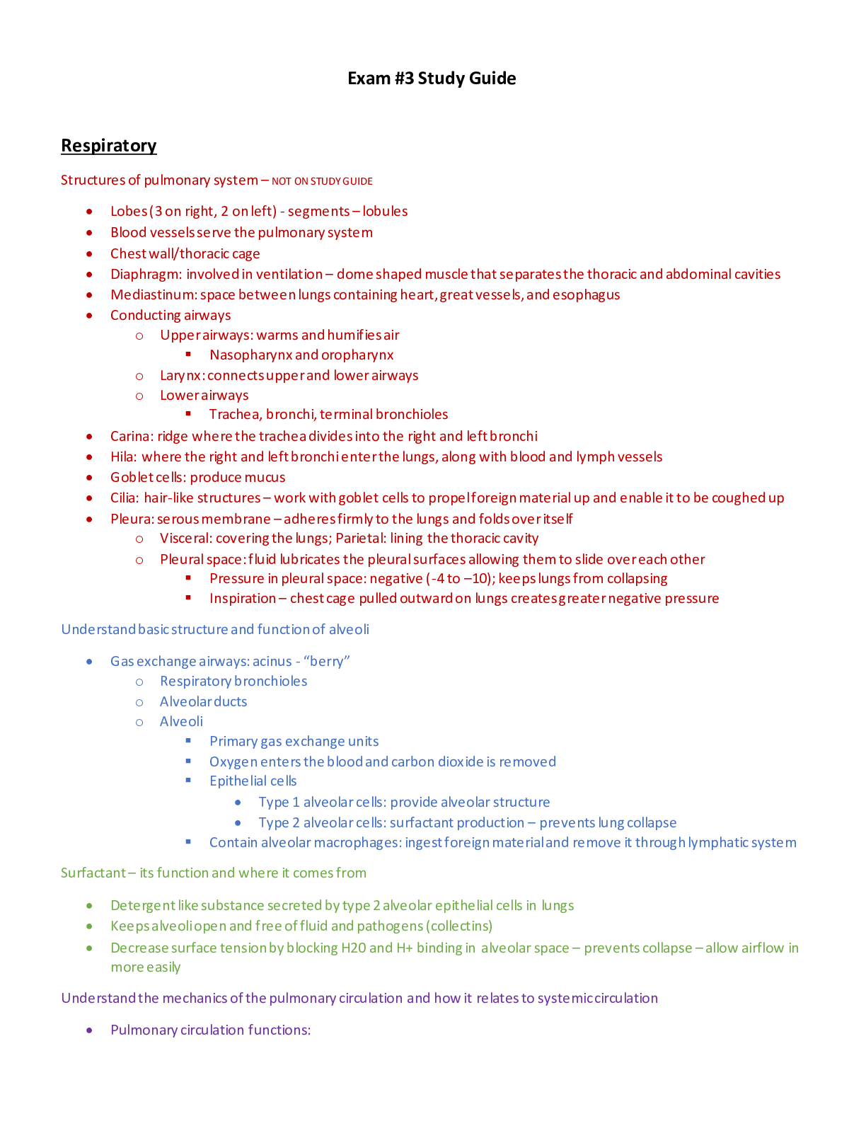
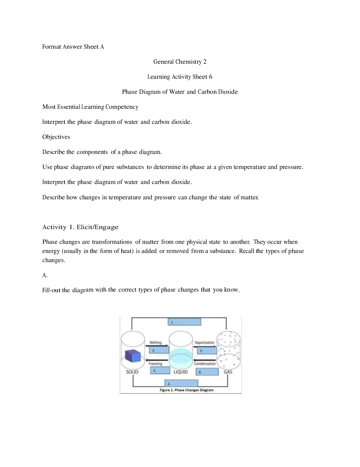
Exam #3 Study Guide Respiratory Structures of pulmonary system – NOT ON STUDY GUIDE • Lobes (3 on right, 2 on left) - segments – lobules • Blood vessels serve the pulmonary system �... �� Chest wall/thoracic cage • Diaphragm: involved in ventilation – dome shaped muscle that separates the thoracic and abdominal cavities • Mediastinum: space between lungs containing heart, great vessels, and esophagus • Conducting airways o Upper airways: warms and humifies air Nasopharynx and oropharynx o Larynx: connects upper and lower airways o Lower airways Trachea, bronchi, terminal bronchioles • Carina: ridge where the trachea divides into the right and left bronchi • Hila: where the right and left bronchi enter the lungs, along with blood and lymph vessels • Goblet cells: produce mucus • Cilia: hair-like structures – work with goblet cells to propel foreign material up and enable it to be coughed up • Pleura: serous membrane – adheres firmly to the lungs and folds over itself o Visceral: covering the lungs; Parietal: lining the thoracic cavity o Pleural space: fluid lubricates the pleural surfaces allowing them to slide over each other Pressure in pleural space: negative (-4 to –10); keeps lungs from collapsing Inspiration – chest cage pulled outward on lungs creates greater negative pressure Understand basic structure and function of alveoli • Gas exchange airways: acinus - “berry” o Respiratory bronchioles o Alveolar ducts o Alveoli Primary gas exchange units Oxygen enters the blood and carbon dioxide is removed Epithelial cells • Type 1 alveolar cells: provide alveolar structure • Type 2 alveolar cells: surfactant production – prevents lung collapse Contain alveolar macrophages: ingest foreign material and remove it through lymphatic system Surfactant – its function and where it comes from • Detergent like substance secreted by type 2 alveolar epithelial cells in lungs • Keeps alveoli open and free of fluid and pathogens (collectins) • Decrease surface tension by blocking H20 and H+ binding in alveolar space – prevents collapse – allow airflow in more easily Understand the mechanics of the pulmonary circulation and how it relates to systemic circulation • Pulmonary circulation functions: o Facilitate gas exchange o Deliver nutrients to lung tissue o Acts as a blood reservoir for the left ventricle o Serves as a filtering system that removes clots, air, and other debris from the circulation o Pulmonary system pressure is 18 mmHg compared to systemic circulation of 90 mmHg o Gas exchange airways are served by the pulmonary circulation Low pressure system, high flow – Supplies venous blood from all parts of the body to the alveolar capillaries where O2 is added and CO is removed; contains 100% of CO o Bronchi and other lung structures are served by systemic circulation – bronchial circulation High pressure system, low flow – supplies blood to trachea, bronchial tree, bronchioles, and out coats (adventia) of pulmonary arteries and veins; contains 1-3% of CO • Pulmonary circulation .....................................................................continued....................................................................................... [Show More]
Last updated: 1 year ago
Preview 1 out of 48 pages
Reviews( 0 )
Document information
Connected school, study & course
About the document
Uploaded On
Jul 12, 2021
Number of pages
48
Written in

Additional information
This document has been written for:
Uploaded
Jul 12, 2021
Downloads
0
Views
83
.png)