*NURSING > Class Notes > NUR 114 Partial Focus Notes Final Fall 2019/2020_complete, A+ guide (All)
NUR 114 Partial Focus Notes Final Fall 2019/2020_complete, A+ guide
Document Content and Description Below
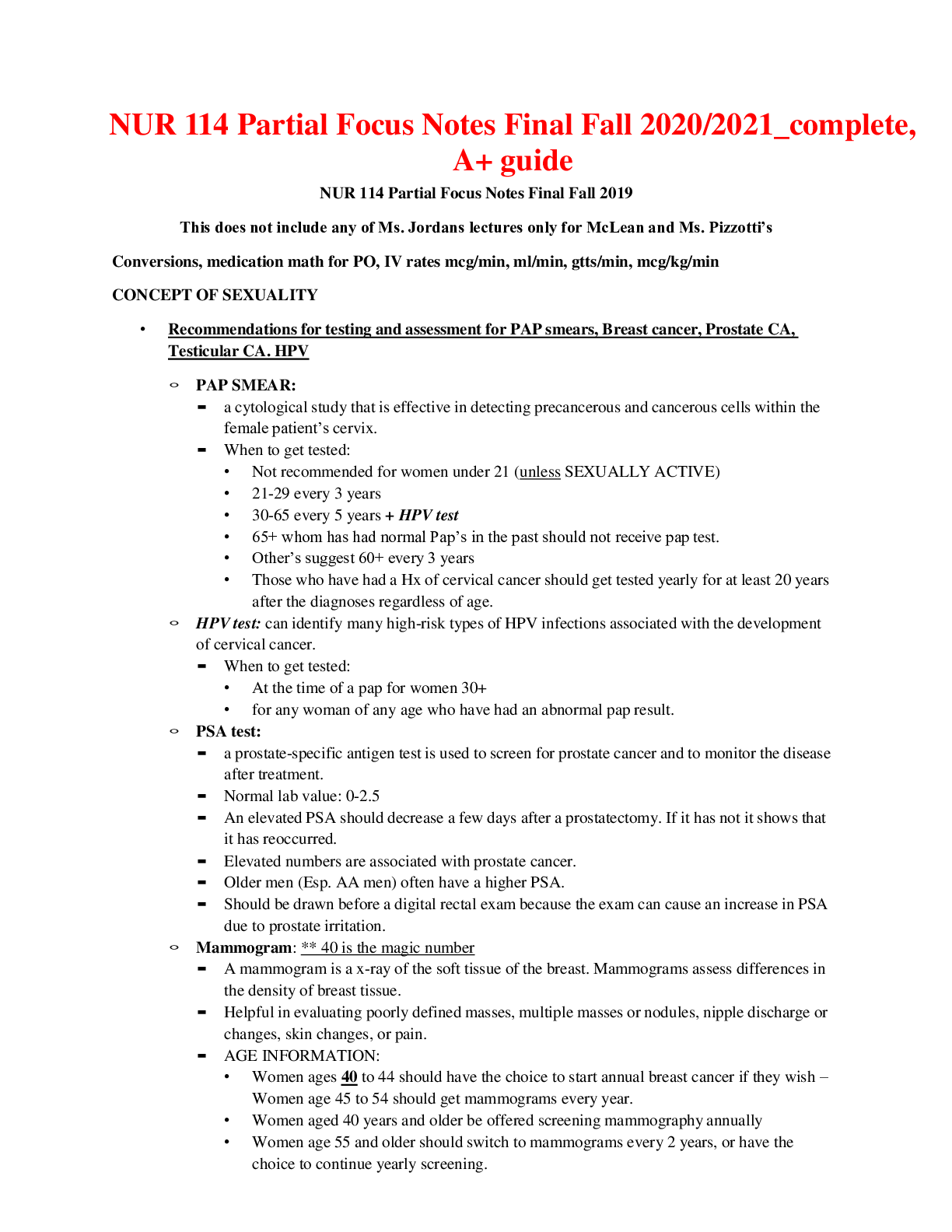
NUR 114 Partial Focus Notes Final Fall 2019 This does not include any of Ms. Jordans lectures only for McLean and Ms. Pizzotti’s Conversions, medication math for PO, IV rates mcg/min, ml/min, gtts/m... in, mcg/kg/min CONCEPT OF SEXUALITY • Recommendations for testing and assessment for PAP smears, Breast cancer, Prostate CA, Testicular CA. HPV ◦ PAP SMEAR: ▪ a cytological study that is effective in detecting precancerous and cancerous cells within the female patient’s cervix. ▪ When to get tested: • Not recommended for women under 21 (unless SEXUALLY ACTIVE) • 21-29 every 3 years • 30-65 every 5 years + HPV test • 65+ whom has had normal Pap’s in the past should not receive pap test. • Other’s suggest 60+ every 3 years • Those who have had a Hx of cervical cancer should get tested yearly for at least 20 years after the diagnoses regardless of age. ◦ HPV test: can identify many high-risk types of HPV infections associated with the development of cervical cancer. ▪ When to get tested: • At the time of a pap for women 30+ • for any woman of any age who have had an abnormal pap result. ◦ PSA test: ▪ a prostate-specific antigen test is used to screen for prostate cancer and to monitor the disease after treatment. ▪ Normal lab value: 0-2.5 ▪ An elevated PSA should decrease a few days after a prostatectomy. If it has not it shows that it has reoccurred. ▪ Elevated numbers are associated with prostate cancer. ▪ Older men (Esp. AA men) often have a higher PSA. ▪ Should be drawn before a digital rectal exam because the exam can cause an increase in PSA due to prostate irritation. ◦ Mammogram: ** 40 is the magic number ▪ A mammogram is a x-ray of the soft tissue of the breast. Mammograms assess differences in the density of breast tissue. ▪ Helpful in evaluating poorly defined masses, multiple masses or nodules, nipple discharge or changes, skin changes, or pain. ▪ AGE INFORMATION: • Women ages 40 to 44 should have the choice to start annual breast cancer if they wish – Women age 45 to 54 should get mammograms every year. • Women aged 40 years and older be offered screening mammography annually • Women age 55 and older should switch to mammograms every 2 years, or have the choice to continue yearly screening.• American College of Obstetricians and Gynecologists recommends annual screening mammography for women age 40 and over • Yearly screening is recommended at age 40 because younger women have higher density in breast tissue and the test is less effective. ▪ In older women, the amount of fatty tissue is higher. Therefore, fatty tissues appear lighter than cancer ▪ Cancer and cysts usually have the same density. However, cysts are smooth borders and cancers are usually star-burst shape ◦ Testicular Cancer: ▪ Rare form of cancer that most often effects men between ages of 20-35 years of age. ▪ With early detection there is a 95% cure rate ▪ Most common type is germ cell tumors arising from sperm-producing cells • Seminoma tumors are usually localized, metastasize late, and respond to treatment. ▪ Least common type is non-germ cell tumors in stromal, interstitial, or Leydig cells • Most do not metastasize • Adroblastomas sometimes secrete estrogen, which causes feminization and gynecomastia ▪ Men with cryptocordism or HIV have higher risk of testicular tumors. ▪ Common tumor markers that confirm testicular cancer are alfa-fetoprotein, hCG, and LDH • What are the types and s/s of benign breast disorders? Benign Breast disorders 1. Fibroadenoma o Most common benign tumor in women during reproductive years o Mass of connective tissue unattached to surrounding breast tissue o Tumors are oval, freely movable, rubbery 2. Fibrocystic Breast Condition (FBC) o Fibrocystic changes of breast (may involve lobules, ducts, stromal tissues) o Common in pre-menopausal women between 20 and 50 years of age o Thought to be caused by imbalance in normal estrogen-to-progesterone ratio o Symptoms: Breast pain, tender lumps, swelling (often before menstrual period) o Two main features of FBC: fibrosis and cysts Fibrosis made up of connective tissue and are hard and firm Cysts are fluid filled and glandular cells – Breast US is used to confirm presence of cysts. o Postmenopausal women taking hormonal replacement may develop FBC or have worsening symptoms. o This does not increase her chances of Breast CANCER; however, if a firm mass arises a mammogram may be done to rule out cancer o A needle biopsy may be done to rule out cancer IF: No fluid is aspirated Mammogram is positive Mass remains palpable after aspiration Aspirated fluid reveals cancer cells o Management of FBC: Analgesics Limit salt intake before menses Wear supportive bra at all times Ice or heat may help Reduce or eliminate caffeine, dairy product Needle aspiration may be necessary diuretics Oral contraceptives or selective estrogen receptor modulators may be prescribed o Explain to women the benefits and risks associated with hormonal drug therapy for FBC, such as stroke, liver disease, and increased intracranial pressure. Teach them to seek medical attention immediately if any signs or symptoms of these complications occur. 3. Ductal Ectasia o Benign breast problem of women approaching menopause o Caused by dilation and thickening of collecting ducts in subareolar area o Hard mass; irregular borders, tender o Greenish-brown nipple discharge, enlarged axillary nodes, redness and edema over mass o Management May improve without treatment Reduce anxiety regarding threat of breast cancer Warm compresses Antibiotics May require surgical removal 4. Intraductal Papilloma o Occurs most often in women 40 to 55 years of age o Benign process in epithelial lining of duct, forming a pedunculated outgrowth of tissue o Trauma and erosion within duct; bloody or serous nipple discharge o Mass is rarely palpableo Must rule out breast cancer Breast cancer types, S/S, High risk groups, risk factors, treatments, diagnostics Invasive VS noninvasive: • There are two broad categories of breast cancer: noninvasive and invasive. Most of these cancers arise from the intermediate ducts. a. Non-invasive cancers account of about 20% of BC – occurs when the cancer remains within the duct. b. Invasive BC is the other 80% – occurs when the BC penetrates the surrounding tissue around the duct. • Metastasis occurs when the cancer cells leave the breast via blood or lymph fluid – permits spread of cancerous cells to distant sites • Common sites of metastasis are lung, bone, brain, and liver. Noninvasive Breast Cancers • Ductal carcinoma in situ (DCIS) ◦ early noninvasive form of breast cancer ◦ cancer cells located within the duct – have not invaded the surrounding fatty breast tissue. ◦ mammography screening and earlier detection has caused the # of women diagnosed with DCIS to > ◦ left untreated, it is estimated that 14% to 53% of DCIS would become invasive and spread into the breast tissue surrounding the ducts over a period of 10 years ◦ Currently there is no way to determine which DCIS lesions will progress to invasive cancer and which ones will remain unchanged – causes anxiety and decisional conflict in many women diagnosed with this ◦ differs from invasive cancer – DCIS cells lack the biologic capacity to metastasizs • lobular carcinoma in situ (LCIS) ◦ rare and occurs as an abnormal cell growth in the lobules (milk-producing glands) of the breast ◦ not a true cancer, but having LCIS increases one's risk for developing a separate breast cancer later ◦ usually diagnosed before menopause in women 40 to 50 years of age. ◦ Traditionally, treated with close observation only ◦ Women with LCIS and other breast cancer risk factors may want to consider prophylactic treatment options such as tamoxifen, raloxifene, or prophylactic mastectomy Invasive Breast Cancers • Infiltrating ductal carcinoma ◦ most common type of invasive breast cancer ◦ originates in the mammary ducts and grows in the epithelial cells lining these ducts ◦ Once invasive, the cancer grows into the tissue around it in an irregular pattern ◦ If a lump is present, it is felt as an irregular, poorly defined mass ◦ As tumor continues to grow, fibrosis (replacement of normal cells with connective tissue and collagen) develops around the cancer▪ fibrosis may cause shortening of Cooper's ligaments – resulting typical skin dimpling – Late sign ▪ another late sign: edematous thickening and pitting of breast skin called peau d'orange (orange peel skin) • inflammatory breast cancer ◦ rare but highly aggressive form of invasive breast cancer ◦ Symptoms include swelling, skin redness, and pain in the breasts. ◦ IBC seldom presents as a palpable lump and may not show up on a mammogram ◦ usually diagnosed at a later stage than other types of breast cancer – often harder to treat successfully Mutations 1. BRCA1 and BRCA2, are related to hereditary breast cancer 2. People who have specific mutations in either one of these genes are at a high risk for developing breast cancer as well as ovarian cancer 3. only 5% to 10% of all breast cancers are hereditary 4. Only women with a strong family history and a reasonable suspicion that a mutation is present have genetic testing for BRCA mutations. Other BC information • Estrogen and progesterone receptor-negative cancers: ◦ harder to treat because the drug choices and treatment options are limited at they will not affect the cancer. • HER2 receptors ◦ Human epithelial receptors are in normal breast and help them grow. ◦ In some breast cancers there are way too many and the cancer grows rapidly • REMEMBER these are Signs in LATE stages cancers: • dimpling may occur due to shortening of cooper’s ligaments ◦ caused by fibrosis which is the replacement of normal cells with connective tissue and collagen • Peau d’orange is visible with edema of the breast (peeling of orange skin) ◦ Edematous thickening and pitting of the breast skin How to preform Self breast exam. Patient teaching. o Self-Breast EXAM steps: Lie down on your back and place your right arm behind your head Use the finger pads of the three middle fingers on your left hand to feel for lumps in the right breast. Use an overlapping dime shaped circular motion to feel for lumps Use three different levels of pressure to feel. Light feels closest to the skin, medium is a little deeper, and firm pressure feels close to the chest wall. It is normal to feel a curved ridge in the lower curve of each breast Move around the breast in an up and down pattern starting at an imaginary line drawn straight down your side from the underarm and moving across the breast to the middle of the chest bone. Be sure to check the entire breast area going down until you feel the ribs and up to the neck. Repeat on the left side While standing, exam the size, shape, contour, dimpling, and look at nipples. Stand with arms by your side in front of a mirror. Exam under the arms with your arm elevated only slightly.o Assessing a breast mass Identify the location of the mass by using the face of a clock method Describe the shape, size, and consistency of the mass Assess whether the mass is fixed or movable Note any skin changes ( dimpling, peau d’orange) Assess axillary and supraclavicular lymphatic nodes Ask patient if they experience pain or soreness in the area of the mass Care of the patient pre and post op Mastectomy and breast reconstruction postoperative care for a Mastectomy: o Sign in room and inform staff to not use the affected arm o Assess vitals every 30 mins for 2 hrs and every hour for 2 hours, and then every 4 hours. o Check dressing for bleeding o Monitor for amount and color of drainage o Assess position to make sure drainage system is not being pulled nor kinked o HOB 30 degrees o Elevated affect arm o Basic comfort measures o Analgesics o Ambulation and regular diet begin day after surgery o During a modified radical mastectomy the surgeon places one or two drainage tubes, usually Jackson-Pratt drains, under the skin flaps and attaches the tubes to a small collection chamber. Gentle suction is exerted, and fluid that would accumulate under the flaps and delay healing is collected. When taking vital signs, monitor for the amount and color of drainage. Add this information to the intake and output record. o Patients undergoing a lumpectomy may also have drainage tubes (usually Jackson-Pratt drains) placed if the lump is large or if axillary node dissection is performed. o Assess the incision and flap of the post-mastectomy patient for signs of bleeding, infection, and poor tissue perfusion. o With short hospital stays, drainage tubes are usually removed about 1 to 3 weeks after hospital discharge when the patient returns for an office visit. The drainage amount should be less than 25 mL in a 24-hour period Inform the patient that tube removal may be uncomfortable although these tubes lie just under the skin Provide or suggest analgesia before they are removed Document all findings, and report any abnormalities to the surgeon immediately. o Instruct the patient to avoid the hunched-back position with the arm flexed because of the risk for elbow contractures o Beginning exercises that do not stress the incision can usually be started on the first day after surgery. These exercises include squeezing the affected hand around a soft, round object (a ball or rolled washcloth) and flexion/extension of the elbow. Postoperative mastectomy exercises: o Hand-wall climbing: Face the wall and place hands on wall at shoulder level Flex your fingers so that your finger walk up the wall Stop when your arms feel fully exerted Slowly “walk” back down the wall o PULLEY: drape a 6 ft long rope over a curtain or door. Grab the ends of the rope and extend your arms out to the sides Keeping your arms straight go down with the left and up with the right o ROPE turning: tie rope to knob of closed door Hold rope with arm extended out in front of you Swing the rope in a circular motion starting with small circles and increasing in size. Postoperative Care of the Patient After Breast Reconstruction o Assess the incision and flap for signs of infection (excessive redness, drainage, odor) during dressing changes. o Assess the incision and flap for signs of poor tissue perfusion (duskiness, decreased capillary refill) during dressing changes. o Avoid pressure on the flap and suture lines by positioning the patient on her nonoperative side and avoiding tight clothing. o Monitor and measure drainage in collection devices, such as for Jackson-Pratt drains. o Teach the patient to return to her usual activity level gradually and to avoid heavy lifting o Remind the patient to avoid sleeping in the prone position o Teach the patient to avoid participation in contact sports or other activities that could cause trauma to the chest. o Teach the patient to minimize pressure on the breast during sexual activity. o Remind the patient to refrain from driving until advised by the physician. o Remind the patient to ask at the 6-week postoperative visit when full activity can be resumed. o Reassure the patient that optimal appearance may not occur for 3 to 6 months postoperatively. o If implants have been inserted, teach the proper method of breast massage to enhance expansion and prevent capsule formation (consult with the physician). o Emphasize breast self-awareness; if the patient performs breast self-examination (BSE), review her technique. o Remind the patient of the importance of clinical breast examination and follow-up surveillance by her physician. Endometriosis s/s treatments diagnostics o Care: Assessment: menstrual history, sexual history, bleeding characteristics PAIN IS THE MOST COMMON SYMPTOM OF ENDOMETRIOSIS Usually peaks just before the menstrual flow; is usually located in the abdomen and causes a sense of rectal pressure Degree of pain not related to the extent, but it is related to the site Other s/s include: painful intercourse, painful defecation, low backache, infertility, nausea, diarrhea are also common Pelvic assessment may reveal pelvic tenderness, tender nodules in the posterior vagina, and limited movement of the uterus. Psychosocial assessment may reveal anxiety Diagnostic tests to rule out PID caused by chlamydia or gonorrhea. Serum CA-125 (cancer antigen) may be positive Transvaginal ultrasound-determine if masses are malignant or endometriosis a laparoscopy is the most definitive diagnosis in endometriosis o Interventions: Hormonal and surgical management may be used Collaborations consists of interventions that: Reduce pain Restore sexual function Alleviate anxiety r/t the disease and the uncertainty of the diagnosis Educate the pt about the disease and its treatment Alleviate fear r/t the possibility of laparoscopy or surgery Prevent self-concept disturbance r/t infertility Nonsurgical Management: Oral contraceptive to control menstrual cycle or progestins such as: o Oral medroxyprogesterone acetate and norethindrone acetate o Injectable forms of progestins: medroxyprogesterone acetate (depoprovera) Continuous low-level heat using wearable heat packs Relaxation techniques, yoga, massage, biofeedback Calcium and Magnesium supplements may relieve cramping Surgical Management: Ablation- a laprascopic removal of endometrial implants and adhesions; sameday surgery – unless you are like me and had complications :) Surgeon may use a laser to treat endometriosis by vaporizing adhesions and endometrial implants. Teach pt that temporary postop pain from CO2 used during laparoscopy to better visualize internal organs, can occurs in the shoulders or chest Toxic Shock syndrome s/s treatments diagnostics o Can result from menstruation and tampon use o Other conditions associated with TSS: Surgical wound infection Nonsurgical infections Gynecologic surgeries Use of internal contraceptives o Menstrual blood provides a growth medium for Staphylococcus aureus exotoxins produced from the bacteria cross the vaginal mucosa to the bloodstream via microabrasions from tampon insertion or prolonged useo Can be fatal o Usually develops within 5 days after the onset of menstruation o Most common s/s: Fever Rash Myalgias Sore throat Edema Hypotension Rash often looks like sunburn Broken capillaries in eyes and skin o Treatment: Removal of infection source (tampon, usually) Restore fluid and electrolyte balance Administer drugs to manage hypotension IV antibiotics Transfusion to reverse low platelet counts Corticosteroids to treat skin changes o Prevention: Wash hands before inserting a tampon Do not use a dirty tampon Inset the tampon carefully to avoid injuring the delicate tissue in your vagina Change your tampon every 3-6 hours Do not use superabsorbent tampons Use sanitary napkins at night Call HCP if you suddenly experience a high temperature, vomiting or diarrhea Do not use tampons at all if you have had TSS Not using tampons almost guarantees that you will not get toxic shock syndrome Menopause and Osteoporosis Menstrual disorders (amenorrhea, dysmenorrhea) Infertility Family Planning Contraception and Sterilization Premenstrual Dysphoric Disorder (PMDD) Leiomyomas s/s treatments diagnostics, Uterine Fibroids • fibroids that are benign, slow-growing solid tumors of uterine myometrium • Intramural leiomyomas ◦ are contained in the uterine wall within the myometrium • Submucosal leiomyomas ◦ protrude into the cavity of the uterus and can cause bleeding and disrupt pregnancy • Subserosal leiomyomas ◦ protrude through the outer surface of the uterine wall and may extend to the broad ligament, pressing other organs • Pedunculated leiomyomas◦ are attached by a pedicle (stalk) to the outside of the uterus and occasionally break off and attach to other tissues • Leiomyomas are the most common reason for hysterectomies. Prostatectomy Pre and post op care Surgical Management: ◦ Surgery is the most common intervention for a cure ◦ Minimally invasive surgery (MIS) or, less commonly, an open surgical technique for radical prostatectomy (prostate removal) is usually performed ◦ A bilateral orchiectomy (removal of both testicles) is another palliative surgery that slows the spread of cancer by removing the main source of testosterone Preoperative Care: ◦ Preoperative care depends on the type of surgery that will be done ◦ Minimally invasive surgery (MIS) ▪ most appropriate for localized prostate cancer and is used as a curative intervention ◦ The most common procedure is the laparoscopic radical prostatectomy (LRP) ▪ most often with robotic assistance ◦ Patients who qualify for LRP must have a PSA less than 10 ng/mL and have no previous hormone therapy or abdominal surgerie. ◦ Other newer procedures include transrectal high-intensity focused ultrasound (HIFU) and cryosurgery Operative Procedures: ◦ For the LRP procedure, the patient is placed in lithotomy positioning with steep Trendelenburg ▪ Nurses in the OR ensure that the patient maintains a balanced body temperature and positions the patient to prevent injury ▪ The urologist makes one or more small punctures or incisions into the abdomen ▪ A laparoscope with a camera on the end is inserted through one of the incisions while other instruments are inserted into the other incisions ▪ The robotic system may be used to control the movement of the instruments by a remote device ▪ The prostate is removed along with nearby lymph nodes, but perineal nerves are not affected. ◦ The open radical prostatectomy can be performed via several surgical approaches, depending on the patient's desired outcomes and the staging of the disease ◦ The transperineal and retropubic (nerve-sparing) approaches are most commonly use ▪ The surgeon removes the entire prostate gland along with the prostatic capsule, the cuff at the bladder neck, the seminal vesicles, and the regional lymph nodes ▪ The remaining urethra is connected to the bladder neck ▪ The removal of tissue at the bladder neck allows the seminal fluid to travel upward into the bladder rather than down the urethral tract, resulting in retrograde ejaculations Postoperative Care: ◦ Nursing interventions include all the typical care for a patient undergoing major surgery ▪ Maintaining hydration ▪ caring for wound drains (open procedure)▪ managing pai ▪ , and preventing pulmonary complications are important aspects of nursing care. Care of the Patient After an Open Radical Prostatectomy • Encourage the patient to use patient-controlled analgesia (PCA) as needed. • Help the patient get out of bed into a chair on the night of surgery and ambulate by the next day. • Maintain the sequential compression device until the patient begins to ambulate. • Monitor the patient for deep vein thrombosis and pulmonary embolus. • Keep an accurate record of intake and output, including Jackson-Pratt or other drainage device drainage. • Keep the urinary meatus clean using soap and water. • Avoid rectal procedures or treatments. • Teach the patient how to care for the urinary catheter because he may be discharged with the catheter in place. • Teach the patient how to use a leg bag. • Emphasize the importance of not straining during bowel movement. Advise the patient to avoid suppositories or enemas. • Remind the patient about the importance of follow-up appointments with the physician to monitor progress. Pelvic Inflammatory Disease: PID Clinical Diagnostic Criteria for PID: ◦ One or more of the following minimum criteria must be present on pelvic examination to diagnose PID: ▪ Cervical motion tenderness ▪ Uterine tenderness ▪ Adnexal tenderness ◦ The following criteria can improve the specificity of the diagnosis: ▪ Oral temperature > 101°F (> 38.3°C) ▪ Abnormal cervical or vaginal mucopurulent discharge ▪ Presence of abundant numbers of white blood cells on saline microscopy of vaginal fluid Elevated erythrocyte sedimentation rate ▪ Elevated C-reactive protein level ▪ Laboratory documentation of cervical infection with gonorrhea or chlamydia ◦ The following test results are the most specific criteria for diagnosing PID: ▪ Endometrial biopsy with histopathologic evidence of endometritis ▪ Transvaginal sonography or magnetic resonance imaging techniques showing thickened, fluid-filled tubes with or without free pelvic fluid or tubo-ovarian complex, or Doppler studies suggesting pelvic infection (e.g., tubal hyperemia) ▪ Laparoscopic abnormalities consistent with PID Treatment: ◦ Antibiotic therapy, self-management measures and rarely surgery. If oral antibiotic therapy is not successful, hospitalization and IV antibiotic therapy is needed.▪ Option 1 Ceftriaxone (Rocephin) 250 mg IM in a single dose plus Doxycycline 100 mg orally twice per day for 14 days with or without Metronidazole (Flagyl) 500 mg orally twice per day for 14 days ▪ Option 2 Cefoxitin plus 2 g IM in a single dose administered concurrently with probenecid (1 g orally) Doxycycline 100 mg orally twice per day for 14 days with or without Metronidazole 500 mg orally twice per day for 14 days ▪ Option 3 Other parenteral thirdgeneration cephalosporin (e.g., ceftizoxime [Cefizox], cefotaxime [Claforan]) plus Doxycycline 100 mg orally twice per day for 14 days with or without Metronidazole 500 mg orally twice per day for 14 days ◦ Teach: Patient to finish all of the antibiotics and to avoid sexual intercourse until treatment and symptoms are gone. Sexual partners should be treated regardless of their lack of symptoms. ◦ Screening for chlamydia and gonorrhea in young women has been shown to decrease the incidence of PID in high-risk populations STI’S (STD’s) • Syphilis, Herpes, Genital warts, HPV, Candidiasis, Trichomoniasis, chlamydia, Gonorrhea, s/s treatments diagnostics • Syphilis ◦ Syphilis is a complex sexually transmitted disease (STD) that can become systemic and cause serious complications, including death. ◦ The causative organism is a spirochete called Treponema pallidum ◦ The infection is usually transmitted by sexual contact and blood exposure, but transmission can occur through close body contact such as kissing. ◦ Syphilis progresses through four stages: primary, secondary, latent, and tertiary ◦ 1st stage: ▪ The appearance of an ulcer called a chancre is the first sign of primary syphilis ▪ It develops at the site of entry (inoculation) of the organism from 10 to 90 days after exposure (3 weeks is average ▪ Chancres may be found on any area of the skin or mucous membranes but occur most often on the genitalia, lips, nipples, and hands and in the mouth, anus, and rectum ▪ During this highly infectious stage, the chancre begins as a small papule ▪ Within 3 to 7 days, it breaks down into its typical appearance: a painless, indurated, smooth, weeping lesion ▪ Regional lymph nodes enlarge, feel firm, and are not painful. ▪ Without treatment, the chancre usually disappears within 6 weeks; however, the organism spreads throughout the body and the patient is still infectious. ◦ 2nd stage: ▪ Secondary syphilis develops 6 weeks to 6 months after the onset of primary syphilis ▪ During this stage, syphilis is a systemic disease because the spirochetes circulate throughout the bloodstream ▪ Commonly mistaken for influenza, manifestations include flu-like symptoms (malaise, lowgrade fever, headache, muscular aches, sore throat) and a generalized rash ▪ There is no typical appearance of this rash except for its presence on the palms and soles of the feet and on mucous membranes▪ It can appear as diffuse macules (reddish brown), papules (usually less than 5 mm) or pustules, scaly psoriasis-like lesions or gray-white wart-like lesions (condylomata lata) ▪ All of these lesions are highly contagious and should not be touched without gloves ▪ Patchy alopecia on the scalp or facial hair (missing part of the eyebrow, “moth-eaten” appearance) is another symptom ▪ The rash subsides without treatment in 4 to 12 weeks. ◦ 3rd stage: ▪ After the second stage of syphilis, there is a period of latency ▪ Early latent syphilis occurs during the first year after infection, and infectious lesions can recur ▪ Late latent syphilis is a disease of more than 1 year's duration after infection ▪ This stage is not infectious except to the fetus of a pregnant woman ▪ Patients with latent syphilis may or may not have reactive serologic test (e.g., Venereal Disease Research Laboratory [VDRL]) findings) ◦ 4th stage: ▪ Tertiary, or late, syphilis occurs after a highly variable period, from 4 to 20 years ▪ This stage develops in untreated cases and can mimic other conditions because any organ system can be affected ▪ Manifestations of late syphilis include: • Benign lesions (gummas) of the skin, mucous membranes, and bones • Cardiovascular syphilis, usually in the form of aortic valvular disease and aortic aneurysms • Neurosyphilis, causing central nervous system symptoms (e.g., meningitis, hearing loss, generalized paresis [weakness]) ◦ Conduct a physical examination, including inspection and palpation, to identify manifestations of syphilis. Wear gloves while palpating any lesions because of the highly contagious treponemes that are present. Observe for and document rashes of any type because of the variable presentation of secondary syphilis. ◦ After the physical examination, the health care provider obtains a specimen of the chancre for examination under darkfield microscope ▪ Diagnosis of primary or secondary syphilis is confirmed if T. pallidum is present. ◦ Blood tests are also used to diagnose syphilis. ▪ The usual screening and/or diagnostic nontreponemal tests are the Venereal Disease Research Laboratory (VDRL) serum test and the more sensitive rapid plasma reagin (RPR). ▪ These tests are based on an antibody-antigen reaction that determines the presence and amount of antibodies produced by the body in response to an infection by T. pallidum. ▪ They become reactive 2 to 6 weeks after infection ▪ VDRL titers are also used to monitor treatment effectiveness ▪ The antibodies are not specific to T. pallidum, and false-positive reactions often occur from such conditions as viral infections, hepatitis, and systemic lupus erythematosus ▪ If a VDRL result is positive, the health care provider requests or the laboratory may automatically perform a more specific treponemal test, such as the fluorescent treponemal antibody absorption (FTA-ABS) test or the microhemagglutination assay for T. palladum (MHA-TP), to confirm the infection Drug Therapy for Syphilis ◦ Benzathine penicillin G given IM as a single 2.4 million-unit dose is the evidence-based treatment for primary, secondary, and early latent syphilis◦ Patients in the late latent stage receive the same dose every week for 3 weeks ◦ A different regimen, found in the CDC's STD Treatment Guidelines, is recommended for patients who are HIV-infected or pregnant. Genital Herpes Pathophysiology: ◦ Genital herpes (GH) is an acute, recurring, incurable viral diseases ◦ Two serotypes of herpes simplex virus (HSV) affect the genitalia: ▪ type 1 (HSV-1) and type 2 (HSV-2) ▪ Most nongenital lesions such as cold sores are caused by HSV-1, transmitted via oral-oral contact. ▪ Historically, HSV-2 caused most of the genital lesions; however, this distinction is academic because the transmission, symptoms, diagnosis, and treatment are nearly identical for the two types ▪ Either type can produce oral or genital lesions through oral-genital or genital-genital contact with an infected person. ▪ HSV-2 recurs and sheds asymptomatically more often than HSV-1 ▪ Most people with GH have not been diagnosed because they have mild symptoms and shed virus intermittently ◦ The incubation period of genital herpes is 2 to 20 days, with the average period being 1 week. Many people do not have symptoms during the primary outbreak. When subsequent outbreaks of genital herpes occur, they are usually more severe and occasionally require hospitalization. ASSESSMENT OF GH ◦ The diagnosis of GH is based on the patient's history and physical examination ◦ Ask the patient if he or she felt itching or a tingling sensation in the skin 1 to 2 days before the outbreak, known as the prodrome ◦ These sensations are usually followed by the appearance of vesicles (blisters) in a typical cluster on the penis, scrotum, vulva, vagina, cervix, or perianal region at the site of inoculation ◦ The blisters rupture spontaneously in a day or two and leave painful ulcerations that can become extensive ◦ Assess for other symptoms such as headaches, fever, general malaise, and swelling of inguinal lymph nodes. ◦ Ask if urination is painful. External dysuria is a painful symptom when urine passes over the eroded areas. Patients with urinary retention may need to be catheterized. ◦ After the lesions heal, the virus remains in a dormant state in the sacral nerve ganglia. Periodically, the virus may activate and symptoms recur. ▪ These recurrences may be triggered by many factors, including stress, fever, sunburn, poor nutrition, menses, and sexual activity. Antiviral drugs are used to treat GH Acyclovir (Zovirax, Avirax image), famciclovir (Famvir), or valacyclovir (Valtrex) may be prescribed Condylomata Acuminata (Genital Warts): • Encourage women to have a Pap test annually, starting at age 21 years; after they have had three normal smears, they should have a Pap test every 3 years if no new risk factors are present (e.g., new partner, other STDs) • The presence of warts should increase suspicion that the patient may have had exposure to other STDs, which warrants additional testing • VACCINES:◦ Gardasil protection from human papilloma virus cervical cancer, genital warts, and cancer of the anus, vagina, and vulva ▪ Approved for use in male and female patients ages 9–26 ◦ Gardasil 9 same protection as the original Gardasil, plus protection against five additional types of high-risk HPV ▪ Approved for use in male patients ages 9–15 and female patients ages 9–26 ◦ Cervarix protects against only the types of HPV that cause cervical cancer ▪ Approved for use solely in female patients ages 9–25 Chlamydia Trachomatis • an intracellular bacterium & causative agent of genital chlamydia infections ◦ It invades the epithelial tissues in the reproductive tract. ◦ Incubation is 1 to 3 weeks but the pathogens may be presents for months without producing symptoms. ◦ Chlamydia is a reportable condition to health departments in all states. ◦ 20 – 40% of women who become infected with chlamydia develop Pelvic Inflammatory Disease (PID) • Often asymptomatic but some signs and symptoms may include: ◦ *Vaginal or Urethral discharge ◦ *Dysuria (painful urination) ◦ *Frequent Urination ◦ *Pelvic pain ◦ *Irregular bleeding Diagnosis: ◦ Sampling cells from the endocervix, urethra or both. Gold standard is a tissue culture from cervical os or the urethra. ◦ Women ages 25 (sexually active) and younger should be screened yearly and women over 25 with multiple sexual partners should be screened yearly for chlamydia. Treatment: ◦ azithromycin (Zithromax) or doxycycline ◦ Expedited partner therapy (EPT), treating and testing sexual partners for other STD’s shows signs of reducing chlamydia infection rates. Gonorrhea: a sexually transmitted bacterial infection that occurs in both men and women ◦ The causative organism is Neisseria Gonorrhoeae, a gram negative intracellular dipococcus. ◦ It is transmitted by direct sexual contact with mucosal surfaces – (vaginal intercourse, orogenital contact, or anogenital contact) Gonorrhea bacteria can grow in the warm, moist areas of the reproductive tract, including: ◦ the cervix (opening to the womb) ◦ uterus (womb) ◦ fallopian tubes (egg canals) in women ◦ urethra (the tube that carries urine from the bladder outside the body) in women and men ◦ The bacteria can also grow in the mouth, throat, and anus The first symptoms of gonorrhea may appear 3 to 10 days after sexual contact with an infected person◦ The disease can be present without symptoms and can be transmitted or progress without warning ◦ In women, ascending spread of the organism can cause pelvic infection (pelvic inflammatory disease [PID]), endometritis (endometrial infection), salpingitis (fallopian tube infection), and pelvic peritonitis ◦ Rare complications of gonorrhea in adults include arthritis, meningitis, hepatitis, and disseminated infection The infection can be asymptomatic in both men and women, but women have asymptomatic, or “silent,” infections more often than do men. ◦ If symptoms are present, men usually notice dysuria and a penile discharge that can be either profuse, yellowish green fluid or scant, clear fluid. ◦ The urethra is most commonly affected, but infection can extend to the prostate, the seminal vesicles, and the epididymis. ◦ Men seek curative treatment sooner, usually because they have symptoms, and thereby avoid some of the serious complications. ◦ Women may report a change in vaginal discharge (yellow, green, profuse, odorous), urinary frequency, or dysuria. ◦ The cervix and urethra are the most common sites of infection. ◦ Anal manifestations may include itching and irritation, rectal bleeding or diarrhea, and painful defecation ◦ Assess the mouth for a reddened throat, ulcerated lips, tender gingivae, and lesions in the throat If fever ccurs, this may be a sign of an ascending (PID or epididymitis) or systemic infection (disseminated gonococcal infection). ◦ Symptoms could include joint or tendon pain, either in a single joint or as migratory arthralgias, especially of the knees, elbows, fingers or toes, and a rash usually on the palms and soles. The rash can be pustular or maculopapula Pelvic Floor Dysfunction • cystocele: ◦ a protrusion of the bladder into the vaginal wall ◦ leading to urinary tract infection and urinary elimination problems, including stress incontinence. • Rectocele: ◦ protrusion of the rectum through a weakened vaginal wall ◦ leading to constipation, hemorrhoids, fetal impaction, and feelings of rectal or vaginal fullnessnary Incontinence Erectile Dysfunction Sexual and Gender Identity; Care of the transgender patient Concept of Immunity and Cellular Regulation: EXAM 2 & 3Notes from conference • Box 2-1; page 50 • page 56 TNM grading scale ◦ understand how to answer questions related to TNM ◦ C ancer Markers ▪ CEA – lung and colon cancer ▪ PSA – prostate cancer ▪ positive tumor markers do not prove you have cancer ▪ BIOPSY IS THE NUMBER ONE REASON TO DIAGNOSE CANCER • page 97 – chart for leukemia (look at picture made) ◦ primary forms of leukemia is correct ◦ AML is most common in older adults ◦ ALL – younger adults and children – rapid progression ◦ CML – philadelphia ◦ CLL often requires no treatment – depending on rate of progresssion • Lymphomas – look at picture made ◦ non hodgekin – EBV and Burkitt lymphoma ◦ hodgekin – reed sternburg cell know that • colorectal cancer ◦ look at chart page 91 ◦ risk factors Colitis and Chrons and age are risk factors for colorectal cancer ◦ rectal bleeding is a big sign for colorectal cancer ◦ other s/s: change in bowel habits, familal polypoisious, weight loss • lung cancer ◦ know chart on 109 ◦ small cell (oat cell) carcinoma ▪ SIADH – hanging onto to your fluid; can not get rid of it – • cushings syndrome and thrombophlebitis ◦ non small cell ◦ blood in sputum • Brachytherapy ◦ lecture notes ◦ nurses taking care of patients who is getting this should NOT BE PREGNANT ◦ NO X-RAYS TO PREGNANT WOMEN ◦ small pellet of radioactive material that is placed into the cancer or into the nearby airway ◦ very dangerous to bystanders for pts getting this treatment • what is the difference between benign and malignant ◦ NORMAL: ▪ SLOW ▪ fibronectin says hey you are too close to me stop growing – helps keep that tightly together ▪ function ◦ BENIGN: ▪ CONTINUOUS AND SLOW; ▪ LOCALIZED ▪ tight adherence Function ◦ MALIGNANT:▪ AGGRESSIVE GROWTHS ▪ RAPID CELL DIVISION ▪ METASTIZE ▪ INVADE/DESTROY SURROUNDING TISSUE ▪ NO Fibronectin – making them loose ▪ NO FUNCTION ▪ MIGRATE • TLS – tumor lysis syndrome ◦ intracellular contents leak into circulation causing ▪ hyperkalemia • arrythmias, dysrhythmias ▪ hyperuricemia ▪ hyperphosphatemia • superior vena cava syndrome ◦ upper ext. edema ◦ postural hypotension ◦ dizziness, pallor, clamminess ◦ swelling of face – SOB ◦ check pulse ox, BP, and respiratory status – airway too • cancer prevention • cancer stages • 7 signs and symptoms of cancer eye part of test • 10-21 IOP • snellen’s chart – jaeger for up close vision problems – ishy hara chart is for color blindnesschal • do not give tropicamide (a dilation eye med) to someone who has increased IOP • trachoma – chlamydia trachomatis • chemical splash – irrigate check ph for chemical irritant to eye • fluorescent light for trauma to eye to see if abrasion has occurred • no cornea transplant if person has cancer at time of death – CANCER IS BIG ONE (small cell lung cancer) • retinal detachment – DO NOT DO THINGS THAT INCREASE IOP • know vocabulary words in notes • glaucoma • prostaglandins is given for glaucoma • Gonioscopy – TO TEST WHICH TYPE OF GLAUCOMA ◦ know difference between the two ◦ one is chronic – open ▪ not a medical emergency ▪ slightly open canal of schlemn ◦ one is acute – closed ▪ medical emergency ▪ cornea starts to flatten and iris bulges ▪ avoid anticholinerics and mydriatics like tropicamide which cause pupil to dilate > IOP ear part • kidney is something you look for if you have ear problems• sensory hearing losses – vestibuochlear nerve 8 is damaged • never irrigate ear infections – always look in ear before ear drops; warm solutions • ototoxic – aminoglycoside antibiotics CAUTION s/s of cancer Changes in bowel or bladder habits. A sore that does not heal. Unusual bleeding or discharge. Thickening or lump in the breast or any other part of the body. Indigestion or difficulty swallowing. Obvious change in a wart or mole. Nagging cough or hoarseness Benign vs. Cancer cells: Grading and staging of cancer TNM: What does Tissue of origin mean with cancer? s/s of different cancers: EXAM 2 LECTURE NOTES – MADISON’S VERSION Principles of Inflammation and Immunity Overview • Immunity: protection from illness or disease that is maintained by the body’s physiologic defense mechanisms ◦ this protection requires interaction of immunity and inflammation WORKING TOGETHER to control infection and other problems caused by harmful microorganisms or altered cells Self versus Non-Self • Non-self proteins and cells: ◦ infected body cells ◦ cancer cells ◦ cells from other people ◦ invading organisms • recognizing self vs. non-self: self tolerance ◦ this prevents immunity from harming healthy body cells◦ possible because of different proteins on cell membranes • This is where you see your HLAs (human leukocyte antigens) come into play ◦ each person’s cells have unique surface proteins that are specific to that person (HLAs) ◦ serve as “universal product code” ◦ your HLAs are recognized as “foreign” or non self by the immune system of someone else ▪ YOUR cell-surface proteins are non self to another adults immune system – they are antigens – proteins capable of stimulating an immunity response!! ◦ these HLAs are key for recognition and self tolerance ▪ immune system cells constantly come into contact w/other body cells and with any invader that enters the body • at each encounter – immune system cells compare the surface protein HLAs to determine whether the encountered cell belongs to the body • if they match – not attacked • if they do not match – it is non self or foreign ◦ immune system cell takes action to neutralize, destroy, or eliminate foreign invader • a key element for recognition of non self by cells involved in general immunity and those involved in specific immunity is the presence of TOLL-LIKE RECEPTORS (TLRs) on these cells ◦ known to be present on immune system cells of humans ◦ protective purpose – interact with the surface of any invading organism ◦ allows recognition that is non self so actions are taken to rid the invader from the body ◦ TLRs also help immune system cells recognize damaged or unhealthy cells Organization of the Immune System • The three processes needed for human protection through immunity: ◦ inflammation ◦ antibody-mediated immunity (AMI) ◦ cell-mediated immunity (CMI) • optimal function of all three divisions is necessary for complete immunity Chart describing immune functions of specific leukocytes – will talk about each section in detail Variable Leukocyte FunctionInflammation Neutrophil Nonspecific ingestion and phagocytosis of microorganisms and foreign protein – Macrophage Nonspecific recognition of foreign proteins and microorganisms; ingestion and phagocytosis. Assist with antibody-mediated immunity and cell-mediated immunity – Monocyte Destruction of bacteria and cellular debris; matures into MACROPHAGE – Eosinophil Releases vasoactive amines during allergic reactions to limit these reactions – Basophil Releases histamine and heparin in areas of tissue damage Variable Leukocyte Function Antibodymediated immunity B-lymphocyte Becomes sensitized to foreign cells and proteins with the assisstance of macrophages and helper/inducer T-cells – Plasma cell Secretes immunoglobulins (antibodies) in response to the presence of a specific antigen – Memory cell Remains sensitized to a specific antigen and can secrete increased amounts of immunoglobulins (antibodies) specific to the antigen on re-exposure Variable Leukocyte Function Cell-mediated immunity Helper/inducer T-cell Enhances immune activity of all parts of general and specific immunity through secretion of various factors, cytokines, and lymphokines – Cytotoxic/ cytolytic cell Selectively attacks and destroys non self cells, including virally infected cells, grafts, and transplanted organs – Natural killer cell Nonselectively attacks non self cells, especially body cells that have undrgone mutation and become malignant; also attacks grafts and transplanted organs GENERAL IMMUNITY – INFLAMMATION• nonspecific – also called innate-native immunity or natural immunity • with inflammation – general immunity provides IMMEDIATE protection ◦ against the effects of tissue injury and invading foreign proteins • innate-native immunity ◦ any natural protective feature of a human ◦ barrier to prevent organism from entering body ◦ can attack organism that have already entered body • this type of immunity CAN NOT be transferred from one adult to another – not an adaptive response • Lines that compose general immunity: ◦ 1st line ▪ skin ▪ mucosa ▪ antimicrobal on the skins (normal flora) ◦ 2nd line ▪ Inflammation (inflammatory response) ▪ complement ▪ natural killer cells • General immunity and inflammation differ from specific immunity IN TWO WAYS: ◦ inflammatory protection is immediate but short term ▪ it does not provide true immunity on repeated exposure to the same organisms ◦ inflammation is a nonspecific body defense to invasion or injury ▪ can be started quickly by almost any event – regardless of where it occurs/what caused it Infection – • usually accompanied by inflammation; however, inflammation can occur without infection ◦ inflammation does not always mean that an infection is present ◦ inflammation seen where?: ▪ w/out infection can be caused by: • joint sprains • MI • blister formation▪ Non-infectious invasion: • allergic rhinits • contact dermatitis ▪ infection: • otitis media • appendicitis • viral hepatitis Cell types involved in inflammation • leukocytes (WBCs) ◦ neutrophils ▪ mature neutrophils make up between 55%-70% of normal total WBC count ▪ come from stem cells – complete the maturation process in bone marrow ▪ provides protection after invaders, especially BACTERIA, enter the body ▪ they destroy invaders by phagocytosis and enzymatic digestion ▪ THIS IS THE CELL WHERE YOU INDICATE A LEFT SHIFT • the segmented neutrophil (mature cells at the far right) is no longer the most numerous type in circulation – more of the circulating cells are bands – the less mature cell type found farther left on the neutrophil pathway • a left shift indicates: ◦ patients bone marrow cannot produce enough mature neutrophils (segs) ◦ cannot keep the pace w/continuing infection ◦ release immature neutrophils into the blood (bands) ◦ the bands are of minimal benefit ▪ not capable of phagocytosis ◦ also known as bandemia (immature neutrophils in the blood) ◦ macrophages ▪ monocytes move from the blood into body tissues where they mature into macrophages ▪ liver, spleen, and intestinal tract contain large # of these cells ▪ helps stimulate immediate inflammatory responses ▪ also stimulates the longer-lasting immune response of antibody-mediated immunity (AMI) and cell-mediated immunity (CMI)▪ function: • phagocytosis, • repair, • antigen presenting/processing, • secretion of cytokines for immune system control ▪ inflammatory function: PHAGOCYTOSIS ◦ eosinophils ▪ contain many vasoactive chemicals ▪ most active against infestations of parasitic larvae ▪ also limits inflammatory reactions ◦ basophils ▪ these cause s/s of inflammation ▪ acts on blood vessels w/basophil chemicals (vasoactive amines) • histamine is one of the vasoactive amines for example** focus on this one • others include; heparin, serotonin, and leukotrines (if you wanted to know) ▪ when ALLERGENS bind to IgE on the basophils • basophil membrane opens • releases the vasoactive amines into blood • most of the vasoactive amines act on smooth muscle and blood vessels walls • **histamine (which is one of the vasoactive amines released) dilates arterioles and constricts small veins ◦ slowing blood flow and decreasing venous return ◦ causing blood to collect in capillaries and arterioles ▪ basophils stimulates both general inflammation and the inflammation of allergic reactions • additional cell type important in inflammation ◦ mast cells – release histamine ▪ have binding sites for the stems of IgE molecules ▪ are involved in allergic reactions ▪ respond to inflammatory products released by T-lymphoctes ▪ tissue mast cells maintain and prolong inflammation and allergic reactionsAnti-microbial substances: ◦ Some books include the complement system, Interleukins, Interferons, lysozymes found in sweat saliva and tears (there are many many more) ◦ Basically these are proteins made by tissues or cells in the body that…The complement system is a part of innate immunity ▪ activate the immune system ▪ promote resistance to viruses ▪ oxidize bacteria ▪ inhibit growth ▪ lyse bacteria cell walls ▪ ect….. • complement system: ◦ can kill bacteria and participate in the inflammatory process ◦ Group of proteins that are activated in sequential order to provide a link to the humoral response ◦ IgG and Igm are responsible for activating the complement cascade ◦ Complement assays diagnose immunodeficiency and autoimmune disease • Interferons: ◦ proteins that come from virus laden cells and lymphocytes that help with resistance to the virus. • Interleukins: ◦ proteins that come from macrophages and lymphocytes that cause fever and activation of the immune system Phagocytosis: • key process in inflammation • neutrophils and macrophages – most efficient at phagocytosis • 7 STEPS: ◦ exposure and invasion: ▪ first step in injury or invasion ▪ leukocytes engage in phagocytois and stimulate inflammation• present in blood and ECF ◦ attraction: ▪ brings the WBC into direct contact with target (antigen, invader, foreign protein) ▪ damaged tissues – secrete chemotaxins which do the following: • attracts neutrophils and macrophages • release debris that binds to the surface of invading proteins ◦ adherence ▪ binds the phagocytic cell to the surface of target ▪ opsonins – • substances that increase contact with the cell w/target by coating the target cell • during inflammation coating the target makes it easier for phagocytic cells to stick to the antigen • substances that are opsonins ◦ dead neutrophils ◦ antibodies ◦ activated complement components ◦ recognition: ▪ when phagocytic cell sticks to target cell • recognizes it as non self • phagocytic cell examines the HLAs (universal product codes) or whatever they encounter ▪ phagocytic cells only START phagocytosis when the target cell is RECOGNIZED as non self or foreign debris ◦ cellular ingestion: ▪ engulfment of the target cell (phagocytosis) ◦ degradation: ▪ final step ▪ enzymes in phagosome digest engulfed target Sequence of inflammation • five cardinal symptoms of inflammation: ◦ warmth◦ redness ◦ swelling ◦ pain ◦ decreased function • 3 stages ◦ stage 1: ▪ vascular response ◦ stage 2: ▪ cellular exudate part of response ◦ stage 3: ▪ features tissue repair and replacement SPECIFIC IMMUNITY – THIS INCLUDES ANTIBODY-MEDIATED IMMUNITY AND CELL MEDIATED IMMUNITY antibody-mediated immunity (AMI): • our humoral immunity • Uses antigen-antibody interactions to neutralize, eliminate, or destroy foreign proteins. ◦ Antibodies are produced by sensitized B-lymphocytes (B-CELLS) ◦ b-cells become sensitized to a specific foreign protein (antigen) and produce antibodies directed SPECIFICALLY AGAINST THAT ANTIGEN ◦ b-cells have the most direct roll in AMI • T-lymphocytes and macrophages (main players in cell mediated immunity) work with the B-cells of our humoral immunity to generate antigen-antibody interactions describing the antigen-antibody interaction: • the body learns to make enough of any specific antibody to provide long-lasting immunity against specific organism or toxins • antigen recognition is the recognition of the antigen by unsensitized B-cells ◦ this action requires the help of T-Cells and macrophages • recognition is started by the macrophages of innate immunity ◦ macrophages recognize the invading antigen as non self and attach to antigen ◦ once attached – macrophages may now present the attached antigen (foreign protein) to the helper/inducer T-cells • The CD4 cell (your helper/inducer T-cells) and the macrophages TOGETHER process the antigen to expose the antigens recognition sites (universal product code) • after processing the antigen◦ CD4 T cell bring antigen into contact with the B-CELL ▪ now the B-cell is able to recognize the antigen as non self – now sensitization can occur • the b-cell is now “sensitized” to this antigen ◦ an un-sensitized b-cell can only be sensitized once (SO TO ONLY ONE TYPE OF ANTIGEN) • immediately after it is sensitized ◦ b-cell divides into two types of b-lymphocytes (each one remaining sensitized to that specific agent) ▪ plasma cell • starts immediately to produce antibodies against the sensitizing antigen • antibodies are produced by plasma cells • produces antibodies specific only to the antigen that originally sensitized the parent b-cell ▪ memory cell • antigen presenting cells/MHC2 • a sensitized b-cell but DOES NOT PRODUCE antibodies until the next exposure to the same antigen • sustained immunity (memory) provides us with long-lasting true immunity to a specific antigen ◦ this results from memory b-cells made during lymphocyte sensitization Acquiring antibody-mediated immunity • Antibody-mediated immunity (AMI) ◦ also known as Acquired immunity◦ is a type of adaptive immunity in which a person's body learns to make as an adaptive response to invasion by organisms or foreign proteins. ◦ Adaptive immunity occurs: ▪ either naturally or artificially through lymphocyte responses ▪ can be either active or passive ▪ look at chart above • ACTIVE IMMUNITY: ◦ occurs when antigens enter a person's body and it responds by making specific antibodies against the antigen ◦ this type of immunity is active because the body takes an active part in making antibodies. ◦ active immunity occurs under natural or artificial conditions ◦ Natural ACTIVE immunity: ▪ immunity occurs when an antigen enters the body naturally without human assistance and the body responds by actively making antibodies against that antigen (e.g., influenza A virus) ▪ Usually, the invasion that triggers antibody production also causes the disease. However, processes occurring in the body at the same time as infection create immunity to that antigen. • Thus the person will not become ill after a second exposure to the same antigen. • Natural active immunity is the most effective and the longest lasting. ◦ Artificial ACTIVE immunity: ▪ protection developed by vaccination or immunization ▪ This type of immunity is used to prevent serious and potentially deadly illnesses (e.g., tetanus, diphtheria, polio) ▪ Small amounts of specific antigens are placed as a vaccination into a person ▪ The person's immune system responds by actively making antibodies against the antigen • Because antigens used for this procedure have been specially processed to make them less likely to grow in the body (attenuated), this exposure usually does not cause the disease • Artificial active immunity lasts many years, although repeated but smaller doses of the original antigen are required as a “booster” to retain the protection. • PASSIVE IMMUNITY: ◦ occurs when the antibodies against an antigen are transferred to a person's body after first being made in the body of another person or animal◦ Because these antibodies are foreign to the receiving person, they are recognized as non-self and eliminated quickly ▪ For this reason, passive immunity provides only immediate, short-term protection against a specific antigen ▪ It is used when a person is exposed to a serious disease for which he or she has little or no actively acquired immunity ▪ Instead, the injected antibodies are expected to inactivate the antigen. ◦ Artificial PASSIVE immunity: ▪ may be used to prevent disease or death for patients exposed to rabies, tetanus, and poisonous snake bites. ◦ Natural PASSIVE immunity: ▪ occurs when antibodies are passed from the mother to the fetus via the placenta or to the infant through colostrum and breast milk. • REMEMBER: ◦ AMI (antibody-mediated immunity) works with inflammation to protect against infection ◦ it provides effective long-lasting immunity ONLY when the actions are combine with those of CMI (cell-mediated immunity) Cell-mediated immunity (CMI): • also known as cellular immunity • Cell mediated immunity is responsible for the mediation of transplant rejection • Cell-mediated immunity (CMI), or cellular immunity, involves many white blood cell (WBC) actions and interactions. CMI is another type of adaptive or acquired true immunity that is provided by lymphocyte stem cells that mature in the secondary lymphoid tissues of the thymus and pericortical areas of lymph nodes. Certain CMI responses influence and regulate the activities of antibodymediated immunity (AMI) and inflammation by producing and releasing cytokines. For total or full immunity, CMI must function optimally. • involve your T-cells • 2 types of T cells: ◦ Helper/Inducer t-cells ◦ cytotoxic/cytolytic (will kill infected cells or abnormal cell) ◦ natural killer cells (NK cell) also contributes to CMI • T-lymphocytes are made in the bone marrow but mature in the thymus • T-lymphocytes recognize a specific histocompatibility complex (a group of proteins that participate in auto immune recognition and tissue rejection)◦ bind them to elicit an immune response • Protein markers on the surface of T-cell ◦ help define specific function receptor sites • T-lymphocytes help protect the body from ◦ bacteria, viral and fungal infections (Humoral immunity is considered the long term process) • T-Helper cells ◦ “raises the Flag” and cries out “help” ◦ Dendritic cells ( a type of phagocytic cell) works very well with helper t-cells to raise that alarm ◦ Dendritic cell will have a MHC 2 complex and present it to the t-helper cell with a specific receptor to the MHC2 complex on the outside of this phagocytic dendritic cell ◦ T-helper cells get specific the same way B-cells doc-- by making many many many differentiated copies of itself ◦ Some become memory T-cells (looks just like the parent) and some become effector T-cells ▪ Effector T-cells start releasing cytokines when they want to sound the “alarm” ▪ The cytokines enter the immune cells that have been activated already and makes them proliferate (divide) more (b-cells and cytotoxic-cells) ▪ ** Remember some B-cells will also have that same MHC2 complex on the outside. This is a double check system and prevents mutated immune cells from attacking healthy cells. Cell Types Involved in Cell-Mediated Immunity • the WBCs with the most important roles in CMI include several specific T-lymphocytes (T-cells) along with a special population of cells known as natural killer (NK) cells ◦ T-cells have a variety of subsets, each of which has a specific function. • Different T-cell subsets can be identified by the presence of “marker proteins” (antigens) on the cell membrane's surface ◦ More than 200 different T-cell proteins have been identified on the cell membrane, and some of these are commonly used clinically to identify specific cells ◦ Most T-cells have more than one antigen on their cell membrane. • The names used to identify specific T-cell subsets include the specific membrane antigen and the overall actions of the cells in a subset ◦ The three T-lymphocyte subsets that are critically important for the development and continuation of CMI are… ▪ helper/inducer T-cells ▪ suppressor T-cells ▪ cytotoxic/cytolytic T-cells ▪ An additional cell, the natural killer cell, although not a true T-cell, also contributes to CMI • Helper/inducer T-cells: ◦ have the T4 protein on their membranes ◦ these cells are usually called T4+ cells or TH cells – most correct name for helper/inducer T-cells is CD4+ ◦ easily recognize self cells versus non-self cells ▪ When they recognize non-self (antigen): ▪ helper/inducer T-cells secrete cytokines that can enhance the activity of other WBCs and increase overall immune function ▪ These cytokines increase bone marrow production of stem cells and speed up their maturation ▪ Thus helper/inducer T-cells act as organizers in “calling to arms” various squads of WBCs involved in inflammatory, antibody, and cellular protective actions• Suppressor T-cells: ◦ have the T8-lymphocyte antigen on membrane surfaces ◦ These cells are commonly called T8+ cells, CD8+ cells, or TS-cells ◦ help regul [Show More]
Last updated: 1 year ago
Preview 1 out of 169 pages
Reviews( 0 )
Document information
Connected school, study & course
About the document
Uploaded On
Dec 11, 2020
Number of pages
169
Written in

Additional information
This document has been written for:
Uploaded
Dec 11, 2020
Downloads
0
Views
59


_removed.png)