Physiology > TEST BANK > BIOL 2050UTest Bank Principles of Anatomy and Physiology, 12th Edition, by Bryan Derrickson, Gerald (All)
BIOL 2050UTest Bank Principles of Anatomy and Physiology, 12th Edition, by Bryan Derrickson, Gerald Tortora
Document Content and Description Below
BIOL 2050UTest Bank Principles of Anatomy and Physiology, 12th Edition, by Bryan Derrickson, Gerald Tortora Testbank Chapter 1. An Introduction to the Human Body Multiple Choice 1. This is the stud... y of the functions of body structures. a. Anatomy b. Physiology c. Dissection d. Histology e. Immunology Ans: B Difficulty: easy Feedback: 1.1 2. This is defined as a group of cells with similar structure and function. a. Tissue b. Organ c. Molecules d. Compounds e. Organism Ans: A Difficulty: easy Feedback: 1.2 3. Using your fingers to find your pulse on your wrist is an example of a. Auscultation b. Palpation c. Responsiveness d. Gross anatomy e. Physiologist Ans: B Difficulty: medium Feedback: 1.24. Percussion techniques can be used to determine a. Heart beats b. Pulse rate c. Amplify sounds d. Fluid in the lungs e. Enlarged organs Ans: D Difficulty: medium Feedback: 1.2 5. This is the sum of all cellular processes that occur in the body. a. Metabolism b. Anabolism c. Catabolism d. Auscultation e. Palpation Ans: A Difficulty: hard Feedback: 1.3 6. List the basic processes of life. Ans: The basic processes of life include metabolism, responsiveness, movement, growth, differentiation and reproduction. Difficulty: medium Feedback: 1.3 7. This is the regulation of body conditions within normal limits. a. Palpation b. Percussion c. Homeostasis d. Autopsy e. Histology Ans: C Difficulty: easy Feedback: 1.48. The systems that provide homeostasis are: a. Cardiovascular and Integumentary b. Nervous system and Endocrine c. Cardiovascular and respiratory systems d. Respiratory and muscular systems e. Urinary and integumentary systems Ans: B Difficulty: easy Feedback: 1.4 9. This body fluid directly affects the proper functioning of cells. a. Lymph b. Blood c. Interstitial fluid d. Aqueous humor e. Vitreous body Ans: C Difficulty: medium Feedback: 1.4 10. Name the differences between a positive and a negative feedback system. Ans: A positive feedback system will strengthen or reinforce a change in one of the body’s controlled conditions while a negative feedback system will reverse a change in a controlled condition. Difficulty: medium Feedback: 1.4 11. This is the structure of a feedback system that receives output from the control center. a. Receptor b. Body fluids c. Brain d. Effector e. AfferentAns: D Difficulty: medium Feedback: 1.4 12. This is the structure of a feedback system that provides input to the control center. a. Receptor b. Muscle c. Brain d. Effector e. Efferent Ans: A Difficulty: medium Feedback: 1.4 13. A condition NOT regulated by a negative feedback loop would be: a. Childbirth b. Body temperature c. Blood pressure d. Heart rate e. Blood sugar Ans: A Difficulty: medium Feedback: 1.4 14. This is a change in body function that can be measured objectively. a. Symptom b. Disorder c. Disturbance d. Disease e. Sign Ans: E Difficulty: medium Feedback: 1.4Essay 15. Describe the anatomical position. Ans: In the anatomical position the subject stands erect facing the observer with the head level and the eyes facing forward. The feet are flat on the floor and directed forward and the arms are at the sides with the palms turned forward. Difficulty: medium Feedback: 1.5 Multiple Choice 16. In which cavity is the brain located? a. Cranial cavity b. Vertebral cavity c. Abdominal cavity d. Pericardial cavity e. Pleural cavity Ans: A Difficulty: Easy Feedback: 1.5 17. In which cavity are the lungs located? a. Cranial cavity b. Vertebral cavity c. Abdominal cavity d. Pericardial cavity e. Pleural cavity Ans: E Difficulty: Easy Feedback: 1.5 18. In which cavity is the stomach located? a. Cranial cavityb. Vertebral cavity c. Abdominal cavity d. Pericardial cavity e. Pleural cavity Ans: C Difficulty: Easy Feedback: 1.5 19. This cavity is inferior to the abdominopelvic cavity. a. Vertebral canal b. Cranial cavity c. Abdominal cavity d. Pericardial cavity e. Pelvic cavity Ans: E Difficulty: medium Feedback: 1.5 20. Which cavity would include the heart? a. Cranial cavity b. Vertebral cavity c. Abdominal cavity d. Pericardial cavity e. Pleural cavity Ans: D Difficulty: Easy Feedback: 1.5 21. The function of the secretions of the serous membrane is to: a. Separate the thoracic and abdominal cavities b. Protect the central nervous system c. Prevent infection d. Reduce friction between organs e. Carry nervous impulses Ans: D Difficulty: MediumFeedback: 1.5 22. This plane divides the body into right and left halves. a. Frontal b. Sagittal c. Transverse d. Oblique e. Coronal Ans: B Difficulty: medium Feedback: 1.5 23. This plane divides the body into anterior and posterior halves. a. Frontal b. Sagittal c. Transverse d. Oblique e. Midsagittal Ans: A Difficulty: medium Feedback: 1.5 24. A transverse plane will cut a body or organ into a. Anterior and posterior b. Left and right c. Superior and inferior d. At an angle e. Unequal left and right sides Ans: C Difficulty: medium Feedback: 1.5 25. This directional term means farthest from the midline. a. Medial b. Anteriorc. Proximal d. Deep e. Lateral Ans: E Difficulty: medium Feedback: 1.5 26. This directional term means farther from the attachment of a limb to the trunk or farther from the origination of a structure. a. Deep b. Contralateral c. Lateral d. Cephalic e. Distal Ans: E Difficulty: medium Feedback: 1.5 27. This directional term is the opposite of deep. a. Superficial b. Superior c. Inferior d. Distal e. Proximal Ans: A Difficulty: medium Feedback: 1.6 28. Choose the directional term that would make the sentence correct. The heart is _____ to the liver. a. Inferior b. Anterior c. Contralateral d. Superior e. Superficial Ans: DDifficulty: medium Feedback: 1.5 29. Choose the directional term that would make the sentence correct: The sternum is ____ to the heart. a. Posterior b. Anterior c. Inferior d. Superior e. Lateral Ans: B Difficulty: medium Feedback: 1.5 30. Which of the following organs is not found in the abdominal cavity? a. Stomach b. Spleen c. Liver d. Gallbladder e. Diaphragm Ans: E Difficulty: medium Feedback: 1.5 31. This covers the viscera within the thoracic and abdominal cavities and lines the walls of the thorax and abdomen. a. Pericardium b. Pleura c. Mediastinum d. Diaphragm e. Serous membrane Ans: E Difficulty: medium Feedback: 1.532. Where on the diagram is the femoral area? a. D b. E c. F d. J e. K Ans: A Difficulty: medium Feedback: 1.5 33.Where on the diagram is the sacral area? a. C b. D c. E d. I e. J Ans: D Difficulty: medium Feedback: 1.5 34.Where on the diagram is the cervical area? a. C b. E c. J d. K e. A Ans: E Difficulty: medium Feedback: 1.5 35.Where on the diagram is the brachial area? a. C b. E c. I d. K e. D Ans: C Difficulty: medium Feedback: 1.5 36.Where on the diagram is the popliteal area? a. H b. I c. J d. D e. E Ans: C Difficulty: medium Feedback: 1.5 37.The liver is ______ to the sternum. a. Lateral b. Medial c. Proximal d. Distal e. Superior Ans: A Difficulty: medium Feedback: 1.5 38.The stomach is ____ to the bladder. a. Lateral b. Medial c. Distal d. Inferior e. Superior Ans: E Difficulty: medium Feedback: 1.5 39.The humerus is _____ to the scapula. a. Proximal b. Distal c. Medial d. Superior e. Anterior Ans: B Difficulty: medium Feedback: 1.5 40.Which plane is parasagittal? a. A b. B c. C d. D e. E Ans: B Difficulty: medium Feedback: 1.5 41.Which plane is frontal? a. A b. B c. C d. D e. E Ans: E Difficulty: medium Feedback: 1.5 42.Which plane is transverse? a. A b. B c. C d. D e. E Ans: C Difficulty: medium Feedback: 1.5 43.Which plane is oblique? a. A b. B c. C d. D e. E Ans: D Difficulty: medium Feedback: 1.5 Essay 44. Name the cavities of the trunk and the serous membranes that line them.Ans: The four cavities are the pericardial, pleural, abdominal and pelvic. The pericardial membrane is lined by the pericardium. The pleura lines the pleural cavity. The abdominal and pelvic cavities are lined by the peritoneum. Difficulty: medium Feedback: 1.5 45. List the eleven systems of the human body. Ans: The eleven system of the human body include the integumentary, skeletal, muscular, nervous, digestive, urinary, respiratory, immune and lymphatic, cardiovascular, endocrine, and reproductive systems. Difficulty: medium Feedback: 1.2 46. Name the structural levels of the body and describe each level. Ans: The chemical level consists of atoms and molecules which are formed from the atoms. The cell level consists of cells which are the smallests form of life. The tissue level consists of groups of cells that work to provide a single function. The organ level consists of organs, constructed of different types of tissue, that can provide several different specific functions. The systems consist of one to many organs that are interlinked in general functions. The organism is made up of all of the systems which work to provide homeostasis. Difficulty: Hard Feedback: 1.2 47. List and briefly describe the six basic life processes. Ans: The six basic life processes include metabolism, which is the sum of all chemical processes in the body. Responsiveness is the body’s ability to detect and respond to internal and external stimuli. Movement includes motion of an individual cell to the entire body. Growth means an increase in body size or an increase in the number of cells. Differentiation is the process from taking a cell from unspecialized to specialized. Reproduction refers to formation of new cells for growth and repair or production of a new individual. Difficulty: hard Feedback: 1.348. Describe a feedback system and list the components. Ans: A feedback loop is a cycle of events in which the status of the body condition is monitored, evaluated and changed to maintain homeostasis. A feedback system will include a receptor that detects the stimuli, a control central that receives the input from the receptor and generates an output and an effector that that produces a response. Difficulty: medium Feedback: 1.4Testbank Chapter 2. The Chemical Level of Organization Multiple Choice 1. What are the major elements found in the body? a. Nitrogen, oxygen, calcium, sodium b. Hydrogen, carbon, phosphorus, calcium c. Carbon, hydrogen, oxygen and nitrogen d. Oxygen, nitrogen, potassium, calcium e. Potassium, phosphorus, sodium, hydrogen Ans: C Difficulty: easy Feedback: 2.1 2. The subatomic particles that make up atoms include: a. Neutrons, quarks, muons b. Protons, neutrons, electrons c. Muons, positons, neutrons d. Electrons, quarks, protons e. Positons, protons, neutrons Ans: B Difficulty: easy Feedback: 2.1 3. Which of the following particles has a neutral charge? a. Neutron b. Electron c. Proton d. All of the above Ans: A Difficulty: easy Feedback: 2.14. What region of an atom contains the protons and neutrons? a. Cloud b. Nucleus c. Element d. Ring e. Shell Ans: B Difficulty: easy Feedback: 2.1 5. This is the number of protons or electrons. a. Mass number b. Atomic number c. Isotope d. Valence shell e. None of the above Ans: B Difficulty: easy Feedback: 2.1 6. As an atoms nucleus decays, it will emit radiation. This is seen in a. Compounds b. Cations c. Anions d. Isotopes e. Molecules Ans: D Difficulty: medium Feedback: 2.1 7. This refers to the atomic weight of all naturally occurring isotopes of an element. a. Mass number b. Atomic number c. Atomic mass d. Ionic mass e. Covalent massAns: C Difficulty: medium Feedback: 2.1 8. Describe a beneficial use of radiation?. Ans: Radiation can be used for imaging purposes, create tracers and treatment of cancers by destroying cancer cells. Difficulty: medium Feedback: 2.1 Essay 9. Briefly describe the octet rule. Ans: One atom is more likely to interact with another atom if doing so will leave both atoms with eight electrons in their valence shells. Difficulty: medium Feedback: 2.1 Multiple Choice 10. Which of the following particles plays a role in creating chemical bonds? a. Neutron b. Electron c. Proton d. All of the above Ans: B Difficulty: medium Feedback: 2.211. This is a negatively charged atom. a. Superoxide b. Isotope c. Catalyst d. Ion e. Valence Ans: D Difficulty: easy Feedback: 2.2 12. Which of the below provide an electrical current?. a. Isotope b. Ionic molecule c. Compound d. Electrolyte e. Valence molecule Ans: D Difficulty: easy Feedback: 2.5 13. This type of bond requires a sharing of electrons. a. Covalent b. Ionic c. Hydrogen d. Atomic e. Electronic Ans: A Difficulty: easy Feedback: 2.2 14. This is the type of bond between the atoms forming water a. Nonpolar covalent b. Polar covalent c. Hydrogen d. Ionic e. AtomicAns: B Difficulty: medium Feedback: 2.2 Essay 15. Describe a hydrogen bond. Ans: Hydrogen bonds form between the slightly positively charged hydrogen atom and a slightly negatively charged atom, mostly oxygen or nitrogen. Difficulty: medium Feedback: 2.2 Multiple Choice 16. Which of the following bonds provides the three dimensional structure of large molecules like proteins and DNA? a. Nonpolar covalent b. Polar covalent c. Hydrogen d. Ionic e. Atomic Ans: C Difficulty: medium Feedback: 2.5 17. This occurs when new bonds form or old bonds break between atoms. a. Ions b. Electrolytes c. Isotopes d. Chemical reaction e. Compounds Ans: DDifficulty: easy Feedback: 2.2 18. This is defined as the capacity to do work. a. Metabolism b. Electrolytes c. Chemical reaction d. Concentration e. Energy Ans: E Difficulty: medium Feedback: 2.3 Essay 19. Describe the law of conservation of energy. Ans: Energy cannot be created or destroyed but it may be converted from one form to another form. Difficulty: medium Feedback: 2.3 Multiple Choice 20. This type of reaction will absorb more energy that it releases. a. Exergonic b. Endergonic c. Potential d. Kinetic e. Activation Ans: B Difficulty: easy Feedback: 2.321. An enzyme acts to a. Raise the activation energy needed b. Lower the activation energy needed c. Convert the activation energy into potential d. Convert the activation energy into kinetic e. Break a chemical reaction Ans: b Difficulty: medium Feedback: 2.3 Essay 22. Describe three factors that increase the rate of chemical reactions. Ans: Three factors that promote reaction rates are the presence of enzymes (catalysts), increased concentration of reactants and increased temperature. Difficulty: Hard Feedback: 2.3 Multiple Choice 23. This type of reaction will combine reactants to produce larger products. a. Synthesis b. Decomposition c. Potential d. Exchange e. Activated Ans: A Difficulty: medium Feedback: 2.324. This type of reaction will break larger reactants to produce smaller products. a. Synthesis b. Decomposition c. Potential d. Exchange e. Activated Ans: B Difficulty: medium Feedback: 2.7 25. This is the most abundant and most important inorganic compound in the body. a. Water b. Oxygen gas c. Carbon dioxide d. Glucose e. DNA Ans: A Difficulty: easy Feedback: 2.4 26. A solute that dissolves in water is. a. Hydrophobic b. Hydrostatic c. Hydroamoure d. Hydrophillic e. Hydrozone Ans: D Difficulty: easy Feedback: 2.4 27. In a typical body solution, the solvent is. a. Glucose b. Lipids c. Carbon dioxide d. Water e. ElectrolyteAns: D Difficulty: easy Feedback: 2.4 Essay 28. Describe the functions of water in the body. Ans: Water is a solvent that allows transportation of solutes. Water acts in hydrolysis reactions to split reactants. Water can transport heat and, through sweating, releasing heat from the body. Water is used as a lubricant, particularly in serous fluids. Difficulty: hard Feedback: 2.4 Multiple Choice 29. A solution with a pH value smaller than 7 would be a(n) a. Base b. Salt c. Acid d. alkaline e. concentrate Ans: c Difficulty: medium Feedback: 2.4 30. A substance that adds or removes Hydrogen ions from a solution is a(n) a. Base b. Salt c. Acid d. alkaline e. bufferAns: e Difficulty: medium Feedback: 2. 10 31. Which of the following is considered a proton donor? a. Acid b. Base c. Salt d. Organic compound e. Colloid Ans: A Difficulty: medium Feedback: 2.4 32. These are specific arrangements of atoms that confer characteristic chemical properties upon organic molecules. a. Acids b. Bases c. Carbons d. Functional groups e. Isomers Ans: D Difficulty: medium Feedback: 2.5 33. Glucose and fructose both have the chemical formula C6H12O6 so they are considered a. Isotopes b. Isometrics c. Isolates d. Isomers e. Isotonics Ans: D Difficulty: medium Feedback: 2.534. Which of the following is a monosaccaride that is important in producing energy. a. Glucose b. Sucrose c. Lactose d. Ribose e. Deoxyribose Ans: A Difficulty: medium Feedback: 2.5 35. The major energy storage polysaccharide in humans is a. Cellulose b. Ribose c. Lipids d. Fats e. Glycogen Ans: E Difficulty: easy Feedback: 2.5 36. This type of triglyceride contains more than one double bond in the fatty acid carbon atoms. a. Saturated b. Monounsaturated c. Polyunsaturated d. Acylglycerols e. Lipoprotein Ans: C Difficulty: medium Feedback: 2.5 37. This type of lipid is the body’s long term energy storage molecule. a. Steroid b. Phospholipid c. Cholesterol d. Triglyceridee. Lipoprotein Ans: D Difficulty: medium Feedback: 2.5 38. This type of lipid is used by the body to create hormones. a. Cellulose b. Phospholipid c. Cholesterol d. Triglyceride e. Lipoprotein Ans: C Difficulty: medium Feedback: 2.5 39. Which of the following is NOT true about phospholipids? a. They contain an glycerol backbone b. The head group is polar c. The molecule is an important part of cell membranes d. The tail groups are nonpolar e. They are a major energy storage lipid Ans: E Difficulty: hard Feedback: 2.5 40. Prostaglandins and leukotrienes are considered: a. Amphipathic b. Both Lipids and Eicosanoids c. Eicosanoids d. All of the above Ans: D Difficulty: medium Feedback: 2.5Essay 41. Describe what structures comprise an amino acid. Ans: Amino acids contain a central carbon atom, an amino group, an acidic carboxyl group and a side chain. Difficulty: medium Feedback: 2.5 42. List the six major functions of proteins. Ans: Proteins can be used for structure, regulation, contraction, immunology, transport and as a catalyst. Difficulty: medium Feedback: 2.5 Multiple Choice 43. The primary structure of a protein contains a. Alpha helix b. Beta-pleated sheets c. Substrates d. Amino acids e. All of the above Ans: D Difficulty: medium Feedback: 2.5 Essay 44. List the three major properties of enzymes.Ans: Enzymes are highly specific, efficient and subject to a variety of cellular controls. Difficulty: medium Feedback: 2.5 45. Describe what happens to a protein’s structure and function when it is denatured. Ans: The protein will become unraveled and lose its unique shape. Loss of that shape will destroy the proteins’s function. Difficulty: hard Feedback: 2.5 Multiple Choice 46. Which of the following is a purine? a. Cytosine b. Guanine c. Thymine d. Ribose e. Phosphate Ans: B Difficulty: medium Feedback: 2.5 47. Which is the function of RNA? a. Produce electrical impulses b. storage of energy c. transfer information for protein synthesis d. store information for protein synthesis e. transport of fluids Ans: c Difficulty: medium Feedback: 2.548. Which is the function of DNA? a. Produce chemical signals b. storage of energy c. transfer information for protein synthesis d. store information for protein synthesis e. transport of electrolytes Ans: d Difficulty: medium Feedback: 2.5 49. Which is the function of ATP? a. Produce electrical impulses b. Transfers energy for cell functions c. transfer information for protein synthesis d. store information for protein synthesis e. transport of fluids Ans: b Difficulty: medium Feedback: 2.5 50. What monomer is used to build RNA and DNA? a. fatty acid b. amino acid c. Glucose d. glycerol e. nucleotide Ans: E Difficulty: medium Feedback: 2.5 51.In the diagram which particles are negatively charged? a. A b. B c. C d. All of the above e. None of the above Ans: C Difficulty: easy Feedback: 2.1 52.In the diagram, which particles are positively charged? a. A b. B c. C d. All of the above e. None of the above Ans: A Difficulty: easy Feedback: 2. 53.In the diagram, which particles have no charge? a. A b. B c. C d. All of the above e. None of the above Ans: B Difficulty: easy Feedback: 2.1 54.Give a brief description of what this diagram represents? Ans: This diagram represents the octet rule in chemical bonding. The octet rule states that two atoms will tend to bond if doing so means that they will both be left with eight electrons in their valence shells. Difficulty: medium Feedback: 2.2 55.What is this molecule, where can it be found in a eukaryotic cell and what are the special properties of this molecule? Ans: This is a phospholipid found in the plasma membranes of eukaryotic cells. It has a polar hydrophillic head group and a nonpolar hydrophobic tail group making it amphipathic. Difficulty: medium Feedback: 2.5 56.Describe what is happening at places 1,2 and 3 in the following figure. Ans: This figure represents how an enzyme works. At number one, the enzyme and substrate come together at the active site of the enzyme forming the enzymesubstrate complex. At number two, the enzyme catalyzes the reaction and transforms the substrate into products. At number three, the reaction is complete and the enzyme remains unchanged and free to catalyze the same reaction again on a new substrate. Difficulty: hard Feedback: 2.5 Multiple Choice 57.In the diagram, which one is a purine? a. A b. B c. E d. Both a and b e. All of the above Ans: B Difficulty: medium Feedback: 2.5 58. In the diagram, which one is deoxyribose? a. B b. E c. Fd. None of the above e. The entire structure is considered a deoxyribose molecule Ans: B Difficulty: medium Feedback: 2.5 59. In the diagram, where is the pyrimidine? a. A b. B c. E d. F e. None of the above Ans: A Difficulty: medium Feedback: 2.5 Essay 60. What is the difference between atomic mass, mass number and atomic number?Ans: Atomic number is the number of protons found in the nucleus of an atom. Atomic mass is the total mass of all an atoms naturally occurring isotopes. Mass number is the total of a naturally occurring atoms protons and neutrons. Difficulty: medium Feedback: 2.1 Multiple Choice 61. In the diagram, what pH value represents an acidic solution? a. 12 b. 10 c. 8 d. 6 e. None of the above Ans: A Difficulty: medium Feedback: 2.4 62.In the diagram, what would happen to the concentration of C if the concentration of A increases? a. Increases b. Decreases c. No change Ans: A Difficulty: medium Feedback: 2.5Testbank Chapter 3. The Cellular Level of Organization Multiple Choice 1. What are the three main parts of a eukaryotic cell? a. Plasma membrane, organelles, cytoplasm b. Plasma membrane, organelles, nucleus c. Plasma membrane, cytoplasm, organelles d. Plasma membrane, cytoplasm, nucleus e. Plasma membrane, cytosol, organelles Ans: D Difficulty: medium Feedback: 3.1 Essay 2. Briefly describe the fluid mosaic model. Ans: The fluid mosaic model represents the molecular arrangement of the plasma membrane as an ever moving sea of fluid lipids that contain a mosaic of many different proteins. Difficulty: medium Feedback: 3.2 Multiple Choice 3. Plasma membranes consist of what three components? a. Phospholipids, glycoproteins, water b. Proteins, cholesterol, fatty acids c. Cholesterol, fatty acids, glycolipids d. Proteins, phospholipids, cholesterol e. Water, proteins, fatty acidsAns: D Difficulty: medium Feedback: 3.2 4. What are the nonpolar parts of a phospholipid? a. Head group b. Tail group c. Both are nonpolar d. Neither are nonpolar Ans: B Difficulty: medium Feedback: 3.2 5. This is the only polar portion of a cholesterol molecule and it forms hydrogen bonds with the polar heads of phospholipids. a. –OH group b. –CH3 group c. –CH4 group d. –OOH group e. None of the above Ans: A Difficulty: hard Feedback: 3.2 6. This type of membrane protein will extend throughout the entire membrane touching both the Intracellular fluid and the Extracellular fluid. a. Integral proteins b. Transmembrane proteins c. Integral proteins and Transmembrane proteins Ans: D Difficulty: medium Feedback: 3.2 Essay7. Describe five different functions of embedded membrane proteins. Ans: Some proteins, like ion channels and carries, transport substances across the membrane. Proteins act as receptors for signaling chemicals to allow communication between cells. Other proteins are enzymes. There are also linker proteins that anchored cells to other cells. Lastly, some proteins act to help recognize cell identification. Difficulty: hard Feedback: Multiple Choice 8. This portion of the cell membrane enables cell membranes to produce chemical products. . a. Receptors b. Phospholipids c. Cholesterol d. Enzymes e. Ligand Ans: D Difficulty: medium Feedback: 3.2 9. Which of the following is a function of a membrane protein that binds with hormones and neurotransmitters? a. Transporters b. Receptors c. Enzymes d. Linkers e. Cell Identification markers Ans: B Difficulty: medium Feedback: 3.210. This type of membrane protein helps to anchor proteins in the plasma membrane of neighboring cells to one another. a. Transporters b. Receptors c. Ligand d. Ion channels e. Linkers Ans: E Difficulty: easy Feedback: 3.2 11. The characteristic of plasma membranes allowing only some substances to move through is known as… a. Selective permeability b. Concentration gradient c. Electrical gradient d. Solubility e. Transportation Ans: A Difficulty: medium Feedback: 3.2 12. Which of the following does NOT influence the rate of diffusion? a. Steepness of the concentration gradient b. Mass of the diffusing substance c. Charge of the diffusing substance d. Amount of ATP available e. Temperature Ans: D Difficulty: medium Feedback: 3.3 Essay 13. Briefly describe the two ways water molecules pass through a plasma membrane.Ans: Water can pass through a plasma membrane by simple diffusion or via osmosis when an integral membrane protein specific for water, called an aquaporin, acts as a water channel. Difficulty: medium Feedback: 3.3 Multiple Choice 12. This is a measure of a solutions ability to change the volume of cells by altering their water content. a. Hyperness b. Hyponess c. Tonicity d. Pressure e. Facilitation Ans: C Difficulty: medium Feedback: 3.3 14. This is the transport process by which gases move through a membrane. a. Osmosis b. Active transport c. Secondary active transport d. Simple diffusion e. Endocytosis Ans: D Difficulty: medium Feedback: 3.3 15. In this type of transport process a solute, like Glucose, binds to a specific transporter protein on one side of the membrane and is released on the other side after the transporter protein undergoes a change in shape. a. Osmosis b. Active transport c. Secondary active transportd. Facilitated diffusion e. Endocytosis Ans: D Difficulty: medium Feedback: 3.3 16. In this transport process, the energy from hydrolysis of ATP is used to drive substances across the membrane against their own concentration gradients. a. Primary active transport b. Secondary active transport c. Facilitated diffusion d. Passive diffusion e. Osmosis Ans: A Difficulty: medium Feedback: 3.3 17. If the solute concentration is greater in the solution on the inside of the cell, compared to the solute concentration of the solution that is outside the cell, then what direction will water move? a. into the cell b. out of the cell c. no movement Ans: A Difficulty: hard Feedback: 3.2 18. In this transport process, the energy stored in Na+ or H+ concentration gradient is used to drive other substances across the membrane against their own concentration gradients. a. Primary active transport b. Secondary active transport c. Facilitated diffusion d. Passive diffusion e. Osmosis Ans: BDifficulty: medium Feedback: 3.3 19. This is a transport process by which cells secrete materials, within vesicles, into the extracellular fluid. a. Endocytosis b. Exocytosis c. Active transport d. Diffusion e. Osmosis Ans: B Difficulty: medium Feedback: 3.3 20. This is a transport process by which cells take up extracellular substances within vesicles.. a. Endocytosis b. Exocytosis c. Active transport d. Diffusion e. Osmosis Ans: A Difficulty: medium Feedback: 3.3 Essay 21. Briefly list the steps involved in receptor mediated endocytosis of ligands. Ans: In receptor mediated endocytosis of ligands the first step is binding, then vesicle formation, uncoating, fusion with endosome, recycling of receptors to the plasma membrane then degradation in the lysosome. Difficulty: medium Feedback: 3.3Multiple Choice 22. This is an extension of the plasma membrane that will surround a particle outside the cell forming a vesicle. a. Phagocyte b. Exterior vesicle c. Interior vesicle d. Pseudopod e. Pinocytosis Ans: D Difficulty: medium Feedback: 3.3 23. The function of the cytosol is. a. ATP production b. Location of chemical reactions c. Production of polypeptides d. Digestion of decomposing proteins e. Protection of nuclear material Ans: B Difficulty: easy Feedback: 3.4 24. These perform special functions in cellular growth, maintenance and reproduction. a. organelles b. cytoskeletom c. cytoplasm d. cytosol e. nucleus Ans: A Difficulty: easy Feedback: 3.425. This organelle is comprised of microfilaments, intermediate filaments and microtubules. a. cytoskeleton b. cytoplasm c. cilia d. flagella e. ribosome Ans: A Difficulty: easy Feedback: 3.4 26. This is comprised of a pair of centrioles and pericentriolar materials. a. cytoskeleton b. cilia c. centrosome d. flagella e. peroxisomes Ans: C Difficulty: easy Feedback: 3.4 27. One type of human cell (spermatozoa) is capable of movement because it has this type of microtubule. a. cilia b. flagella c. cytoskeleton d. cytosol e. intermediate filament Ans: B Difficulty: easy Feedback: 3.4 28. This organelle contains ribosomes, which synthesis proteins a. rough endoplasmic reticulum b. cytosol c. nucleus d. centriolee. mitochondria Ans: A Difficulty: medium Feedback: 3.4 29. This is an organelle that modifies proteins produced elsewhere. a. endoplasmic reticulum b. Golgi body c. peroxisomes d. nucleus e. proteasome Ans: B Difficulty: medium Feedback: 3.4 Essay 30. Briefly state the functions of the rough and smooth endoplasmic reticulum. Ans: The rough endoplasmic reticulum synthesizes glycoproteins and phospholipids that are transferred into cellular organelles, inserted into the plasma membrane or secreted during exocytosis. The smooth endoplasmic reticulum synthesizes fatty acids and steroids, detoxifies harmful substances, removes phosphate groups from glucose-6- phosphate and stores and releases calcium ions that trigger contractions in muscle fibers. Difficulty: hard Feedback: 3.4 Multiple Choice 31. These are small flattened curved membranous sacs with bulging edges. a. secretory vessels b. membrane vessels c. cisternae d. cis facee. trans face Ans: C Difficulty: medium Feedback: 3.4 Essay 32. Briefly describe how proteins go through the Golgi body. Ans: Proteins are surrounded by a transport vesicle at the endoplasmic reticulum membrane. The vesicle goes into the cis face of the Golgi body, the proteins move through the cisternae, from cis face through to trans face. Some of the proteins will leave the last trans face in secretory vessels, membrane vessels or transport vessels and go onto other organelles in the cell. Difficulty: hard Feedback: 3.4 Multiple Choice 33. Which organelle oxidizes organic molecules? a. peroxisomes b. mitochondria c. proteasome d. ribosomes e. lysosomes Ans: A Difficulty: medium Feedback: 3.4 34. Which organelle recycles worn out organelles? a. peroxisomes b. mitochondria c. proteasome d. ribosomese. lysosomes Ans: E Difficulty: medium Feedback: 3.4 35. Proteasomes a. Perform extracellular digestion b. Perform autophagy c. Perform autolysis d. Degrade faulty cellular proteins e. Degrade ATP Ans: D Difficulty: easy Feedback: 3.4 36. Mitochondria a. move the cell b. generate ATP c. produce proteins d. oxidize organells e. synthesize glycolipids Ans: B Difficulty: medium Feedback: 3.4 37. Which of the following protects the contents of the nucleus? a. Nucleic acids b. nuclear membrane c. nuclear centrosome d. cilia e. golgi apparatus Ans: B Difficulty: easy Feedback: 3.538. What is the major function of a histone protein? a. For helix shape b. Adds a charge to DNA c. Help organize coiling of DNA d. Degrade proteins e. Enzymatic effect Ans: C Difficulty: medium Feedback: 3.5 39. This is a long molecule of DNA that contains genes-there are 46 in each body cell. a. chromatin b. chromatid c. chromosome d. centromere e. centrosome Ans: C Difficulty: medium Feedback: 3.5 40. This is the set of rules that, relating the base triplet sequence of DNA to the corresponding codons of RNA, specifies a sequence of amino acids. a. gene expression b. genomics c. anticodons d. genetic code e. protein synthesis Ans: D Difficulty: easy Feedback: 3.6 Essay41. Briefly explain the difference between transcription and translation. Ans: Transcription is making RNA from DNA and it occurs in the nucleus while translation is making a protein from the information in RNA and happens in the cytosol of the cell. Difficulty: medium Feedback: 3.6 Multiple Choice 42. This binds to an amino acid and holds it in place on a ribosome until it is incorporated into a protein during translation. a. mRNA b. rRNA c. tRNA d. DNA e. cDNA Ans: C Difficulty: medium Feedback: 3.6 43. This portion of a DNA segment does not code for a protein. a. RNA b. Intron c. Exon d. Polyribosome e. Ribosome Ans: B Difficulty: medium Feedback: 3.6 Essay 44. Briefly describe alternative splicing.Ans: Alternative splicing of mRNA is a process in which the pre-mRNA transcribed from a gene is spliced in different ways to produce several different mRNAs. The different mRNAs are used to make different proteins. Difficulty: medium Feedback: 3.6 Multiple Choice 45. This process is division of the cytoplasm. a. mitosis b. meiosis c. cytokinesis d. cytosol e. centrioles Ans: C Difficulty: easy Feedback: 3.7 Essay 46. Define homologous. Ans: homologous means containing similar information arranged in the same order as in homologous chromosomes. Difficulty: easy Feedback: 3.7 Multiple Choice 47. During which phase do organelles duplicate and centrosome replication begin? a. Interphase b. Prophasec. Metaphase d. Anaphase e. Telophase Ans: A Difficulty: medium Feedback: 3.7 48. Normally how long does the G2 phase last? a. 8 hours b. 4-6 hours c. 2 hours d. 24 hours e. 1 day Ans: B Difficulty: medium Feedback: 3.7 49. During this phase the chromatin fibers condense and shorten into chromosomes that are visible under the microscope. a. Interphase b. Prophase c. Metaphase d. Anaphase e. Telophase Ans: B Difficulty: medium Feedback: 3.7 50. The function of mitosis is a. production of gametes b. create more cilia c. form flagella d. production of proteins e. production of new cells Ans. E Difficulty: mediumFeedback: 3.7 51. During this phase a cleavage furrow forms. a. Interphase b. Prophase c. Metaphase d. Anaphase e. Telophase Ans: D Difficulty: medium Feedback: 3.7 52. In the diagram, which organelle is responsible for autophagy and autolysis? a. C b. B c. F d. J e. KAns: A Difficulty: medium Feedback: 3.4 53. In the diagram, which organelle is used to modify, sort and transport proteins? a. D b. I c. J d. K e. G Ans: D Difficulty: medium Feedback: 3.4 54.In the diagram, which organelle aids movement of the cell? a. A b. G c. H d. D e. I Ans: A Difficulty: medium Feedback: 3.4 55.In the diagram, this structure directs cellular activities. a. D b. F c. G d. A e. H Ans: C Difficulty: medium Feedback: 3.4 56.In the diagram, what is used as a medium for chemical reactions? a. D b. F c. G d. H e. K Ans: D Difficulty: medium Feedback: 3.4 57.In the diagram this organelle contains the enzyme oxidase which oxidizes organic substances. a. C b. E c. I d. J e. K Ans: A Difficulty: medium Feedback: 3.4 58.Which of the following represents a carrier protein? a. A b. B c. C d. D e. E Ans: B Difficulty: easy Feedback: 3.2 59.Which of the following represents a receptor? a. B b. C c. D d. E e. F Ans: B Difficulty: easy Feedback: 3.2 60.Which of the following represents a linker? a. B b. C c. D d. E e. F Ans: C Difficulty: easy Feedback: 3.2 61.In the diagram, which one represents facilitated diffusion? a. A b. B c. C d. Both a and c e. Both b and c Ans: E Difficulty: easy Feedback: 3.3 62. In the diagram, which one represents a hypertonic solution? a. A b. B c. C d. Both b and c e. All of the above Ans: C Difficulty: easy Feedback: 3.363. What does this figure represent? a. Fluid mosaic model of the plasma membrane b. Cyotkinesis c. Mitosis d. Protein synthesis e. Organelle digestion Ans: D Difficulty: easy Feedback: 3.6 64.Where would you see this occur? a. Nucleus b. Cytoplasm c. Smooth endoplasmic reticulum d. All of the above e. None of the above Ans: B Difficulty: medium Feedback: 3.6 Essay 65.Describe briefly what is occurring at each step in the figure. Ans: This figure represents protein synthesis. Step one the initiator tRNA attaches to a start codon. In step two the large and small ribosomal subunits join to form a functional ribosome and initiator tRNA fits into P site. In step three the anticodon of an incoming tRNA pairs with the next mRNA codon at A site. Step four the amino acid on the tRNA at P site forma a peptide bond with the amino acid at A site. Step five the tRNA at the P site leaves the ribosome and the ribosome shifts down by one codon. The tRNA previously at the A site is now at the P site. Step six is when protein synthesis stops when the ribosome reaches one of three stop codons on the mRNA strand. Difficulty: hard Feedback: 3.6 Multiple Choice66. In the diagram, which one represents anaphase? a. A b. B c. C d. D e. E Ans: D Difficulty: easy Feedback: 3.7 67.In the diagram, which phase represents the kinetochore microtubules aligning the centromeres in the center of the cell? a. A b. B c. C d. D e. E Ans: C Difficulty: easy Feedback: 3.7 68.In the diagram, which phase has the formation of the centrosome? a. A b. B c. C d. D e. E Ans: A Difficulty: easy Feedback: 3.7 Essay 61. Compare and contrast primary and secondary active transport. Ans: Both are mediated, energy-requiring processes moving substances against their concentration gradients. Energy obtained from hydrolysis of ATP drives primary active transport, while energy stored in an ionic concentration gradient drives secondary active transport. Difficulty: mediumFeedback: 3.3 62. Compare mitosis to meiosis. Ans: Mitosis is the division of nuclear contents of the cell. Mitosis is preceded by replication of DNA. During mitosis the two copies of DNA are separated into two different cells. Those two cells are identical to the original cell. Mitosis occurs in somatic cells and is necessary for growth and repair. Meiosis is the type of cell division that produces the gametes, the ova and sperm. Just as in mitosis, replication of DNA occurs before meiosis so that each chromosome is doubled. However, during meiosis, there are several differences from mitosis. During prophase I of meiosis, the homologous chromosome synapse and may exchange portions in a process called crossing-over. By the end of meiosis I, the homologs have been separated into different cells. These new cells are called haploid because they only contain one of the two homologs. Immediately after meiosis I, another round of division occurs and separates the sister chromatids in each cell. At the end of meiosis II, there are four cells. Each of those cells contains just 1 copy of each chromosome. Difficulty: medium Feedback: 3.7Testbank Chapter 4. The Tissue Level of Organization Multiple Choice 1. Which of the following is not one of the main tissue types found in the human body? a. epithelial b. connective c. myocardial d. muscle e. nervous Ans: C Difficulty: easy Feedback: 4.1 2. This type of cell junction anchors adjacent cells and resists their separation during contractile activities. a. tight and hemidesmosome b. gap and tight c. adherens and desmosome d. desmosome and gap e. hemidesmosome and tight Ans: C Difficulty: medium Feedback: 4.2 3. This is a thin extracellular layer, that commonly consists of basal lamina and reticular lamina, that is found in epithelial tissues. a. basement membrane b. epithelial cells c. connective tissue d. vascular tissuee. gap junctions Ans: A Difficulty: easy Feedback: 4.3 4. Epithelial tissue a. Is used as a covering b. Is used as a lining c. Is used in glands d. Has a free surface e. All of the above Ans: E Difficulty: easy Feedback: 4.3 Essay 5. Name and briefly describe the cell shapes and arrangements of cell layers found in epithelial tissues. Ans: In epithelial tissues the layer arrangements can be simple, which means a single layer of cells, stratified which is two or more layers of cells and pseudostratified which is one layer of cells but looks like more than one layer due to the arrangement of the nuclei in each cell. The four cell shapes in clued squamous is are thin and flat, cuboidal which are similar in width in height, columnar are taller than they are wide and transitional which can change shape from cuboidal to squamous and back again. Difficulty: medium Feedback: 4.3 Multiple Choice 6. This tissue is found lining the heart, blood vessels and lymphatic vessels.a. Simple squamous epithelial b. Simple cuboidal epithelial c. Stratified squamous epithelial d. Stratified cuboidal epithelial e. Simple columnar epithelial Ans: A Difficulty: medium Feedback: 4.3 7. This tissue forms the most superficial layer of the skin. a. Simple squamous epithelial b. Simple cuboidal epithelial c. Stratified squamous epithelial d. Stratified cuboidal epithelial e. Simple columnar epithelial Ans: C Difficulty: medium Feedback: 4.3 8. This tissue forms glands. a. Simple squamous epithelial b. Areolar connective tissue c. Stratified squamous epithelial d. Stratified cuboidal epithelial e. Transitional epithelial Ans: D Difficulty: medium Feedback: 4.3 9. This tissue lines the respiratory tract and the fallopian tubes. a. Simple cuboidal epithelial b. Simple columnar epithelial c. Stratified columnar epithelial d. Stratified cuboidal epithelial e. Transitional epithelial Ans: BDifficulty: medium Feedback: 4.3 10. Where is it most likely to find transitional epithelial cells? a. Lining esophagus b. Outer layer of skin c. Urinary bladder d. Heart e. Covering skull bones Ans: C Difficulty: medium Feedback: 4.3 11. These are categorized by whether the ducts are branched or unbranched. a. Unicellular glands b. Multicellular glands c. Endocrine glands d. Exocrine glands e. All of the above Ans: B Difficulty: medium Feedback: 4.3 12. This type of multicellular gland branching has a rounded secretory part attached to a single unbranched duct and is found mainly in sebaceous glands. a. Simple coiled tubular b. Compound tubular c. Compound tuboloacinar d. Simple branched acinar e. Simple branched tubular Ans: D Difficulty: medium Feedback: 4.3 13. Simple tubular multicellular branching hasa. The tubular secretory portion straight and attaches to a single unbranched duct. b. The tubular secretory part coiled and attaches to a single unbranched duct c. The secretory part is rounded and attaches to a single unbranched duct d. Rounded secretory part is branched and attaches to a single unbranched duct. e. None of the above Ans: A Difficulty: medium Feedback: 4.3 14. How are exocrine glands classified? a. Based on shape b. Based on size c. Based on how their secretion is released d. Based on how they synthesize their secretion e. All of the above Ans: C Difficulty: medium Feedback: 4.3 15. This type of exocrine gland accumulates their product in their cytosol. a. Apocrine b. Exocrine c. Holocrine d. All of the above e. None of the above Ans: C Difficulty: medium Feedback: 4.3 16. In connective tissue, the matrix consists of a. Protein based enzymes and organelles b. Plasma membranes and ground substance c. Blood cells and protein fibers d. Calcified crystals of minerals and enzymes e. Protein fibers and ground substanceAns: E Difficulty: medium Feedback: 4.4 17. Connective tissues do NOT include a. Bone b. Blood cells c. Cartilage d. tendons e. Liver cells Ans: E Difficulty: easy Feedback: 4.4 18. This component of connective tissue is found between the cells and fibers and is used for support and as a medium for chemical reactions. a. Matrix b. Formed elements c. Ground substance d. Basement membrane e. Plasma membrane Ans: C Difficulty: medium Feedback: 4.4 19. Common polysaccharides found in ground substance include a. Hyaluronic acid b. Melatonin c. Cholesterol d. All of the above e. None of the above Ans: A Difficulty: hard Feedback: 4.420. Which fibers are seen in embedded in the matrix of connective tissue? a. Elastic b. Reticular c. Collagen d. All of the above e. None of the above Ans: D Difficulty: easy Feedback: 4.4 21. Reticular fibers help form this, which is used as a supporting framework for many soft organs. a. Matrix b. Ground substance c. Basement membrane d. Stroma e. Visceral layer Ans: D Difficulty: hard Feedback: 4.4 22. Which of the following is classified as loose connective tissue? a. Blood b. Bone tissue c. Areolar connective tissue d. Elastic tissue e. Hyaline connective tissue Ans: C Difficulty: medium Feedback: 4.4 23. Where is the most common location for adipose tissue? a. Subcutaneous layer deep to skin b. Lining bones c. Covering brain d. Inside ear canale. Cartilage Ans: A Difficulty: easy Feedback: 4.4 24. Dense connective tissues main function is a. Storage of mineral b. Forms stroma of organs c. Reducing heat loss d. Strong attachment between structures e. Aids in elasticity Ans: D Difficulty: easy Feedback: 4.4 25. This type of connective tissue is used mainly for support within the skeletal system. a. Adipose b. Elastic c. Fibrocartilage d. Dense irregular e. Reticular Ans: C Difficulty: medium Feedback: 4.4 Essay 26. Name and briefly describe the two types of growth and repair seen in cartilage. Ans: Growth and repair of cartilage can be classified as interstitial or appositional. In interstitial growth the cartilage increases rapidly in size due to the division of existing chondrocytes and the continuous deposition of increasing amounts of matrix by the chondrocytes. In appositional growth, activity of the cells in the inner chondrogenic layer of the perichondrium leads to growth. Deeper layersdivide and cells mature resulting in the matrix accumulating beneath the perichondrium on the outer surface of the cartilage causing it to grow in width. Difficulty: hard Feedback: 4.4 Multiple Choice 27. What is the basic unit of compact bone tissue? a. osteon b. stroma c. lamellae d. chromosome e. lacunae Ans: A Difficulty: easy Feedback: 4.4 28. Spongy bone lacks a. Lamellae b. Lacunae c. Osteocytes d. Osteons e. Blood Ans: D Difficulty: medium Feedback: 4.4 29. The matrix in blood tissue is a. Red blood cells b. White blood cells c. Plasma d. Platelets e. Albumin Ans: C Difficulty: easyFeedback: 4.4 30. What is NOT an epithelial membrane in the human body? a. Mucous membrane b. Serous membrane c. Cutaneous membrane d. Endocardium membrane Ans: D Difficulty: easy Feedback: 4.5 31. This type of membrane lines a body cavity that does NOT open directly to the outside. a. Cutaneous b. Serous c. Mucous d. Basement e. Connective Ans: B Difficulty: easy Feedback: 4.5 32. These are immature, undifferentiated cells that can divide to replace lost or damaged cells. a. Stem cells b. Parenchyma c. Fibrosis d. Granular tissue e. Adhesions Ans: A Difficulty: easy Feedback: 4.9 33.In the diagram, which one is a desmosome? a. A b. B c. C d. D e. E Ans: C Difficulty: easy Feedback: 4.2 34.In the diagram, which one represents a tight junction? a. A b. B c. C d. D e. E Ans: A Difficulty: easy Feedback: 4.2 35.In the diagram, where is the apical surface? a. A b. B c. C d. D e. E Ans: A Difficulty: easy Feedback: 4.3 36.In the diagram, where is the basal surface? a. A b. B c. C d. D e. E Ans: C Difficulty: easy Feedback: 4.3 37.In the diagram, what is composed of basal lamina and reticular lamina? a. A b. B c. C d. D e. E Ans: D Difficulty: easy Feedback: 4.3 38.In the diagram, which one represents simple acinar? a. A b. B c. C d. D e. E Ans: D Difficulty: easy Feedback: 4.3 39. In the diagram, which one represents compound tubular?a. C b. D c. E d. F e. G Ans: D Difficulty: easy Feedback: 4.3 40. In the diagram, which one represents compound tubuloacinar? a. D b. E c. F d. G e. H Ans: E Difficulty: easy Feedback: 4.3 41.Which of the following represents simple columnar epithelium? a. A b. B c. C d. D e. E Ans: C Difficulty: medium Feedback: 4.3 42.Which of the following represents stratified cuboidal epithelium? a. B b. C c. D d. E e. F Ans: D Difficulty: medium Feedback: 4.3 43.Which of the following represents pseudostratified columnar epithelium? a. A b. B c. C d. D e. E Ans: D Difficulty: medium Feedback: 4.3 44.In the figures, which one represents the most abundant connective tissue found in the human body? a. E b. F c. G d. H e. J Ans: G Difficulty: hard Feedback: 4.4 45.In the figures, which one represents areolar tissue? a. A b. B c. C d. D e. E Ans: E Difficulty: medium Feedback: 4.4 46.In the figures, which one represents reticular connective tissue? a. B b. C c. E d. F e. H f. K Ans: B Difficulty: medium Feedback: 4.4 47.In the figures, which one represents fibrocartilage? a. E b. F c. G d. H e. J Ans: D Difficulty: medium Feedback: 4.4 48.In the figures which one represents dense regular connective tissue? a. C b. D c. E d. F e. G Ans: B Difficulty: medium Feedback: 4.4 49.In the figures, which one represents blood? a. K b. J c. H d. G e. F Ans: A Difficulty: easy Feedback: 4.4 50.In the figures, which one represents the hardest matrix found in connective tissue? a. K b. J c. G d. B e. D Ans: B Difficulty: easy Feedback: 4.4 51.In the figures which one represents tissue that is under involuntary control? a. A b. B c. C d. Both a and b e. Both b and c Ans: B Difficulty: easy Feedback: 4.6 52. In the figures, which one represents cardiac muscle? a. A b. B c. C d. All of the above e. None of the above Ans: CDifficulty: easy Feedback: 4.6 Essay 53. Briefly describe the four parts of an osteon. Ans: Each osteon includes lamellae, lacunae, canaliculi and a Haversian canal. The lamellae are concentric rings of matrix that consist of mineral salts and collagen fibers. Lacunae are small spaces between lamellae that contain mature bone cells called osteocytes. Projecting from the lacunae are canaliculi, which are networks of minute canals containing the processes of osteocytes. Canaliculi provide routes for nutrients to reach osteocytes and for wastes to leave them. A central Haversian canal contains blood vessels and nerves. Difficulty: hard Feedback: 4.4 54. Name and describe the types of cells found in connective tissue. Ans: Connective tissue can contain fibroblasts, which are large, flat cells with branching projections that secrete the fibers and ground substance of the matrix. Macrophages develop from monocytes and are irregular shaped with short branching projections and are capable of engulfing bacteria and cellular debris by phagocytosis. Plasma cells secrete antibodies that attack or neutralize foreign substances in the body. Mast cells produce histamine that dilates small blood vessels. Adipocytes store triglycerides. White blood cells migrate from blood to connective tissue in response to certain immune system conditions. Difficulty: hard Feedback: 4.4 Multiple Choice 55. Which of the below tissues is responsible for pumping blood? a. skeletal muscle b. smooth musclec. cardiac muscle d. transitional muscle e. areolar muscle Ans: C Difficulty: easy Feedback: 4.6 56. Which of the below tissues is small, spindle shaped and found in the walls of hollow organs? a. skeletal muscle b. smooth muscle c. cardiac muscle d. transitional muscle e. areolar muscle Ans: B Difficulty: easy Feedback: 4.6 57. Which of the below tissues has more than one nucleus and is voluntary? a. skeletal muscle b. smooth muscle c. cardiac muscle d. transitional muscle e. areolar muscle Ans: A Difficulty: easy Feedback: 4.6 Essay 58. Compare and contrast the functional and structural characteristics of muscular tissue. Ans: Both cardiac and skeletal muscle tissues are striated and smooth muscle is not, hence its name. Skeletal muscle cells are have more than one nucleus, are large andrun parallel to each other. Cardiac muscle cells usually have only one, sometimes two, nuclei and are branched. They also are connected to each other via gap junctions. Smooth muscle cells are small but form thick layers of hollow organs. Like cardiac muscle cells, some smooth muscle cells communicate via gap junctions. Skeletal muscle is voluntarily controlled while both cardiac and smooth muscle tissue are involuntary. Difficulty: hard Feedback: 4.6 Multiple Choice 59. Which cells are excitable and, therefore, able to carry electrical impulses? a. neuroglial and muscular b. muscular and connective c. neuroglial and nervous d. nervous and epithelial e. muscular and nervous Ans: E Difficulty: medium Feedback: 4.8Testbank Chapter 5. The Integumentary System Multiple Choice 1. This is another name for the subcutaneous layer. a. Dermis b. Epidermis c. Corpuscle d. Basale e. Hypodermis Ans: E Difficulty: easy Feedback: 5.1 2. This layer is composed of keratinized stratified squamous epithelium. a. Epidermis b. Dermis c. Hypodermis d. Subcutaneous e. Corpuscle Ans: A Difficulty: easy Feedback: 5.1 3. Keratin a. Is a protein b. Helps protect the bone from viruses c. Is a protein that is made of melanocytes d. Both a and c e. All of the above Ans: A Difficulty: medium Feedback: 5.14. This pigment absorbs ultraviolet light. a. Keratin b. Melanin c. Melatonin d. Carotene e. Nigrosin Ans: B Difficulty: medium Feedback: 5.1 5. This epidermal cell has a role in immunity and disease resistance. a. Langerhans b. Keratinocytes c. Melanocyte d. Merkel cell e. Corpuscle Ans: A Difficulty: medium Feedback: 5.1 6. This layer of epidermis is composed of a single row of cuboidal or columnar keratinocytes. a. Stratum basale b. Stratum spinosum c. Stratum granulosum d. Stratum lucidum e. Stratum corneum Ans: A Difficulty: medium Feedback: 5.1 7. This layer of the epidermis contains layers of flattened keratinocytes that are going through apoptosis. a. Stratum basale b. Stratum spinosum c. Stratum granulosumd. Stratum lucidum e. Stratum corneum Ans: C Difficulty: medium Feedback: 5.1 8. The stratum lucidum a. Contains lamellar granules b. Lies just superficially to stratum basale c. Is the layer used in a skin graft d. Is present only in thick skin e. Consists of 25-30 layers of dead keratinocytes. Ans: D Difficulty: medium Feedback: 5.1 9. Constant exposure of skin to friction stimulates the formation of a callus which is the thickening of which layer? a. Stratum basale b. Stratum spinosum c. Stratum granulosum d. Stratum lucidum e. Stratum corneum Ans: E Difficulty: medium Feedback: 5.1 10. Keratinization a. Is the process of synthesizing pigment in the skin b. Is the process of cells accumulating keratin c. Is only seen in thin skin d. Is the process of synthesizing pigment and accumulating keratin e. None of the above Ans: B Difficulty: medium Feedback: 5.111. This layer is composed mainly of connective tissue with collagen and elastic fibers. a. Hypodermis b. Basement membrane c. Epidermis d. Dermis e. Hypodermis Ans: D Difficulty: easy Feedback: 5.1 Essay 12. Describe how fingerprints are formed and what they are used for. Ans: Epidermal ridges develop during the third or fourth fetal month as the epidermis conforms to the contours of the underlying dermal papillae of the papillary region. The ridges increase the surface area of the epidermis and thus increase the grip of the hand or foot by increasing friction. Because the ducts of sweat glands open on the tops of the epidermal ridges as sweat pores, the sweat and ridges form fingerprints upon touching a smooth object. The epidermal ridge pattern is genetically determined and is unique for each individual. Difficulty: hard Feedback: 5.1 Multiple Choice 13. Albinism a. Is the absence of melanin b. Is the absence of carotene c. Is the absence of both melanin and carotene d. Is the absence of keratin e. Is the absence of thick skinAns: A Difficulty: medium Feedback: 5.1 14. Thick skin a. Is found in the palms, soles of the feet and fingertips b. Does not contain hair follicles c. Contains more sweat glands than thin skin d. Contain epidermal ridges e. All of the above Ans: E Difficulty: medium Feedback: 5.1 15. Which of the following plays an important role in thermoregulation? a. Merkel cell b. Sebaceous gland c. Sweat gland d. Nail e. Fingerprint Ans: C Difficulty: easy Feedback: 5.2 16. This skin structure act to prevent water loss as well as inhibiting bacterial growth on the surface of the skin. a. Thick skin b. nails c. Sweat glands d. Hair e. Oil glands Ans: E Difficulty: medium Feedback: 5.2Essay 17. Describe how an arrector pili works. Ans: Arrector pili are a smooth muscle that extends from the superficial dermis of the skin to the side of the hair follicle. In its normal position, hair emerges at an angle to the surface of the skin. Under physiological or emotional stress, such as cold or fright, autonomic nerve endings stimulate the arrector pili muscle to contract, which pulls the hair shafts perpendicular to the skin surface. This action causes “goose bumps” because the skin around the shaft forms slight elevations. Difficulty: medium Feedback: 5.2 Multiple Choice 18. This is fine nonpigmented hair that covers the body of the fetus. a. Alopecia b. Vellus c. Lanugo d. Papilla e. Hair follicles Ans: C Difficulty: easy Feedback: 5.2 19. This type of gland is a branched acinar gland connected to a hair follicle. a. Sebaceous b. Sudoriferous c. Both sebaceous and sudoriferous d. None of the above Ans: A Difficulty: medium Feedback: 5.220. This is a mixture of triglycerides, cholesterol, proteins and inorganic salts. a. Sweat b. Lanugo c. Vellus d. Sebum e. Mucous Ans: D Difficulty: medium Feedback: 5.2 21. This type of gland is a coiled tubular gland found almost throughout the entirety of the skin. a. Sebaceous b. Eccrine c. Appocrine d. Ceruminous e. None of the above Ans: B Difficulty: medium Feedback: 5.2 Essay 22. Briefly describe the functions of the skin. Ans: The functions of the skin include thermoregulation. The skin thermoregulates in two major ways. The skin does this by liberating sweat at its surface and by adjusting the flow of blood in the dermis. In response to low temperatures, production of sweat is decreased to conserve heat. The skin covers the body and provides protection by keratin, lipids, secretions like oil and sweat and immunological protection. The skin can detect and respond to cutaenous sensations. The skin acts in secretion and absorption. The skin also can synthesize vitamin D. Difficulty: mediumFeedback: 5.4 23. Briefly describe the steps in epidermal wound healing. Ans: In response to an epidermal injury, basal cells of the epidermis surrounding the wound break contact with the basement membrane. The cells then enlarge and migrate across the wound. When epidermal cells encounter each other they stop migrating due to contact inhibition. Migration of the epidermal cells stops completely when each is finally in contact with other epidermal cells on all sides. Difficulty: medium Feedback: 5.5 24. Briefly describe the steps in deep wound healing. Ans: In deep wound healing, a blood clot forms during the inflammation response and loosely unites the wound edges. Then in the migratory phase the clot becomes a scab and epithelial cells migrate beneath the scab to bridge the wound. Fibroblasts migrate along fibrin threads and begin synthesizing scar tissue and damaged blood vessels begin to regrow. During the maturation phase, the scab sloughs off once the epidermis has been restored to normal thickness. Collagen fibers become more organized, fibroblasts decrease in number and blood vessels are restored to normal. Difficulty: medium Feedback: 5.5 Multiple Choice 25. In this type of scarring, the scar tissue extends beyond the boundary of the injury into normal tissue. a. Hypertrophic b. Keloid c. Proliferative d. Inhibitory e. Granulatory Ans: B Difficulty: medium Feedback: 5.426. In the diagram, where is the sebaceous gland? a. C b. D c. E d. F e. H Ans: A Difficulty: medium Feedback: 5.1 27.In the diagram, where is the apocrine sweat gland? a. C b. D c. A d. F e. H Ans: D Difficulty: medium Feedback: 5.1 28.In the diagram, where are the ridges that serve as fingerprints? a. A b. B c. G d. H e. None of the above Ans: A Difficulty: medium Feedback: 5.1 29.In the diagram, where is the reticular region of the dermis? a. E b. F c. G d. H e. A Ans: D Difficulty: medium Feedback: 5.1 30.In the diagram, where is the arrector pili muscle? a. C b. D c. E d. F e. G Ans: B Difficulty: medium Feedback: 5.1 31.In the photo, which layer is the stratum spinosum? a. A b. B c. C d. D e. E Ans: D Difficulty: medium Feedback: 5.1 32.In the photo, which layer is only seen in thick skin? a. A b. B c. C d. D e. E Ans: B Difficulty: medium Feedback: 5.1 33.In the photo, which layer is the stratum basale? a. A b. B c. D d. E e. F Ans: D Difficulty: medium Feedback: 5.1 34.In the diagram, where is the cortex? a. B b. C c. D d. E e. F Ans: A Difficulty: medium Feedback: 5.2 35.In the diagram, where is the dermal root sheath? a. C b. D c. E d. F e. G Ans: D Difficulty: medium Feedback: 5.2 36.In the diagram, where is the cuticle of the hair? a. A b. B c. C d. D e. E Ans: C Difficulty: medium Feedback: 5.2 37.In the figure, where is the eponychium? a. A b. B c. E d. F e. G Ans: B Difficulty: medium Feedback: 5.2 38. In the figure, where is the hyponychium? a. Bb. C c. E d. F e. G Ans: E Difficulty: medium Feedback: 5.2 39. In the figure, where is the nail root? a. A b. C c. E d. F e. G Ans: A Difficulty: medium Feedback: 5.2 40.In the figure, where is the nail matrix? a. A b. C c. E d. F e. G Ans: C Difficulty: medium Feedback: 5.2 41. In the figure, where is the lunula? a. Ab. B c. C d. D e. E Ans: C Difficulty: medium Feedback: 5.2 42. In the figure, what is happening at B? a. Blood clot is forming b. Collagen fibers are forming c. Damaged blood vessels are regrowing d. Epithelium are migrating across wound e. Scar tissue is formingAns: D Difficulty: medium Feedback: 5.5 43. In the figure, where are the cells that have phagocytic properties? a. B b. C c. D d. E e. G Ans: D Difficulty: hard Feedback: 5.544. In the figure, where is the blood clot? a. A b. C c. D d. G e. H Ans: A Difficulty: easy Feedback: 5.5 Essay45. Describe the structural characteristics of the epidermis that relate to its protection function. Ans: Multiple layers of cells in stratified squamous epithelium help resist friction. Keratin of intermediate filaments provides strength to tissue by binding cells tightly together and to underlying tissue, and by producing a barrier to microbes. Lamellar granules of keratinocytes produce lipid-rich, water-repellent sealant to protect from dehydration and entry of foreign materials. Melanin, produced by melanocytes, protects underlying tissue from UV light. Sebum secreted onto the surface helps protect from dehydration and microbial invasion. Langerhans cells participate in immune response to microbial invasion. Difficulty: medium Feedback: 5.1 46. Compare and contrast the locations and structure of thin and thick skin. Ans: Thick skin is found on palms and palmar surfaces of digits and soles, while thin skin is found in all other areas but not these. Thick skin is 4-5x thicker than thin skin. The stratum lucidum is present in thick skin but not thin, and strata spinosum and corneum are thicker. Thick skin exhibits epidermal ridges, more sweat glands and denser sensory receptors. Thin skin has hair follicles and sebaceous glands, while thick skin does not. Difficulty: medium Feedback: 5.3 47. List and briefly discuss the functions of skin. Ans: 1. regulation of body temperature via sweat production and changes in blood flow 2. protection from mechanical injury, bacterial invasion (via keratin), dehydration (via product of lamellar granules), and UV light (via melanin) 3. sensory reception via receptors for temperature, touch, pressure, pain 4. excretion via sweat 5. immunity via Langerhans cells 6. blood reservoir for 8-10% of total blood flow 7. synthesis of vitamin D from precursors in skin activated by UV light Difficulty: medium Feedback: 5.4 48. John has just been brought into the emergency room following a fiery explosion at a chemical plant. He is diagnosed with third degree burns over the anterior surfaces of hisarms and trunk. What specific structural damage has occurred to his skin? What risks to John's life have resulted from this damage? Ans: John has lost approximately 36% of his skin's surface area (according to the Rule of Nines), which leads to severe systemic effects. The epidermis, dermis and associated structures have been destroyed. Sensory function is lost. Loss of epidermis (and so, lost keratin and Langerhans cells) leaves John open to microbial invasion. Loss of keratinized structures and lamellar granules allows for extreme water loss, leading to dehydration, reduced blood volume and circulation, and decreased urine output. Difficulty: hard Feedback: 5.1Testbank Chapter 6. The Skeletal System: Bone Tissue Essay 1. List and briefly describe the six main functions of the skeletal system. Ans: The skeletal system is used for support for the soft tissues of the body and as attachment sites for tendons of most skeletal muscles. The skeletal system also functions in protection for the internal organs. The skeletal system assists in movement working together with the muscle system. Skeletal system aids in mineral (calcium and phosphorus) homeostasis by storing several minerals and releasing them into the bloodstream on demand. Within some bones is red bone marrow that produces all blood cells. Bones are also a storage site for triglycerides throughout the body. Difficulty: medium Feedback: 6.1 Multiple Choice 2. This is a structure of a long bone that stores energy. a. Diaphysis b. Epiphysis c. Metaphysis d. Periosteum e. Marrow Ans: E Difficulty: easy Feedback: 6.2 3. This is the region of a long bone that articulates with other bones. a. Diaphysis b. Epiphysis c. Metaphysis d. Periosteum e. MarrowAns: B Difficulty: easy Feedback: 6.2 4. This is the shaft of a long bone. a. Diaphysis b. Epiphysis c. Metaphysis d. Periosteum e. Marrow Ans: A Difficulty: easy Feedback: 6.2 5. This is a layer of hyaline cartilage that reduces friction between bones involved in the joint. a. Periosteum b. Distal epiphysis c. Nutrient foramen d. Articular cartilage e. Epiphyseal plate Ans: D Difficulty: medium Feedback: 6.2 6. This is a layer of hyaline cartilage that allows the Diaphysis to grow in length. a. Periosteum b. Distal epiphysis c. Nutrient foramen d. Articular cartilage e. Epiphyseal plate Ans: E Difficulty: medium Feedback: 6.27. This is the region in mature bone where the diaphysis and the epiphysis join. a. Epiphyseal plate b. Epiphyseal line c. Metaphysis d. Diaphyseal line e. Diaphyseal plate Ans: C Difficulty: easy Feedback: 6.2 8. This is a lining found in bone that promotes bone growth in width a. Periosteum b. Endosteum c. Marrow d. Epiphysis e. Metaphysis Ans: A Difficulty: medium Feedback: 6.2 9. Put the bones cells in order of their maturation from unspecialized to specialized. a. Osteogenic, osteoblast, osteocytes b. Osteogenic, osteoblast, osteocytes c. Osteogenic, osteocytes, osteoblast d. Osteogenic, osteocytes, osteoclast e. None of the above Ans: B Difficulty: medium Feedback: 6.3 10. These are considered bone-building cells. a. Osteogenic b. Osteoclast c. Oscteocytes d. Osteoblasts e. All of the aboveAns: D Difficulty: medium Feedback: 6.3 11. These are considered bone-dissolving cells. a. Osteogenic b. Osteoclast c. Oscteocytes d. Osteoblasts e. All of the above Ans: B Difficulty: medium Feedback: 6.3 12. Which of the following structures contains osteocytes? a. Haversian b. Volkmann’s c. Concentric d. Lacunae e. Canaliculi Ans: D Difficulty: medium Feedback: 6.3 13. These are extensions of the lacunae and are filled with extracellular fluid. a. Volkmann’s canals b. Haversian canals c. Osteons d. Canaliculi e. Periosteum Ans: D Difficulty: medium Feedback: 6.3 14. Osteons in compact bone tissue are aligned alonga. Horizontal to the metaphysis b. Parallel to the epiphysis c. Lines of stress d. Randomly between the Epiphyseal plate e. Parallel to blood vessels Ans: C Difficulty: medium Feedback: 6.3 Essay 15. Describe the two major differences between compact and spongy bone. Ans: Spongy bone is light, which reduces the overall weight of a bone so that it moves more readily when pulled by a skeletal muscle. The trabeculae of spongy bone tissue support and protect the red bone marrow. Compact bone is usually found in the exterior of bone, is heavy and dense and made up of osteons. Its function is to resist breakage and provide support of the body. Difficulty: medium Feedback: 6.3 16. Name and describe the major arteries found in bone. Ans: The periosteal arteries accompany nerves that enter the diaphysis through Volkmann’s canals and supply the periosteum and outer portion of compact bone. The nutrient artery passes through the nutrient foramen. The metaphyseal arteries enter the metaphysis of a long bone and supply the red bone marrow and bone tissue of the metaphyses. Difficulty: medium Feedback: 6.4 17. Briefly describe the steps in endochondral ossification. Ans: Intramembraneous ossification begins with the development of the ossification center within cartilage formed from mesenchyme. The cartilage continues to grow, through the action of chondroblasts, while eventually the cartilage is exchanged with bone tissue. When the cartilage becomes surroundedby bone, and nutrients cannot pass through the new bone matrix, it begins to die. Blood vessels will grow into the inside of the bone and transport osteoblasts into the interior of the bone. There is formation of trabeculae to create spongy bone tissue at this first, or primary, ossification center inside the bone. As with intramembranous ossification, calcification occurs by calcium and other mineral salts depositing in the matrix and hardening. As the interior of the cartilage model forms bone it is also remodeled and eventually hollowed out to form the medullary cavity. Cartilage continues to grow lengthwise. A second ossification center forms at each end to form bony epiphyses. This leaves a plate of hyaline cartilage between the new epiphysis and diaphysis and a layer of hyaline cartilage at the proximal and distal ends of the bone (articular cartilage). Ossification continues until the complete development of the periosteum around the outside of the bone. Difficulty: medium Feedback: 6.5 18. Briefly describe the steps in intramembraneous ossification. Ans: Intramembraneous ossification begins with the development of the ossification center within sheets of mesenchyme. At the site where bone will develop, mesenchymal cells cluster together and differentiate into osteoblasts. Then calcification occurs by calcium and other mineral salts depositing in the matrix and hardening. There is a formation of trabeculae to form spongy bones. Blood vessels will grow into the spaces in the trabeculae. It finishes with development of the periosteum around the outside of the bone. Difficulty: medium Feedback: 6.5 Multiple Choice 19. Bone remodeling does NOT a. Occur through out life b. Involve bone resorption c. Involve bone deposition d. Occur at different rates at different locations e. Affect compact bone tissue but does affect spongy bone tissue Ans: E Difficulty: easyFeedback: 6.6 20. The renewal rate for compact bone tissue is a. 4% per year b. 15% per year c. 20% per year d. 25% per year e. No way to measure Ans: A Difficulty: medium Feedback: 6.5 Essay 21. Briefly describe the steps in bone deposition. Ans: During bone deposition, osteogenic cells develop into osteoblasts. The osteoblasts secrete collagen fibers and other organic substances to begin formation of matrix. Within the spaces between fibers, calcification occurs to form a solid matrix that eventually surrounds the osteoblast and some extracellular fluid. The osteoblast in its new lacuna become isolated and become osteocytes. The osteocytes continue to secrete matrix but at a lower level. Difficulty: medium Feedback: 6.5 22. Briefly describe the steps in bone resorption. Ans: During bone resorption, an osteoclast attaches tightly to the endosteum or periosteum of the bone and forms a leak proof seal at the edges. Then it releases protein-digesting lysosomal enzymes and several amino acids into the sealed pocket. The enzymes digest collagen fibers and other organic substances while the acids dissolve the bone materials. Working together several osteoclasts carve out a small tunnel in the old bone. The degraded bone proteins and extracellular matrix minerals enter an osteoclast by endocytosis, cross the cell in vesicles, and undergo exocytosis on the side opposite the ruffled border. Now in the interstitial fluid, the products of bone resorption diffuse into nearby blood capillaries. Once asmall area of bone has been resorbed, osteoclasts depart and osteoblasts move in to rebuild bone in that area. Difficulty: medium Feedback: 6.5 Multiple Choice 23. Which of the following minerals is needed when bones are growing? a. Chlorine b. Sulfur c. Magnesium d. Both Chlorine and Sulfur e. All of the above Ans: C Difficulty: hard Feedback: 6.5 24. The correct sequence of processes that occur during bone elongation at the epiphyseal plate are: a. calcification, resting, proliferation, hypertrophication b. resting, proliferation, calcification, hypertrophication c. proliferation, resting, hypertrophication, calcification d. resting, proliferation, hypertrophication, calcification e. hypertrophication, calcification, proliferation, resting Ans: D Difficulty: medium Feedback: 6.5 25. During adulthood, which of the following does NOT contribute to bone remodeling and growth? a. Calcium b. Vitamins c. Enzymes d. Sex hormones e. Human growth hormoneAns: C Difficulty: hard Feedback: 6.5 26. This type of fracture is considered a partial fracture and is usually seen in children. a. Open b. Comminuted c. Impacted d. Greenstick e. Stress Ans: D Difficulty: easy Feedback: 6.5 27. About 25% of all stress fractures involve which bone? a. Rib b. Clavicle c. Humerus d. Ulna e. Tibia Ans: E Difficulty: hard Feedback: 6.5 28. Bone reduction a. Is the alignment of Epiphyseal plates b. Can be open or closed c. Is followed by a period of mobilization d. Is the alignment of epiphyseal plates and can be open or closed e. All of the above Ans: B Difficulty: medium Feedback: 6.529. Bone mass reduction is promoted by which hormone? a. Calcitriol b. Calcitonin c. Human growth hormone d. Parathyroid e. Insulin Ans: D Difficulty: medium Feedback: 6.6 30. An increase in bone growth is promoted by which hormones? a. Calcitriol and human growth hormone b. Calcitonin and Calcitriol c. Human growth hormone and Parathyroid d. Parathyroid and Insulin e. Insulin and human growth hormone Ans: B Difficulty: medium Feedback: 6.5 Essay 31. Describe the steps in bone repair Answer: After the break, blood vessels are damaged and blood surrounds the fracture. The blood solidifies to form a clot which reduces bleeding. White blood cells migrate in to remove bacteria and damaged cells. Osteoclasts migrate in to remove dead bone matrix. Secondly, fibroblasts migrate into the fracture to form a plate of cartilage to prevent further movement and damage at the fracture site. Thirdly, osteoblasts invade to replace the cartilage with bone matrix in the form of spongy bone. Lastly, the bone matrix will be remodeled to until the bone is returned to normal Ans: After the break, blood vessels are damaged and blood surrounds the fracture. The blood solidifies to form a clot which reduces bleeding. White blood cells migrate in to remove bacteria and damaged cells. Osteoclasts migrate in to remove dead bone matrix. Secondly, fibroblasts migrate into the fracture to form a plate of cartilage to prevent further movement and damage at the fracture site. Thirdly, osteoblasts invade to replace the cartilage with bone matrix in the form of spongy bone. Lastly, the bone matrix will be remodeled to until the bone is returned to normal.Difficulty: Easy Feedback: 6.5 32. Describe the process by which bone increases in length and diameter. Ans: The only means by which bone can increase in length is by activity at the epiphyseal plate. Until full height is reached, the plate consists of layers of chondrocytes, which generate matrix that is then calcified and replaced by bone matrix secreted by osteoblasts on the diaphyseal side of the plate. Around ages 18-20 the cartilage is replaced completely by bone and no more lengthwise growth can occur. Bone increases in diameter via appositional growth as new bone matrix is laid down by osteoblasts in the periosteum. Difficulty: medium Feedback: 6.5 33. Patient X has a tumor of the parathyroid glands that causes a hyper secretion from these glands. Predict the effect on the skeletal system and on the secretion of calcitonin. Ans: High levels of PTH would cause high levels of osteoclast activity, thus removing calcium from bones. Bones would become weak and soft. Excess phosphate would be lost from the kidneys. High levels of calcium ions in blood may disrupt nerve and muscle function. Calcitonin levels would probably be high, trying to restore homeostasis by increasing deposition of calcium into bone. Difficulty: hard Feedback: 6.5 34. An archeologist and an anthropologist are studying two skeletons from an ancient tomb that had apparently been looted in an earlier time. Both skeletons are females; both are approximately the same height. However, the anthropologist is absolutely certain that one skeleton is the remains of someone from the privileged class while the other skeleton is the remains of a servant or slave. How could the anthropologist be so sure of the economic status of the individuals based solely on their skeletal remains? Ans: A person who is from the privileged class probably has better nutrition but doesn’t have to do as much physical labor (exercise) a person from the servant or slave class. The anthropologist probably notes a difference in bone density in the two skeletons— especially on parts of bones where muscles attached. The skeleton of the servant probably has more density than the skeleton of the privileged class at those points. It isalso possible that the servant suffered from malnutrition during life. If so, her skeleton might show evidence of rickets or osteopenia. Difficulty: hard Feedback: 6.7 – 6.8 35. Describe the signs and symptoms of osteoporosis and describe the risk factors for developing osteoporosis. Ans: In osteoporosis, bone resorption outpaces bone deposition so that bone mass is depleted, sometimes to the point of spontaneous fracture. Pain and height loss may occur as vertebrae shrink. Postmenopausal women are especially at risk due to lower initial bone mass than men due to dramatically reduced estrogen levels. Family history may play a role, as does ethnicity (white and Asian women have a higher rate of disease), inactivity, cigarette smoking, excessive alcohol consumption, and a diet low in calcium and vitamin D. Difficulty: medium Feedback: 6.8 Multiple Choice 36.Where in the diagram is the distal epiphysis? a. A b. B c. C d. D e. E Ans: D Difficulty: easy Feedback: 6.2 37.Where in the diagram can you find the medullary cavity? a. A b. B c. C d. D e. E Ans: C Difficulty: medium Feedback: 6.2 38.Where in the diagram can you find red bone marrow in an adult? a. A and B b. B and D c. A and D d. C e. E Ans: C Difficulty: medium Feedback: 6.2 39.Where in the diagram is the metaphysis? a. A b. B c. C d. D e. E Ans: B Difficulty: easy Feedback: 6.2 40.Where in the diagram is the only place not to have a periosteum? a. A b. B c. C d. D e. E Ans: E Difficulty: easy Feedback: 6.2 41.Which of the following cells starts forming the bone matrix? a. A b. B c. C d. D Ans: B Difficulty: medium Feedback: 6.3 42. Which of the following cells is an osteoclast? a. A b. B c. C d. D Ans: D Difficulty: easy Feedback: 6.3 43.Which of the following cells helps the most to maintain bone tissue? a. A b. B c. C d. D Ans: C Difficulty: easy Feedback: 6.3 44. Which of the following cells is an osteogenic cell? a. A b. B c. C d. D Ans: A Difficulty: easy Feedback: 6.3 45.In the diagram, where is the Haversian canal? a. C b. A c. E d. F e. D Ans: C Difficulty: medium Feedback: 6.3 46. In the diagram, where is the Volkman’s canal?a. A b. B c. C d. E e. F Ans: E Difficulty: medium Feedback: 6.3 47. In the diagram, where is the osteon? a. A b. B c. C d. D e. E Ans: C Difficulty: easy Feedback: 6.3 48.In the diagram, where is the trabeculae? a. B b. C c. D d. E e. F Ans: A Difficulty: easy Feedback: 6.3 49.In the diagram which of the following vessels supply the periosteum? a. A and B b. B and C c. C and D d. D and E e. E and F Ans: E Difficulty: easy Feedback: 6.4 50.In the diagram, where is the epiphyseal vein? a. A b. B c. C d. D e. E Ans: A Difficulty: medium Feedback: 6.4 51.In the diagram, where is the nutrient artery? a. D b. E c. F d. G e. H Ans: E Difficulty: medium Feedback: 6.4 Essay 52.Briefly describe what is happening in each of the stages above. Ans: The diagram is depicting intramembraneous ossification in a child’s skull. In number 1 there is the development of an ossification center. In picture 2 calcification begins. In picture 3 trabeculae begins to form. In picture 4 the periosteum develops Difficulty: medium Feedback: 6.5 Multiple Choice 53.In the diagram, where is the zone of hypertrophic cartilage? a. A b. B c. C d. D Ans: B Difficulty: easy Feedback: 6.5 54. In the diagram, this zone contains dead chondrocytes with a calcified matrix. a. A b. B c. C d. D Ans: A Difficulty: mediumFeedback: 6.5 55. In the diagram, where is the zone of resting cartilage? a. A b. B c. C d. D Ans: D Difficulty: easy Feedback: 6.5 Essay 56.Briefly describe what is happening in the above diagram. Ans: The diagram illustrates repair of a bone fracture. In step one there is formation of a fracture hematoma. In step two a fibrocartilaginous callus is formed. In step three a bony callus is formed and in step four bone remodeling begins. Difficulty: medium Feedback: 6.5Testbank Chapter 7. The Skeletal System: The Axial Skeleton Multiple Choice 1. The branch of medicine that deals with correction of disorders of the musculoskeletal system is called a) Rheumatics b) Podiatry c) Orthopedics d) Cardiology e) Interologist Ans: C Difficulty: easy Feedback: Chapter Opener 2. How many bones are found in the adult human skeleton? a) 200 b) 206 c) 212 d) 227 e) 250 Ans: B Difficulty: easy Feedback: 7.1 3. Which of the following is not true? a) The axial skeleton has 80 bones b) The appendicular skeleton has 126 bones c) The axial skeleton is composed of the bones that run through the axis of the body. d) Children have more bones than adults. e) The appendicular skeleton does not include the girdles. Ans: E Difficulty: easy Feedback: 7.14. Which of the following is NOT found in the axial skeleton? a) Hyoid b) Ribs c) Vertebrae d) Carpals e) Sternum Ans: D Difficulty: medium Feedback: 7.1 5. Which of the following is found in the axial skeleton? a) Tarsal b) Tibia c) Sphenoid d) Scapula e) Clavicle Ans: C Difficulty: medium Feedback: 7.1 Essay 6. Briefly describe the types of bones found in the human body. Ans: The human body contains five types of bones. Long bones have greater length than width and consist of a shaft and a variable number of extremities. Short bones are somewhat cube-shaped because they are nearly equal in length and width. Flat bones are generally thin and composed of two nearly parallel plates of compact bones tissue enclosing a layer of spongy bone tissue. Irregular bones have complex shapes and cannot be grouped into any of the previous categories. Sesamoid bones develop in certain tendons where there is considerable friction, tension and physical stress and are shaped similarly to a sesame seed. Difficulty: medium Feedback: 7.2Multiple Choice 7. Which type of bone is the femur? a) Long bone b) Short bone c) Flat bone d) Irregular bone e) Sesamoid bone Ans: A Difficulty: medium Feedback: 7.2 8. Which type of bone is the occipital? a) Long bone b) Short bone c) Flat bone d) Irregular bone e) Sesamoid bone Ans: C Difficulty: medium Feedback: 7.2 9. This is a bone located within ankles or wrists. a) Long bone b) Sutural bone c) Irregular bone d) Sesamoid bone e) Short bone Ans: E Difficulty: medium Feedback: 7.2 10. Which of the following is NOT true of surface markings on bone.a) They allow the passage of nerves and blood vessels. b) They provide attachments for muscles. c) They provide movement within the bone. d) They help form joints. e) They can be a depression or a projection. Ans: C Difficulty: medium Feedback: 7.3 11. Bones in the following area protect the brain. a) Cranium b) Vertebral column c) Sacrum d) Face e) Ribcage Ans: A Difficulty: medium Feedback: 7.4 12. Which of the following is not a facial bone? a) Parietal b) Nasal c) Maxillae d) Zygomatic e) Palatine Ans: A Difficulty: medium Feedback: 7.4 13. Which is not true of the skull bones? a) They include mucous membranes b) The only movable bone in the skull is the mandible c) The skull contains foraminas but no fissures d) The facial bones provide support for entrance into the digestive system e) They include sinus cavities Ans: CDifficulty: medium Feedback: 7.4 14. Which of the following bones is not visible from the anterior view of the skull? a) Parietal b) Frontal c) Mandible d) Occipital e) Maxilla Ans: D Difficulty: medium Feedback: 7.4 15. These bones form the inferior lateral aspects of the cranium and part of the cranial floor. a) Frontal b) Temporal c) Parietal d) Occipital e) Nasal Ans: B Difficulty: medium Feedback: 7.4 16. These bones include the organs of hearing and balance and articulate with the mandible. a) Frontal b) Temporal c) Parietal d) Occipital e) Nasal Ans: B Difficulty: medium Feedback: 7.417. The mastoid process a) Is a rounded projection of the parietal bones b) Is the point of attachment for several neck muscles c) Is anterior to the external auditory meatus. d) Both a and b e) All of the above Ans: B Difficulty: hard Feedback: 7.4 18. These projections on either side of the foramen magnum articulate with depressions on the first cervical vertebrae. a) Mastoid processes b) Temporomandibular joint c) Foramen magnum d) Occipital condyles e) Sella turcica Ans: D Difficulty: hard Feedback: 7.4 19. Which cranial bone articulates with every other cranial bone? a) Occipital b) Frontal c) Ethmoid d) Nasal e) Sphenoid Ans: E Difficulty: easy Feedback: 7.4 20. This cranial bone is anterior to the sphenoid and posterior to the nasal bones. It contains foramina for the olfactory cranial nerve. a) Ethmoid b) Frontal c) Palatine d) Maxillae) Temporal Ans: A Difficulty: medium Feedback: 7.4 21. This facial bone articulates with teeth. a) Lacrimal b) Palatine c) Vomer d) Maxillae e) Nasal Ans: D Difficulty: medium Feedback: 7.4 22. Which of the following is not a facial bone? a) Vomer b) Palatine c) Lacrimal d) Occipital e) Mandible Ans: D Difficulty: easy Feedback: 7.4 Essay 23. Briefly describe the bones of the of the eye orbits. Include the areas formed by each bone. Ans: Parts of the frontal and sphenoid bones comprise the roof of the orbit. Parts of the zygomatic and sphenoid bones form the lateral wall of the orbit. Parts of the maxilla, zygomatic and palatine bones make up the floor of the orbit. Parts of the maxilla, lacrimal, ethmoid, and sphenoid bones form the medial wall of the orbit. Difficulty: mediumFeedback: 7.4 24. Briefly describe the location each of the four sutures including relevant bones. Ans: The coronal suture unites the frontal bon and both parietal bones. The sagittal suture unites the two parietal bones on the superior midline of the skull. The lambdoid suture unites the two parietal bones to the occipital bone. The squamous suture unites the parietal and temporal bones on the lateral aspects of the skull. Difficulty: medium Feedback: 7.4 25. Briefly describe the function of the fontanels. Ans: The fontanels are important in allowing flexibility during childbirth and allow brain growth. Difficulty: medium Feedback: 7.4 26. Briefly describe the function of the paranasal sinuses. Ans: The sinuses lighten the skull and contain a mucus membrane that cleans inspired air. Difficulty: medium Feedback: 7.4 27. Briefly describe the six fontanels of the infant skull including their location, shape and time of closure. Ans: The unpaired anterior fontanel is located at the midline between the two parietal bones and the frontal bone. It is roughly diamond shaped and is the largest fontanel. It usually closes 18-24 months after birth. The unpaired posterior fontanel is located at the midline between the two parietal bones and the occipital bone. It is smaller than the anterior fontanel and closes about 2 months after birth. The paired anterolateral fontanels are located laterally between the frontal, parietal, temporal and sphenoid bones and are smaller in shape. They close about 3 months after birth. The paired posterolateral fontanels are located laterally between the parietal, occipital and temporal bones and are irregularly shaped.They begin to close 1 to 2 months after birth, but closure is not complete until 12 months. Difficulty: hard Feedback: 7.4 Multiple Choice 28. Which bone of the axial skeleton does NOT articulate with any other bone? a) Vertebrae b) Ethmoid c) Sternum d) Hyoid e) Ilium Ans: D Difficulty: easy Feedback: 7.5 29. Joe was found dead. His hyoid bone was broken. What was the most likely cause of death? a) Natural causes b) Cardiac arrest c) Gun shot d) Strangulation e) Choking Ans: D Difficulty: easy Feedback: 7.5 30. Describe all the regions of the adult vertebral column including how many bones are in each region. Ans: There are seven cervical vertebrae in the neck region. There are twelve thoracic vertebrae posterior to the thoracic cavity. The five lumbar vertebrae support the lower back. There is one fused sacrum vertebrae consisting of five fused bones. There is one coccyx consisting of four fused bones. Difficulty: mediumFeedback: 7.6 31. What is the purpose of the nucleus pulposus? a) To compress the vertebral bones b) To absorb vertical shock c) Calcium storage d) Spinal fluid reservoir e) Muscle attachment Ans: B Difficulty: medium Feedback: 7.6 32. Primary curves of the vertebrae include a) Thoracic curve b) Sacral curve c) Lumbar curve d) Cervical curve e) All of the above Ans: E Difficulty: medium Feedback: 7.6 33. The function of vertebral processes is a) Attachment site for muscles b) Calcium storage c) To support the body of the vertebrae d) To hold the hyoid in place e) To allow passage of the spinal cord Ans: A Difficulty: medium Feedback: 7.6 34. What bone feature of the second cervical vertebrae articulates with the first cervical vertebrae? a) Primary projectionb) Occular process c) Odontoid process d) Cervical projection e) Cervix Ans: C Difficulty: easy Feedback: 7.6 35. Of the following, which is the largest individual vertebrae? a) C5 b) L4 c) T4 d) C7 e) T12 Ans: B Difficulty: medium Feedback: 7.6 36. These are the inferior articular processes of the fifth sacral vertebrae. a) Sacral cornua b) Sacral hiatus c) Lateral sacral crest d) Anterior sacral foramina e) Median sacral crest Ans: A Difficulty: hard Feedback: 7.6 37. Which gender shows the coccyx pointed inferiorly? a) Females b) Males c) Both genders d) The coccyx does not point inferiorly in either gender Ans: A Difficulty: easy Feedback: 7.638. What is the junction between the manubrium and the body of the sternum called? a) Suprasternal notch b) Xiphoid process c) Sternal angle d) Sternoclavicular joint e) Manubrium joint Ans: C Difficulty: hard Feedback: 7.7 39. What is inflammation of the costal cartilage called? a) Costochondritis b) Floating ribs c) Cartilaginous distension d) Costal angle e) Intercostals space distension Ans: A Difficulty: hard Feedback: 7.7 40.In the diagram, which bone is the flat bone? a) A b) B c) C d) D e) E Ans: C Difficulty: easy Feedback: 7.2 41.In the diagram, which bone is the short bone? a) A b) B c) C d) D e) E Ans: B Difficulty: easy Feedback: 7.2 42.In the diagram, where is the ethmoid bone? a) A b) B c) C d) D e) E Ans: D Difficulty: medium Feedback: 7.4 43.In the diagram, where is the lacrimal bone? a) E b) F c) G d) H e) I Ans: A Difficulty: medium Feedback: 7.4 44.In the diagram, which bone primarily forms the roof of the cranial cavity? a) A b) B c) C d) D e) G Ans: B Difficulty: hard Feedback: 7.4 45.In the diagram, where is the zygomatic bone? a) G b) H c) I d) J e) None of the above Ans: B Difficulty: medium Feedback: 7.4 46.In the diagram, where is the temporal bone? a) A b) B c) C d) G e) None of the above Ans: E Difficulty: medium Feedback: 7.4 47.In the diagram, where is the vomer? a) D b) E c) F d) I e) J Ans: C Difficulty: medium Feedback: 7.4 48.In the diagram, which bone includes the foramen magnum? a) B b) C c) D d) F e) G Ans: B Difficulty: hard Feedback: 7.7 49.In the diagram, which bone is considered the keystone of the cranial floor? a) E b) B c) G d) H e) I Ans: A Difficulty: medium Feedback: 7.4 50.In the diagram, where is the ethmoid bone? a) J b) K c) G d) H e) I Ans: C Difficulty: medium Feedback: 7.4 51.In the diagram, where is the styloid process? a) J b) K c) G d) H e) I Ans: B Difficulty: medium Feedback: 7.4 52.In the diagram, where is the mastoid process? a) J b) K c) G d) H e) I Ans: A Difficulty: medium Feedback: 7.4 53.In the diagram, where is the pterygoid process? a) A b) B c) C d) D e) E Ans: C Difficulty: medium Feedback: 7.4 54.In the diagram, where is the palatine bone? a) A b) B c) C d) D e) E Ans: E Difficulty: medium Feedback: 7.4 55.In the diagram, where is the nasal bone? a) A b) B c) C d) D e) E Ans: D Difficulty: medium Feedback: 7.4 56.In the diagram, where is the mastoid process? a) A b) B c) C d) D e) G Ans: B Difficulty: medium Feedback: 7.4 57.In the diagram, where is the superior nuchal line? a) A b) E c) F d) H e) I Ans: B Difficulty: medium Feedback: 7.4 58.In the diagram, where is the foramen magnum? a) C b) D c) G d) H e) I Ans: C Difficulty: medium Feedback: 7.4 59.In the diagram, where is the inferior nuchal line? a) B b) E c) F d) G e) I Ans: C Difficulty: medium Feedback: 7.4 60.In the diagram, where is the crista galli? a) A b) B c) C d) D e) None of the above Ans: E Difficulty: medium Feedback: 7.4 61.In the diagram, where is the mandibular fossa? a) B b) C c) D d) E e) F Ans: C Difficulty: medium Feedback: 7.4 62.In the diagram, where is the foramen ovale? a) C b) D c) E d) F e) G Ans: A Difficulty: medium Feedback: 7.4 63.In the diagram, where is the foramen spinosum? a) A b) B c) C d) G e) H Ans: C Difficulty: medium Feedback: 7.4 64.In the diagram, where is the cribiform plate? a) D b) A c) F d) G e) H Ans: C Difficulty: medium Feedback: 7.4 65.In the diagram, where is the foramen rotundrum? a) A b) B c) C d) E e) F Ans: A Difficulty: medium Feedback: 7.4 66.In the diagram, where is the olfactory foramina? a) C b) D c) E d) H e) G Ans: C Difficulty: medium Feedback: 7.4 67.In the diagram, these are the only bones involved in the sense of smell. a) C b) D c) E d) F e) G Ans: B Difficulty: hard Feedback: 7.4 68. In the diagram, where is the lateral mass?a) A b) B c) C d) D e) E Ans: C Difficulty: medium Feedback: 7.4 69. In the diagram, where is the perpendicular plate? a) A b) B c) C d) F e) G Ans: B Difficulty: medium Feedback: 7.4 70.Where is the mental foramen in the diagram? a) C b) D c) E d) F e) None of the above Ans: A Difficulty: medium Feedback: 7.4 71.Where is the mandibular notch in the diagram? a) A b) B c) E d) F e) None of the above Ans: B Difficulty: medium Feedback: 7.4 72.Where is the coronoid process in the diagram? a) A b) B c) E d) F e) None of the above Ans: D Difficulty: medium Feedback: 7.4 73.This fontanel usually closes within 18 to 24 months after birth. a) A b) B c) C d) D e) Both A and C Ans: A Difficulty: medium Feedback: 7.4 74.This fontanel usually closes about two months after birth. a) A b) B c) C d) D e) None of the above Ans: B Difficulty: medium Feedback: 7.4 75.These are the smallest fontanels in size at birth. a) A b) B c) C d) D e) Both A and D Ans: D Difficulty: medium Feedback: 7.4 76.These fontanels begin closing within the first month or two after birth but do not completely close until close to one year after birth. a) A b) B c) C d) D e) All of the above Ans: C Difficulty: medium Feedback: 7.4 77.Where is the inferior articular process in the diagram? a) C b) D c) E d) F e) None of the above Ans: D Difficulty: medium Feedback: 7.6 78.In the diagram, where is lamina of the vertebral arch? a) A b) B c) C d) D e) F Ans: B Difficulty: medium Feedback: 7.6 79.Where is the spinous process in the diagram? a) B b) C c) D d) E e) F Ans: C Difficulty: medium Feedback: 7.6 80.Which of the cervical vertebrae are responsible for the ability to move your head from side to side signifying “no”? a) A b) B c) C d) D e) E Ans: B Difficulty: medium Feedback: 7.6 81.Which of the cervical vertebrae are responsible for permitting the movement of the head seen when saying “yes”? a) A b) B c) C d) D e) E Ans: A Difficulty: medium Feedback: 7.6 82.Where is the superior vertebral notch? a) A b) B c) F d) G e) K Ans: C Difficulty: medium Feedback: 7.6 83.Where is the facet for articular part of the tubercle of the rib? a) B b) C c) D d) H e) I Ans: B Difficulty: medium Feedback: 7.6 84.Where is the pedicle? a) B b) H c) I d) K e) D Ans: C Difficulty: medium Feedback: 7.6 85.Where is the superior demifacet? a) A b) B c) F d) G e) E Ans: D Difficulty: medium Feedback: 7.6 86.Where is the lamina? a) C b) D c) H d) I e) J Ans: B Difficulty: medium Feedback: 7.6 87.In the diagram, which part is most often broken during CPR? a) A b) B c) D d) D e) E Ans: B Difficulty: hard Feedback: 7.7 88.Where is the clavicular notch? a) A b) C c) D d) B e) F Ans: B Difficulty: medium Feedback: 7.7 89.Where is the sternal angle? a) A b) C c) D d) E e) F Ans: C Difficulty: medium Feedback: 7.7Principles of Anatomy and Physiology Chapter 8: Appendicular Skeleton Multiple Choice 1. This is the anterior bone that articulates with the manubrium of the sternum at the sternoclavicular joint. a. Scapula b. Clavicle c. Xiphoid d. Sternum Ans: b Difficulty: medium Feedback: 8.1 2. This bone’s shape comes from the medial half of the bone being convex anteriorly and the lateral half is concave anteriorly. a. Sternum b. Scapula c. Humerus d. Clavicle Ans: d Difficulty: medium Feedback: 8.1 3. Which of the following do the pelvic girdles articulate with? a. Vertebral column b. Humerus c. Sacrum d. None of the above Ans: c Difficulty: medium Feedback: 8.34. This part of the clavicle is rounded and articulates with the manubrium of the sternum. a. Acromial end b. Conoid tubercle c. Sternal extremity d. Costal tuberosity Ans: C Difficulty: medium Feedback: 8.1 5. The following is/are attachment sites on the clavicle. a. Conical tuberosity b. Acromial tuberosity c. Costal extremity d. All of the above e. None of the above Ans: E Difficulty: medium Feedback: 8.1 6. Why is the clavicle one of the most broken bones in the human body? a. Due to the pressure from the lungs b. Due to the weakness of the Acromial joint c. Due to weakness of the junction of the curves d. Due to its position to the humerus e. Due to rubbing against the first rib Ans: C Difficulty: medium Feedback: 8.1 7. Which part of the clavicle articulates with the manubrium?a. A b. B c. C d. D e. None of the above Ans: D Difficulty: medium Feedback: 8.1 8. Which is the only part of the clavicle that articulates with the scapula? a. A b. B c. C d. D e. None of the above Ans: A Difficulty: medium Feedback: 8.1 9. Which part of the clavicle is an attachment site for the costoclavicular ligament? a. A b. Bc. C d. D e. None of the above Ans: C Difficulty: medium Feedback: 8.1 10. Which site labeled on the diagram is considered the weakest point of the clavicle? a. A b. B c. C d. D e. None of the above Ans: E Difficulty: hard Feedback: 8.1 11. Which of the following bones is located in the posterior thorax between the second and seventh vertebrae? a. Sternum b. Clavicle c. Pelvis d. Scapula e. None of the above Ans: D Difficulty: hard Feedback: 8.1 12. What is found in the glenoid cavity? a. Subscapular fossa b. Acromion c. Supraspinous fossa d. Spine e. Humerus Ans: E Difficulty: mediumFeedback: 8.2 13. This is the edge of the scapula closest to the humerus. a. Axillary border b. Medial border c. Infraspinous fossa d. Coracoid process e. Acromion Ans: A Difficulty: hard Feedback: 8.1 14. Which is more superior on the scapula. a. Infraspinous fossa b. Supraspinous fossa c. Acromion d. Subscapular fossa e. Scapular notch Ans: C Difficulty: medium Feedback: 8.1 15. Which of the following is found anteriorly on the scapula? a. A) Infraspinous fossa b. B) Supraspinous fossa c. C) Subscapular fossa d. Both a and b e. None of the above Ans: C Difficulty: medium Feedback: 8.1 16. What is the scapular notch used for? a. A) Ligament attachment b. B) Tendon attachmentc. C) Passageway for a nerve d. Both a and b e. None of the above Ans: C Difficulty: medium Feedback: 8.1 17. Which of the following is fully visible with the scapula in a lateral orientation. a. A) Coracoid process b. B) Glenoid cavity c. C) Acromion d. Both a and b e. All of the above Ans: E Difficulty: medium Feedback: 8.1 18. Which of the following is true? a. There are 5 carpals, 8 metacarpals and 14 phalanges. b. There are 8 carpals, 6 metacarpals and 14 phalanges c. There are 8 carpals, 5 metacarpals and 12 phalanges d. There are 8 carpals, 5 metacarpals and 14 phalanges e. There are 5 carpals, 8 metacarpals and 14 phalanges Ans: D Difficulty: medium Feedback: 8.2 19. The glenohumeral joint includes: a. The humerus, radius and ulna b. The humerus and radius c. The humerus and clavicle d. The humerus and ulna e. The humerus and scapula Ans: E Difficulty: Medium Feedback: 8.220. The anatomical neck of the humerus includes the: a. Epiphyseal line b. Greater tubercle c. Intertubercular sulcus d. Surgical neck e. Both a and c Ans: A Difficulty: easy Feedback: 8.2 21. The lesser tubercle of the humerus: a. Articulates with the ulna b. Articulates with the radius c. Is found in the glenohumeral joint d. Projects anteriorly e. Is distal to the glenohumeral joint Ans: D Difficulty: hard Feedback: 8.2 22. The deltoid tuberosity of the humerus is found: a. At the distal end b. At the proximal end c. At the midpoint of the shaft d. At the Epiphyseal plate e. None of the above Ans: C Difficulty: medium Feedback: 8.2 23. The capitulum: a. A) Articulates with the head of radius b. B) Is a rounded knob on the lateral aspect of the humerus c. C) Projects anteriorlyd. Both a and b e. All of the above Ans: D Difficulty: medium Feedback: 8.2 24. On the proximal end of the humerus is found: a. The capitulum b. The radial fossa c. The trochlea d. All of the above e. None of the above Ans: E Difficulty: medium Feedback: 8.2 25. This is a spool shaped surface of the humerus medial to the capitulum that articulates with the ulna. a. Coronoid fossa b. Trochlea c. Medial epicondyle d. Lateral epicondyle e. Lesser tubercle Ans: B Difficulty: medium Feedback: 8.2 26. The medial and lateral epicondyle are found on the distal end of the humerus and are used for a. A) Muscle attachment b. B) Tendon attachment c. C) Groove for nerves d. Both a and b e. All of the above Ans: B Difficulty: mediumFeedback: 8.2 27. Which is the longer bone? a. Radius b. Phalange c. Clavicle d. Ulna e. 1st rib Ans: D Difficulty: medium Feedback: 8.2 28. Which is found in the elbow? a. A) Acromion b. B) Surgical neck c. C) Olecranon d. Lesser tubercle e. Both a and c Ans: C Difficulty: easy Feedback: 8.2 29. Which of the following receives the trochlea of the humerus? a. A) Olecranon b. B) Coronoid process c. Both a and b d. None of the above And: D Difficulty: easy Feedback: 8.2 30. Which notch is found between the olecranon and coronoid process? a. Ulnar notch b. Radial notch c. Olecranal notchd. Trochlear notch e. Epicondyle notch Ans: D Difficulty: medium Feedback: 8.2 31. Where does the biceps brachii muscle attach itself to the ulna? a. Radial tuberosity b. Styloid process c. Ulnar tuberosity d. Coronoid process e. None of the above Ans: E Difficulty: hard Feedback: 8.2 32. The ulna and radius connect with each other at how many points? a. 1 b. 2 c. 3 d. 4 e. 5 Ans: C Difficulty: easy Feedback: 8.2 33. What is the function of the interosseous membrane between the ulna and radius? a. A) Joins the shafts of both bones b. B) Tendon attachment c. C) Site or bone repair d. Both a and b e. None of the above Ans: D Difficulty: medium Feedback: 8.234. This depression is found laterally and inferior to the trochlear notch. a. Radial notch b. Elbow notch c. Proximal radioulnar joint d. Ulnar notch e. Styloid process Ans: A Difficulty: hard Feedback: 8.2 35. The distal end of the radius articulates with how many bones of the wrist? a. 1 b. 2 c. 3 d. 4 e. 5 Ans: C Difficulty: medium Feedback: 8.2 36. How are the carpal bones arranged? a. 2 transverse rows of 5 b. 2 parallel rows of 5 c. 2 transverse rows of 4 d. 2 parallel rows of 4 e. Randomly And: C Difficulty: hard Feedback: 8.2 37. Which of the following carpal bones is found most distally? a. Lunate b. Scaphoid c. Triquetrum d. Hamatee. Pisiform And: D Difficulty: hard Feedback: 8.2 38. What is included in the carpal tunnel? a. A) Pisiform b. B) Trapezium c. C) Deep fascia d. Both a and b e. All of the above Ans: E Difficulty: medium Feedback: 8.2 39. The carpometacarpal joint consists of: a. Base of metacarpal bone and distal end of carpal bone b. Base of carpal bone and proximal end of metacarpal bone c. Head of metacarpal bone and distal end of carpal bone d. Head of carpal bone and proximal end of metacarpal bone e. None of the above Ans: A Difficulty: medium Feedback: 8.2 40. How many phalanges in each hand? a. 10 b. 12 c. 14 d. 16 e. 20 Ans: C Difficulty: easy Feedback: 8.241. The coxal bones unite posteriorly at a joint called: a. Pubic symphysis b. Bony pelvis c. Pelvic girdle d. Acetabulum e. None of the above Ans: E Difficulty: medium Feedback: 8.3 42. What is the function of the pelvic girdle? a. A) Support for vertebral column b. B) Attachment site for lower limbs c. C) Aids in height d. Both a and b e. All of the above Ans: D Difficulty: ease Feedback: 8.3 43. Which of the following hipbones is superior? a. A) Ilium b. B) Pubis c. C) Ischium d. Both a and c e. All of the above Ans: A Difficulty: medium Feedback: 8.3 44. The auricular surface a. Is a point of attachment for the sacroiliac ligament b. Is a point of attachment for tendons of the iliacus muscles c. Articulates with the sacrum d. Is a point of attachment for the gluteal muscles e. Articulates with the sacrumAns: C Difficulty: medium Feedback: 8.3 45. This is the largest foramen in the skeleton a. Acetabulum b. Obturator foramen c. Vertebral foramen d. Cranial foramen e. None of the above Ans: B Difficulty: easy Feedback: 8.3 46. This extends superiorly and laterally along the superior ramus to merge with the arcuate line of the ilium. a. Iliopectineal line b. Ischial tuberosity c. Anterior gluteal line d. Inferior gluteal line e. Greater sciatic notch Ans: B Difficulty: hard Feedback: 8.3 47. The hip joint is comprised of the a. A) Femur b. B) Pelvic girdle c. C) Acetabulum d. Both a and b e. Both a and c Ans: E Difficulty: easy Feedback: 8.348. This is bordered by the lumbar vertebrae posteriorly, the upper portion of the hip bones laterally and the abdominal wall anteriorly. a. A) False pelvis b. B) Greater pelvis c. C) True pelvis d. Both a and b e. All of the above Ans: D Difficulty: hard Feedback: 8.3 49. The pelvic inlet a. A) Is the superior opening of the pelvic cavity b. B) Does not contain pelvic organs c. C) Begins posteriorly at the sacral promontory d. Both a and b e. All of the above Ans: A Difficulty: medium Feedback: 8.3 50. The female pelvis is a. Wider b. Shallower c. Larger in the pelvic inlet d. Larger in the pelvic outlet e. All of the above Ans: E Difficulty: easy Feedback: 8.4 51. Each lower limb has a. 30 bones in 3 location b. 30 bones in 4 locations c. 32 bones in 3 locations d. 32 bones in 3 locationse. 34 bones in 4 locations Ans: B Difficulty: easy Feedback: 8.5 52. The shaft of the femur is slightly angled a. Medially b. Laterally c. Anteriorly d. Posteriorly e. It is not angled at all Ans: A Difficulty: easy Feedback: 8.5 53. Which ridge serves as an attachment point for tendons of the thigh muscles? a. A) Gluteal tuberosity b. B) Linea aspera c. C) Intertrochanteric crest d. Both a and b e. Both b and c Ans: D Difficulty: hard Feedback: 8.5 54. Which is found medially? a. Lesser trochanter b. Greater trochanter c. Scaphoid d. Radius e. Styloid process of radius Ans: A Difficulty: hard Feedback: 8.555. This is a bone that develops in the tendon of the quadriceps femoris muscle. a. Ischium b. Ilium c. Pubis d. Patella e. Femur Ans: D Difficulty: hard Feedback: 8.5 56. The medial and lateral condyles of the femur fit into what part of the patella? a. Articular facets b. Patellofemoral joint c. Tibiofemoral joint d. Apex e. None of the above Ans: A Difficulty: hard Feedback: 8.5 57. The tibiofemoral joint includes a. A) Femur condyles b. B) Condyles of the tibia c. C) Condyles of the fibula d. Both a and b e. All of the above Ans: D Difficulty: easy Feedback: 8.5 58. The shin is also known as the a. Anterior border b. Tibial tuberosity c. Medial condyle d. Tibiofemoral joint e. Intercondylar eminenceAns: A Difficulty: medium Feedback: 8.5 59. The lateral malleolus is found on the distal end of what bone? a. Tibia b. Fibula c. Tarsal d. Metatarsals e. None of the above Ans: B Difficulty: easy Feedback: 8.5 60. Which is not a tarsal bone? a. Talus b. Calcaneus c. Navicular d. Cuneiform e. Capitate Ans: E Difficulty: easy Feedback: 8.5 61. Which is not found in the foot? a. Pollex b. Hallux c. Interphalangeal joints d. Longitudinal arch e. Transverse arch Ans: A Difficulty: easy Feedback: 8.562. This is the sharp ridge called a spine that runs across the posterior surface of the scapula. a. A b. B c. C d. D e. E Ans: E Difficulty: medium Feedback: 8.1 63. Which of the following in the diagram serve as attachment sites for tendons of the shoulder muscles?a. A,B b. B,C,D c. A,B,C, d. A,B,C,D, e. A,B,C,D,E Ans: E Difficulty: hard Feedback: 8.1 64. Which in the diagram is the coracoid process? a. A b. B c. C d. D e. E Ans: A Difficulty: easy Feedback: 8.1 65. Where is the Supraspinous fossa in the diagram? a. A b. B c. C d. D e. E Ans: C Difficulty: easy Feedback: 8.1 66. In the diagram of the humerus, which is the lateral epicondyle?a. A b. B c. F d. G e. H Ans: B Difficulty: medium Feedback: 8.2 67. In the diagram of the humerus, this receives the head of the head of the radius when the forearm is flexed. a. A b. B c. C d. D e. F Ans: A Difficulty: mediumFeedback: 8.2 68. In the diagram of the humerus, where is the anatomical neck? a. D b. E c. F d. G e. None of the above Ans: E Difficulty: easy Feedback: 8.2 69. In the diagram of the humerus, where do most of the tendons of the forearm attach? a. G, H b. H, B c. B, E d. E, H e. H Ans: C Difficulty: hard Feedback: 8.2 70. In the diagram of the humerus, where is the olecranon fossa? a. A b. B c. C d. F e. None of the above Ans: E Difficulty: medium Feedback: 8.271. In the diagram of the humerus, where is the trochlea? a. B b. C c. D d. E e. F Ans: C Difficulty: easy Feedback: 8.2 72. In the diagram of the ulna and radius, where is the styloid process of the radius? a. A b. B c. Ed. F e. None of the above Ans: C Difficulty: medium Feedback: 8.2 73. In the diagram of the ulna and radius, where are attachment sites for tendons of the deep skeletal muscles of the forearm? a. A b. B c. D d. E e. F Ans: C Difficulty: hard Feedback: 8.2 74. In the diagram of the ulna and radius, where is the radial tuberosity? a. A b. B c. E d. F e. None of the above Ans: B Difficulty: medium Feedback: 8.2 75. In the diagram of the ulna and radius, this is where the head of the ulna articulates with the radius. a. C b. E c. F d. B e. None of the above Ans: E Difficulty: mediumFeedback: 8.2 76. In the diagram of the ulna and radius, where is the ulnar tuberosity? a. A b. B c. C d. D e. E Ans: A Difficulty: easy Feedback: 8.2 77. In the diagram of the hand, where is the capitate bone? a. D b. E c. F d. G e. HAns: D Difficulty: medium Feedback: 8.2 78. In the diagram of the hand, where is the trapezoid bone? a. A b. B c. C d. D e. E Ans: C Difficulty: medium Feedback: 8.2 78. In the diagram of the hand, where is the pisiform bone? a. C b. D c. E d. F e. G Ans: D Difficulty: medium Feedback: 8.2 79. In the diagram of the hand, where is the scaphoid bone? a. A b. B c. C d. E e. G Ans: A Difficulty: medium Feedback: 8.280. In the diagram of the hand, where is the lunate bone? a. C b. D c. E d. F e. G Ans: B Difficulty: medium Feedback: 8.2 81. In the diagram of the hipbones, where is the posterior inferior iliac spine? a. C b. Dc. E d. F e. G Ans: E Difficulty: medium Feedback: 8.3 82. In the diagram of the hipbones, where do the tendons of the gluteal muscles attach? a. B b. E c. F d. G e. H Ans: A Difficulty: hard Feedback: 8.3 83. In the diagram of the hipbones, where do the tendons of the iliacus muscles attach? a. C b. D c. G d. F e. H Ans: C Difficulty: hard Feedback: 8.3 84. In the diagram of the hipbones, this is where the longest nerve in the body passes through. a. C b. D c. F d. G e. HAns: C Difficulty: hard Feedback: 8.3 85. In the diagram of the hipbones, where is the Ischial tuberosity? a. E b. F c. G d. H e. None of the above Ans: D Difficulty: medium Feedback: 8.3 86. In the diagram of the hipbones, this ends anteriorly in a blunt anterior superior iliac spine. a. A b. B c. C d. D e. H Ans: A Difficulty: medium Feedback: 8.3 87. In the diagram of the femur, this serves as points of attachment for the tendons of some of the thigh and buttocks muscles.a. A,B b. A,B,C c. A,D d. A,D,M e. D,M,L Ans: C Difficulty: hard Feedback: 8.5 88. In the diagram of the femur, where is the intertrochanter line? a. B b. C c. D d. M e. None of the aboveAns: A Difficulty: easy Feedback: 8.5 89. In the diagram of the femur, the gluteal tuberosity blends into the linea aspera; where are they in the diagram? a. B and C b. A and D c. M and L d. J and K e. E and H Ans: C Difficulty: medium Feedback: 8.5 90. In the diagram of the femur, where is the medial condyle? a. E b. F c. G d. H e. I Ans: D Difficulty: medium Feedback: 8.5 91. In the diagram of the femur, where is the Intercondylar fossa? a. H b. I c. J d. K e. L Ans: C Difficulty: medium Feedback: 8.592. In the diagram of the femur, where is the lateral epicondyle? a. F and I b. G and H c. E and K d. I and J e. None of the above Ans: C Difficulty: medium Feedback: 8.5 93. In the diagram of the tibia and fibula, where is the tibial tuberosity? a. B b. C c. E d. Fe. G Ans: B Difficulty: medium Feedback: 8.5 94. In the diagram of the tibia and fibula, where is the lateral condyle? a. A b. B c. C d. D e. E Ans: A Difficulty: medium Feedback: 8.5 95. In the diagram of the tibia and fibula, what articulates with the condyles of the femur to form the tibiofemoral joint? a. A and B b. B and C c. A and C d. E and F e. None of the above Ans: A Difficulty: medium Feedback: 8.5 96. In the diagram of the tibia and fibula, this forms an articulation with the talus bone and forms a protrusion on the medial surface of the ankle. a. E b. F c. G d. Both E and F e. All of the above Ans: BDifficulty: hard Feedback: 8.5 97. In the diagram of the tibia and fibula, this forms the prominence on the lateral surface of the ankle. a. E b. F c. G d. Both E and F e. All of the above Ans: A Difficulty: medium Feedback: 8.5 98. In the diagram of the foot, where is the first cuneiform?a. B b. C c. D d. E e. F Ans: E Difficulty: medium Feedback: 8.5 99. In the diagram of the foot, where is the navicular? a. A b. B c. C d. D e. E Ans: C Difficulty: medium Feedback: 8.5 100. In the diagram of the foot, this is the largest and strongest tarsal bone. a. A b. B c. C d. Both a and b e. All of the above Ans: A Difficulty: medium Feedback: 8.5 101. In the diagram of the foot, between these bones is where intertarsal joints are found. a. B b. C c. D d. Ee. All of the above Ans: E Difficulty: medium Feedback: 8.5 102. In the diagram of the foot, this is the only bone of the foot that articulates with the fibula and tibia. a. A b. B c. C d. All of the above e. None of the above Ans: B Difficulty: medium Feedback: 8.5 Essay 103. Compare and contrast the male and female pelvis. Ans: Male pelvis is heavier, deeper false pelvis, smaller pelvic inlet, rounder Obturator foramen and a pubic arch less than 90 degrees. The female pelvis is lighter, shallower false pelvis, oval shaped pelvic inlet, and a pubic atch of over 90 degrees. Difficulty: medium Feedback: 8.4 104. Name the bones that are included in each lower limb. Ans: 30 bones: femur, patella, tibia, fibula, seven tarsals, 5 metatarsals and 14 phalanges. Difficulty: medium Feedback: 8.5105. Name the bones that are included in each upper limb. Ans: 30 bones; humerus, ulna, radius, 8 carpals, 5 metacarpals and 14 phalanges. Difficulty: medium Feedback: 8.2Testbank Chapter 9. Joints Essay 1. Briefly describe the structural classifications of joints. Ans: Joints can be classified structural based on two criteria: 1) the presence or absence of a space between the articulating bones called a synovial cavity and 2) the type of connective tissue that binds the bones together. The structural types of joints include fibrous, cartilaginous and synovial. Difficulty: medium Feedback: 9.1 Multiple Choice 2. This type of joint is held together by a fibrous connective tissue. a) A. Fibrous joint b) B. Cartilaginous joint c) C. Synovial joint d) Both a and b e) All of the above Ans: A Difficulty: medium Feedback: 9.1 3. This type of joint lacks a synovial cavity. a) A. Fibrous b) B. Cartilaginous c) C. Synovial d) Both A and B e) All of the above Ans: D Difficulty: medium Feedback: 9.1Essay 4. Briefly describe the three types of functional classifications of joints. Ans: Joints can be classified functionally as synarthrosis, which is totally immovable, amphiarthrosis that has slight movement, and diarthrosis, which is a freely moveable joint. Difficulty: medium Feedback: 9.1 Multiple Choice 5. This type of joint is freely movable. a) A. Synarthrosis b) B. Amphiarthrosis c) C. Diarthrosis d) Both b and c e) All of the above Ans: C Difficulty: medium Feedback: 9.1 6. This is a fibrous joint composed of a thin layer of dense fibrous connective tissue. a) A. Syndesmoses b) B. Gomphosis c) C. Suture d) Both a and b e) None of the above Ans: C Difficulty: medium Feedback: 9.27. How is a suture joint classified functionally? a) Synarthrosis b) Amphiarthrosis c) Diarthrosis d) Synovial e) Cartilaginous Ans: A Difficulty: medium Feedback: 9.2 8. In this type of fibrous joint, the connective tissue is arranged either as a bundle or as a sheet. a) A. Suture b) B. Gomphoses c) C. Syndesmoses d) Both a and b e) Both b and c Ans: C Difficulty: medium Feedback: 9.2 9. Another term for this joint is dentoalveolar joint. a) Syndesmoses b) Gomphoses c) Synchondroses d) Symphyses e) Suture Ans: B Difficulty: medium Feedback: 9.2 10. Which of the following can be classified as synarthrosis joints? a) A. Gomphosis b) B. Synchondrosis c) C. Suture d) Both a and c e) All of the aboveAns: E Difficulty: medium Feedback: 9.2, 9.3 11. An example of this type of joint is the epiphyseal plate. a) Gomphosis b) Suture c) Symphysis d) Synovial e) Synchondrosis Ans: E Difficulty: medium Feedback: 9.3 12. The joint between the first rib and the manubrium of the sternum is classified as a) Synchondrosis b) Synarthrosis c) Cartilaginous joint d) All of the above e) None of the above Ans: D Difficulty: medium Feedback: 9.3 13. Where do symphyses occur? a) Upper limbs b) Lower limbs c) Axial skeleton d) Ankles e) Knees Ans: C Difficulty: medium Feedback: 9.314. Synovial joints do NOT a) Include a cavity b) Are freely moveable c) Have bones covered in hyaline cartilage d) include elastic cartilage e) have ligaments Ans: D Difficulty: easy Feedback: 9.4 15. Which of the following is made from dense regular connective tissue? a) Ligaments b) Fibrous capsule of articular cartilage c) Articular fat pads d) Synovial membrane e) Synovial fluid Ans: A Difficulty: easy Feedback: 9.4 Essay 16. Briefly describe what is meant by a person being “double-jointed”. Ans: A double-jointed person does not have extra joints. They have greater flexibility in their articular capsules and ligaments; the resulting increase in range of motion allows them to do such moves as touching their thumbs to their wrists and putting their ankles or elbows behind their necks. Unfortunately, such flexible joints are less structurally stable and are more easily dislocated. Difficulty: medium Feedback: 9.4 17. Describe the functions of synovial fluid Ans: Synovial fluid reduces friction and absorbs shock. Difficulty: easyFeedback: 9.4 18. Briefly describe what is happening when a person “cracks their knuckles”. Ans: When the synovial cavity expands, the pressure of the synovial fluid decreases, creating a partial vacuum. The suction draws carbon dioxide and oxygen out of the blood vessels in the synovial membrane, forming bubbles in the fluid. When the bubbles are forced to burst, as when the fingers are hyperflexed, the cracking or popping sound is heard. Difficulty: medium Feedback: 9.4 Multiple Choice 19. Examples of these include the fibular and tibial collateral ligaments of the knee joint. a) Synovial membranes b) Articular fat pads c) Menisci d) Extracapsular ligaments e) Tendon sheath Ans: D Difficulty: hard Feedback: 9.4 20. Another term for menisci are a) Articular fat pads b) Articular discs c) Articular spaces d) Capsular fat pads e) Capsular discs Ans: B Difficulty: easy Feedback: 9.421. Articular discs a) A. Maintain the stability of a joint b) B. Direct flow of the synovial fluid c) C. Are made of hyaline cartilage d) D. Are only found in the vertebral column e) Both a and b Ans: E Difficulty: medium Feedback: 9.4 22. This is used to reduce friction in joints. a) A. Bursae b) B. Synovial fluid c) C. Accessory ligaments d) D. Elastic fibers e) Both a and b Ans: E Difficulty: medium Feedback: 9.4 23. Which of the following is not used to reduce friction at joints? a) Tendon sheaths b) Synovial fluid c) Bursae d) Menisci e) All of the above reduce friction Ans: D Difficulty: hard Feedback: 9.4 24. This type of motion results from relatively flat bone surfaces moving back and forth and from side to side with respect to one another. a) Flexion b) Extension c) Gliding d) Circumductione) Hyperextension Ans: C Difficulty: easy Feedback: 9.5 25. This is a type of movement where there is a decrease in the angle between articulating bones. a) Flexion b) Extension c) Gliding d) Circumduction e) Rotation Ans: A Difficulty: easy Feedback: 9.5 26. Bending the trunk forward at the intervertebral discs is an example of what type of angular movement? a) Flexion b) Extension c) Lateral flexion d) Hyperextension e) None of the above Ans: A Difficulty: medium Feedback: 9.5 27. Flexion and extension usually occur along which plane? a) Frontal b) Transverse c) Sagittal d) Oblique e) None of the above Ans: C Difficulty: medium Feedback: 9.528. Examples of this type of movement include moving the humerus laterally at the shoulder joint. a) Lateral flexion b) Hyperextension c) Adduction d) Abduction e) Gliding Ans: D Difficulty: medium Feedback: 9.5 29. This type of movement is a continuous sequence of flexion, abduction, extension, and adduction. a) Gliding b) Lateral flexion c) Hyperextension d) Circumduction e) Elevation Ans: D Difficulty: medium Feedback: 9.5 30. Which of the following is not considered a special movement? a) Depression b) Protraction c) Elevation d) Supination e) All of the above are special movements Ans: E Difficulty: easy Feedback: 9.5 31. Which of the following bones can NOT be elevated or depressed? a) Hyoidb) Clavicle c) Ribs d) Maxilla e) Mandible Ans: D Difficulty: hard Feedback: 9.5 32. What type of movement can be seen in your clavicles at your acromioclavicular and sternoclavicular joints by crossing your arms? a) Protraction b) Retraction c) Inversion d) Eversion e) Supination Ans: A Difficulty: medium Feedback: 9.5 33. This special movement occurs when you stand on your heels. a) Inversion b) Eversion c) Dorsiflexion d) Plantar flexion e) Supination Ans: C Difficulty: medium Feedback: 9.5 34. This special movement includes the ability of your thumb to move across the palm to touch the tips of the fingers on the same hand. a) Pronation b) Supination c) Eversion d) Retraction e) None of the aboveAns: E Difficulty: medium Feedback: 9.5 35. Which of the following is not a structural category of synovial joint? a) Planar b) Hinge c) Condyloid d) Inversion e) Saddle Ans: D Difficulty: easy Feedback: 9.6 36. In this type of joint, the rounded or pointed surface of one bone articulates with a ring formed partly by another bone and partly by a ligament. a) Pivot joint b) Planar joint c) Hinge joint d) Ball-and-socket joint e) Saddle joint Ans: A Difficulty: medium Feedback: 9.6 37. A condyloid joint a) Is also called ellipsoidal b) Is biaxial c) Can have flexion–extension or abduction–adduction d) Found in the wrist and metacarpophalangeal joints for the second through fifth digits e) All of the above Ans: E Difficulty: medium Feedback: 9.638. This refers to the range through which the bones of a joint can be moved. a) Axial b) Biaxial c) Multiaxial d) Range of motion e) Disuse Ans: D Difficulty: easy Feedback: 9.7 39. Which of the following is not a factor in affecting range of motion? a) Structure of articulating bones b) Tension of the muscles c) Hormones d) Disuse e) Mineral homeostasis Ans: E Difficulty: easy Feedback: 9.7 40.In the above diagram of a suture joint, where is the spongy bone? a) A b) B c) C d) Both a and c e) Both a and b Ans: B Difficulty: easy Feedback: 9.2 41.In the diagram, which type of joint is represented? a) Suture b) Syndesmosis c) Symphysis d) Cartilaginous e) Synovial Ans: B Difficulty: easy Feedback: 9.2 42.In the figure, which represents a saddle joint? a) A b) B c) C d) D e) E Ans: E Difficulty: easy Feedback: 9.6 43.In the diagram, which one represents a pivot joint? a) A b) B c) C d) D e) E Ans: C Difficulty: easy Feedback: 9.6 44.In the figure, which one represents a triaxial joint? a) B b) C c) D d) E and F Ans: E Difficulty: medium Feedback: 9.6 45.In the figure, which represents a biaxial joint? a) B b) C c) D d) E e) Both D and E Ans: E Difficulty: medium Feedback: 9.6 46.In the figure, which one is not monoaxial? a) A b) B and C c) E d) All are monoaxial Ans: D Difficulty: medium Feedback: 9.6 47.In the figure shown, which one represents a condyloid joint? a) B b) C c) D d) E e) F Ans: C Difficulty: medium Feedback: 9.6 48.In the figure, which one permits only side to side movement? a) A b) B c) C d) Both a and b e) Both b and c Ans: A Difficulty: medium Feedback: 9.6 49.Which type of joint permits this type of movement? a) A. Synovial b) B. Cartilaginous c) C. Fibrous d) Both a and b e) Both b and c Ans: A Difficulty: medium Feedback: 9.5 50.Which of the following shows a movement only seen in the intervertebral joints? a) B b) F c) G d) D e) A Ans: C Difficulty: easy Feedback: 9.5 Essay 52. Describe the affects of aging on joints.Ans: Changes include: decreased production of synovial fluid; thinning of articular cartilage; ligaments shorten and lose flexibility; osteoarthritis develops from wear and tear; bone exposed at joints, spurs form and synovial membrane becomes inflamed, all affecting range of motion. Difficulty: medium Feedback: 9.9 53. Name and briefly describe the common types of arthritis. Ans: 1. Rheumatoid arthritis is an autoimmune disease in which the immune system attacks cartilage and joint linings, causing swelling, pain, and loss of function. Bones may fuse, making them immovable. 2. Osteoarthritis results from deterioration of articular cartilage due to wear and tear. Bone spurs form, which restricts movement. 3. Gouty arthritis occurs due to deposition of uric acid crystals in soft tissues of joints, eroding cartilage and causing inflammation. Difficulty: medium Feedback: 9.9Testbank Chapter 10. Muscle Tissue Multiple Choice 1. How much of the total body weight of the average adult is muscle tissue? a) 20-30% b) 30-40% c) 40-50% d) 50-60% e) 60-70% Ans: C Difficulty: medium Feedback: Chapter Opener Essay 2. Compare and contrast the functional and structural characteristics of muscular tissue. Ans: Both cardiac and skeletal muscle tissues are striated and smooth muscle is not, hence its name. Skeletal muscle cells have more than one nucleus, are large and run parallel to each other. Cardiac muscle cells usually have only one, sometimes two, nuclei and are branched. They also are connected to each other via gap junctions. Smooth muscle cells are small but form thick layers of hollow organs. Like cardiac muscle cells, some smooth muscle cells communicate via gap junctions. Skeletal muscle is voluntarily controlled while both cardiac and smooth muscle tissue are involuntary. Difficulty: hard Feedback: 10.1 Multiple Choice 3. The different types of muscle tissue differ from each other by: a) A. Microscopic anatomyb) B. Location c) C. Type of Control d) Both a and b e) All of the above Ans: E Difficulty: medium Feedback: 10.1 4. Which of the following is NOT a major function of muscle tissue. a) Moving blood throughout the body b) Generating heat through contractions c) Stopping the movement of joints d) Promoting movement of body structures e) Storing energy Ans: E Difficulty: medium Feedback: 10.1 5. This is the property of muscle that gives it the ability to stretch without damage. a) Electrical excitability b) Contractility c) Extensibility d) Elasticity e) Thermogenesis Ans: C Difficulty: easy Feedback: 10.1 6. In an isometric contraction the muscle develops tension but does not a) Lengthen b) Widen c) Shorten d) Conduct electrical current e) Exhibit elasticity Ans: C Difficulty: mediumFeedback: 10.5 7. This is a band of connective tissue that surrounds muscles. a) Tendon b) Ligament c) Endomysium d) Epimysium e) Perimysium Ans: D Difficulty: Easy Feedback: 10.2 8. When connective tissue extends as a broad flat layer, the tendon is referred to as a) Perimysium b) Deep fascia c) Fascicle d) Aponeurosis e) Endomysium Ans: D Difficulty: easy Feedback: 10.2 9. For every nerve that penetrates a skeletal muscle there are general how many arteries and veins? a) One artery and one or two veins b) Two arteries and two veins c) Three arteries and two veins d) One artery and three veins e) One artery and a varied amount of veins Ans: A Difficulty: hard Feedback: 10.2 10. Axon terminal clusters at the ends of neuromuscular junctions are referred to as: a) Myelin bulbsb) Neuromuscular bulbs c) Synaptic end bulbs d) Axon collateral bulb e) Tubule bulb Ans: C Difficulty: easy Feedback: 10.3 11. After the fusion of myoblasts, the muscle fiber loses its ability to do what? a) Grow b) Lengthen c) Contract d) Go through mitosis e) All of the above Ans: D Difficulty: easy Feedback: 10.2 12. The sequence that muscle action potentials must go through to excite a muscle cell a) Sarcolemma, axon of neuron, T tubules b) T tubules, sarcolemma, myofilament c) Muscle fiber, axon of neuron, myofibrils d) Axon of neuron, sarcolemma, T tubules e) Myofibrils, myofilaments, mitochondria Ans: D Difficulty: medium Feedback: 10.2 13. The mitochondria in muscle fiber are arranged a) Randomly b) In circles c) Around the nuclei d) In rows e) Closest to the sarcolemma Ans: EDifficulty: easy Feedback: 10.2 14. These are the contractile organelles of the muscle fiber. a) Myofibrils b) Myoglobin c) Mitochondria d) Z disc e) M line Ans: A Difficulty: easy Feedback: 10.2 15. This part of the skeletal muscle cell releases calcium when stimulated by the T tubules. a) Myofibrils b) Sarcoplasm c) Terminal cisterns of sarcoplasmic reticulum d) Sarcomeres e) None of the above Ans: C Difficulty: easy Feedback: 10.2 16. The sarcoplasmic reticulum is used for storing a) Oxygen b) ATP c) PO4- d) Na+ e) Ca+ Ans: E Difficulty: easy Feedback: 10.2 17. Which of the following contain thin filaments.a) A. I band b) B. A band c) C. H zone d) Both a and b e) All of the above Ans: D Difficulty: easy Feedback: 10.2 18. Which of the following contain thick filament? a) A. Zone of overlap b) B. A band c) C. H zone d) Both b and c e) All of the above Ans: E Difficulty: easy Feedback: 10.2 19. Myofibrils contain a) Contractile proteins b) Regulatory proteins c) Structural proteins d) All of the above e) None of the above Ans: D Difficulty: medium Feedback: 10.2 20. Which of the following functions as a motor protein in all three types of muscle tissue? a) Actin b) Myosin c) Troponin d) Titin e) TropomyosinAns: B Difficulty: medium Feedback: 10.2 21. What regulatory proteins can be found on an actin molecule? a) Troponin and Titin b) Tropomyosin and Troponin c) Myosin and Titin d) Titin and Tropomyosin e) Titin and Actin Ans: B Difficulty: easy Feedback: 10.2 22. Titin is found in a sarcomere a) In the A band only b) In the H zone only c) From Z disc to Z disc d) From M line to Z disc e) In the I band only Ans: D Difficulty: easy Feedback: 10.2 23. Which of the following is used to reinforce the sarcolemma? a) Troponin b) Tropomyosin c) Myosin d) Actin e) Dystrophin Ans: E Difficulty: easy Feedback: 10.2 24. In the sliding filament mechanism, the thin filament is being pulled towards thea) Z disc b) sarcolemma c) M line d) A band e) I band Ans: C Difficulty: easy Feedback: 10.2 25. The sarcoplasmic reticulum releases calcium ions into the cytosol a) At the beginning of a contraction b) Throughout the entire contraction c) During the midpoint only of the contraction d) After the contraction ends e) All of the above Ans: A Difficulty: hard Feedback: 10.3 26. What energizes the myosin head? a) The actin filaments b) Calcium ions c) Potassium ions d) ATP hydrolysis reaction e) ADP synthesis Ans: D Difficulty: medium Feedback: 10.3 27. What is needed for the contraction cycle to continue? a) Ca b) ATP c) ACh d) All of the above Ans: D Difficulty: mediumFeedback: 10.3 28. This results from a muscle action potential propagating along the sarcolemma and into the T tubules. a) Summation b) Relaxation c) Excitation d) Release channel e) ATP hydrolysis Ans: C Difficulty: medium Feedback: 10.3 29. The signal to excite a muscle cell must cross the neuromuscular junction by the diffusion of acetylcholine across the a) Motor neuron axon b) Synaptic cleft c) Sarcolemma d) Synaptic vesicles e) Myofibril Ans: B Difficulty: easy Feedback: 10.3 30. How many molecules of acetylcholine need to bind to open the ion channel of the ACh receptor? a) 100 b) More than 1000 c) 10 d) An unknown amount e) None of the above Ans: B Difficulty: hard Feedback: 10.331. How do muscles produce ATP? a) Creatine phosphate b) Anaerobic cellular respiration c) Aerobic cellular respiration d) All of the above e) None of the above Ans: D Difficulty: easy Feedback: 10.4 32. Creatine phosphate and ATP together create enough energy for a muscle to contract for a) 15 seconds b) 15 minutes c) 1.5 minutes d) Less than 5 seconds e) One minute Ans: A Difficulty: easy Feedback: 10.4 Essay 33. Describe the factors that lead to muscle fatigue Ans: Increased fatigue is the result of a build up of lactic acid and inadequate levels of creatine phosphate and other nutrients, oxygen and calcium, Difficulty: medium Feedback: 10.4 Multiple Choice 34. This consists of a somatic motor neuron plus all the skeletal muscle fibers it stimulates.a) Sarcomere b) Motor unit c) Neuromuscular junction d) Somatic motor neuron e) Muscle fiber Ans: B Difficulty: easy Feedback: 10.5 35. This is a brief contraction of all muscle fibers in a motor unit in response to a single action potential. a) Recovery oxygen uptake b) Motor unit movement c) Muscle fatigue d) Refractory period e) Twitch contraction Ans: E Difficulty: medium Feedback: 10.5 36. This is also referred to as the period of lost excitability. a) Refractory period b) Contraction period c) Latent period d) Relaxation period e) Wave summation Ans: A Difficulty: medium Feedback: 10.5 37. A sustained contraction is a) Myogram b) Muscle tone c) Flaccid d) Tetanus e) TwitchAns: D Difficulty: medium Feedback: 10.5 38. Increasing the number of active motor units is called a) Wave summation b) Fused tetanus c) Motor unit recruitment d) Muscle tone e) Flaccid Ans: C Difficulty: medium Feedback: 10.5 39. This is the least powerful type of muscle fiber. a) Slow oxidative fiber b) Fast oxidative fibers c) Fast glycolytic fiber d) Slow glycolytic fibers e) None of the above Ans: A Difficulty: easy Feedback: 10.6 40. Which of the below structures is found in cardiac muscle tissue but not skeletal muscle tissue? a) Myosin b) Tropomysoin c) Sarcomeres d) Intercalated discs e) Striations Ans: D Difficulty: easy Feedback: 10.841. Which of the following exhibits autorhythmicity? a) Cardiac muscle fibers b) Actin fibers c) Mutli-unit smooth muscle fibers d) Intermediate fibers e) All of the above Ans: A Difficulty: medium Feedback: 10.8, 10.9 42. Smooth muscle tone is due to the prolonged presence of what in the cytosol? a) ATP b) Calcium ions c) Phosphate ions d) Myoglobin e) None of the above Ans: B Difficulty: medium Feedback: 10.9 43. Hyperplasia is a) An increase in the size of muscle fibers b) A decrease in the size of muscle fibers c) An increase in the number of muscle fibers d) A decrease in the number of muscle fibers e) None of the above Ans: C Difficulty: medium Feedback: 10.2 44.On the diagram, where is the deep fascia? a) A b) B c) C d) D e) E Ans: B Difficulty: Easy Feedback: 10.2 45.In the diagram, what all comes together as one broad sheet of connective tissue to make the tendon? a) C b) D c) H d) Both C and D e) All of the above Ans: E Difficulty: hard Feedback: 10.2 46.In the diagram, what is made from dense irregular connective tissue? a) F b) G c) I d) All of the above e) None of the above Ans: E Difficulty: hard Feedback: 10.2 47.In the diagram, where is the muscle fiber located? a) E b) F c) G d) H e) I Ans: B Difficulty: easy Feedback: 10.2 48.In the diagram, what is made from dense regular connective tissue? a) A b) B c) C d) D e) All of the above Ans: A Difficulty: hard Feedback: 10.2 49.In the diagram, where is the epimysium located? a) B b) C c) D d) G e) H Ans: C Difficulty: easy Feedback: 10.2 50. In the diagram, which parts make up the neuromuscular junction? a) A,B,E b) C,A,B,Ec) B,E d) A,B,C,D,E, e) C,E Ans: C Difficulty: easy Feedback: 10.2 51. In the diagram, where is the axon collateral? a) B b) C c) D d) E e) None of the above Ans: B Difficulty: easy Feedback: 10.3 52.In the diagram, where is the axon terminal? a) A b) B c) C d) D e) E Ans: A Difficulty: easy Feedback: 10.3 53.In the diagram, where would you find stored Ca2+? a) B b) D c) E d) F e) K Ans: D Difficulty: hard Feedback: 10.2 54.In the diagram, there would you the largest amount of glycogen? a) A b) D c) F d) M e) B Ans: D Difficulty: hard Feedback: 10.2 55.In the diagram, what structure is filled with mostly interstitial fluid? a) G b) E c) D d) F e) B Ans: C Difficulty: medium Feedback: 10.2 56.In the diagram, where is the mitochondria? a) A b) B c) D d) C e) G Ans: B Difficulty: easy Feedback: 10.2 57.In the diagram, where is the endomysium? a) K b) J c) L d) M e) A Ans: C Difficulty: easy Feedback: 10.2 58.In the diagram, where is the thick filament? a) G b) H c) I d) J e) None of the above Ans: B Difficulty: easy Feedback: 10.2 59.In the diagram, what is the basic functional unit of a myofibril? a) B b) C c) F d) Both a and b e) All of the above Ans: C Difficulty: medium Feedback: 10.2 60.In the diagram, which is only a structural protein? a) B b) C c) E d) Both a and b e) All of the above Ans: C Difficulty: medium Feedback: 10.2 61.In the diagram, which are regulatory proteins? a) B b) C c) E d) All of the above e) None of the above Ans: E Difficulty: medium Feedback: 10.2 62.In the diagram, which are motor proteins? a) C b) A c) E d) D e) None of the above Ans: A Difficulty: medium Feedback: 10.2 63.In the diagram, where is the A band? a) G b) H c) I d) J e) None of the above Ans: D Difficulty: easy Feedback: 10.2 64.In the diagram, where is the I band? a) G b) H c) I d) J e) D Ans: A Difficulty: easy Feedback: 10.2 65.In the diagram, where is the M line? a) A b) D c) H d) J e) I Ans: B Difficulty: easy Feedback: 10.2 66.In the diagram, which parts move to the center of the sarcomere (closer together) when the fiber contracts? a) A b) D c) J d) Both a and b e) None of the above Ans: A Difficulty: medium Feedback: 10.2 67.In the diagram, where is the zone of overlap? a) E b) F c) G d) H* e) I Ans: D Difficulty: easy Feedback: 10.2 68.In the diagram, where is thin filament found? a) C b) E c) B d) Both a and b e) All of the above Ans: D Difficulty: easy Feedback: 10.3 69.In the diagram, where is thick filament found? a) C b) E c) A d) B e) All of the above Ans: B Difficulty: easy Feedback: 10.2 70.In the diagram, where are myosin proteins found? a) A b) B c) D d) E e) None of the above Ans: D Difficulty: hard Feedback: 10.3 71.In the diagram, where is the H zone? a) C b) E c) D d) A e) B Ans: C Difficulty: easy Feedback: 10.3 Essay 72. Name the four steps in order of the contraction cycle and what is needed for the steps to continuously repeat. Ans: ATP hydrolysis, attachment of myosin to actin forming crossbridges, the powerstroke, then detachment of myosin from actin. The cycle will continue as long as ATP and calcium ions are available. Difficulty: medium Feedback: 10.3 73. How dies a nerve impulse elicit a muscle action potential?Ans: Single unit smooth muscle tissue has one neuron that synapses with several muscle fibers that are connected via gap junctions so they contract as a unit. In multi unit smooth muscle tissue each fiber has its own neuron. Difficulty: medium Feedback: 10.3 74. What are the three ways that ATP can be produced in muscle fibers? Ans: ATP can be produced by creatine phosphate, anaerobic cellular respiration and aerobic cellular respiration. Difficulty: easy Feedback: 10.4 75. What is the major difference between the two types of smooth muscle tissue? Ans: Single unit smooth muscle tissue one neuron synapses with several muscle fibers, in multi unit smooth muscle tissue each fiber has its own neuron. Difficulty: medium Feedback: 10.9 76. In embryonic muscle development, what happens to the somites? Ans: Somite cells differentiate into myotome, dermatome, and sclerotome. Difficulty: easy Feedback: 10.11Testbank Chapter 11. The Muscular System Multiple Choice 1. Most muscles cross at least one a. tendon b. joint c. bone d. ligament e. body plane Ans: B Difficulty: easy Feedback: 11.1 2. The attachment of a muscles tendon to the stationary bone is called the ______; the attachment of the muscles other tendon to the movable bone is called ______. a. origin, action b. insertion, action c. origin, insertion d. insertion, origin e. insertion, action Ans: C Difficulty: easy Feedback: 11.1 3. This is an inflammation of the synovial membrane surrounding a joint. a. tenosynovitis b. tendonitis c. synovitis d. tendon inflammation e. capsule inflammation Ans: A Difficulty: medium Feedback: 11.14. A lever is acted on at two different points by two different forces which are a. fulcrum and resistance b. leverage and load c. lever and resistance d. effort and load e. lever and effort Ans: D Difficulty: medium Feedback: 11.1 5. Motion will occur in a muscle when the ______ supplied exceeds the _______. a. effort, load b. resistance, lever c. load, effort d. load, resistance e. lever, effort Ans: A Difficulty: medium Feedback: 11.1 Essay 6. Briefly describe the differences between a mechanical advantage and a mechanical disadvantage. Ans: A lever operates at a mechanical advantage when a smaller effort can move a heavier load. The effort must move a greater distance or have a longer range of motion and be faster than the load. A lever operates at a mechanical disadvantage when a larder effort moves a lighter load. The effort must move more slowly and for a shorter distance than the load. Difficulty: medium Feedback: 11.17. Briefly describe the three categories of levers, an example of each and if each one works at an advantage or disadvantage. Ans: If the fulcrum is between the effort and the load it is a first class lever. Scissors are an example of a first class lever. A first class lever can produce either a mechanical advantage or disadvantage depending on whether the effort or load is closer to the fulcrum. If the load is between the fulcrum and the effort it is a second class lever. A wheel barrow is an example of a second class lever. They operate at a mechanical advantage because the load is always closer to the fulcrum than the effort. If the effort is between the fulcrum and the load it is a third class lever. Forceps are an example of a third class lever. They operate at a mechanical disadvantage because the effort is always closer to the fulcrum than the load. Difficulty: medium Feedback: 11.1 Multiple Choice 8. Which of the following is not a fascicle arrangement? a. pennate b. triangular c. oval d. parallel e. fusiform Ans: C Difficulty: medium Feedback: 11.1 9. This type of fascicle arrangement has the fascicles spread over a broad area and converges at a thick central tendon. a. triangular b. pennate c. circular d. fusiform e. multipennate Ans: A Difficulty: medium Feedback: 11.110. What is another name for a prime mover? a. antagonist b. agonist c. synergist d. asynergist e. fixator Ans: B Difficulty: medium Feedback: 11.1 11. This type of muscle works by stabilizing the origin of a prime mover so that it can act more efficiently. a. synergist b. agonist c. antagonist d. fixator e. secondary mover Ans: D Difficulty: medium Feedback: 11.1 Essay 12. Briefly describe four main benefits of stretching before exercising. Ans: Stretching can increase flexibility and a flexible joint has the ability to move through a greater range of motion which improves performance. Stretching decreases resistance in various soft tissues so there is less of a chance of exceeding maximum tissue extensibility during activity. Stretching can reduce some of the muscle soreness that results after exercise. Stretching can help realign soft tissues to improve and maintain good posture. Difficulty: medium Feedback: 11.1Multiple Choice 13. Which of the following is not one of the descriptive ways to name a muscle? a. Size b. Shape c. Number of origins d. Sites of origins e. Number of cells Ans: E Difficulty: medium Feedback: 11.2 Essay 14. Identify the anatomical parts corresponding to the generic components of a lever system. Describe the arrangement of these parts in first, second, and third class lever systems. Ans: The bone is the lever; the joint is the fulcrum; the muscle contraction pulling on its insertion point is the effort; the weight of the part to be moved is the resistance. A first class lever has the fulcrum between the effort and the resistance; a second class lever has the resistance in the middle; the third class lever has the effort in the middle. In all cases, the lever moves around the fulcrum. Difficulty: medium Feedback: 11.1 15. Discuss the roles of prime movers, antagonists, synergists, and fixators in movement. Ans: The prime mover contracts to cause a particular action. The antagonist causes the opposite action, and so, must relax while the prime mover contracts. Synergists prevent unwanted movements during an action, while fixators stabilize the origin of the prime mover. Both allow the prime mover to work more efficiently. Difficulty: medium Feedback: 11.116. Chuck has a rotator cuff injury. Which muscles and associated structures might be involved, and what sorts of activities might have led up to this injury. What movements might be inhibited by this injury? Ans: The tendons of the subscapularis, supraspinatus, infraspinatus, and teres minor make up the rotator cuff. Any activity involving these muscles, from throwing baseballs to shoveling snow, could cause the problem. Inhibited movements depend on specific muscle involved: medial and lateral rotation, adduction, abduction, or extension of arm. Difficulty: medium Feedback: Exhibit 11.12 17. Name the three groups of muscles constituting the intrinsic muscles of the hand. Briefly describe their structures and functions. Ans: 1. Thenar: four muscles that act on the thumb and form the lateral rounded contour of the palm 2. Hypothenar: three muscles that act on the little finger and form the medial rounded contour of the palm 3. Intermediate: 12 muscles acting on all digits except the thumb; subgrouped as lumbricals, palmar interossei, and dorsal interossei, and located between metacarpals; needed for all phalangeal movements Difficulty: medium Feedback: Exhibit 11.15 18. Name and describe the locations and actions of the muscles typically used in breathing. Ans: The diaphragm forms the floor of the thoracic cavity. It flattens during inspiration to increase the size of the thoracic cavity. The external intercostals between the ribs increase the lateral and anteroposterior dimensions of the thorax. Internal intercostals between ribs pull ribs together in the opposite movement during expiration to decrease the size of the thoracic cavity. The diaphragm relaxes during expiration to form a dome and decrease the size of the thoracic cavity. The anterior scalenes and the pectoralis minor may assist during forced inspirations. Difficulty: medium Feedback: Exhibit 11.8 Multiple Choice19. In the diagram, where is the fulcrum? a. a b. B c. C d. All of the above e. None of the above Ans: B Difficulty: easy Feedback: 11.3 20.In the diagram, where is the effort? a. A b. B c. C d. All of the above e. None of the above Ans: A Difficulty: easy Feedback: 11.3 21.Which one represents a second-class lever? a. A b. B c. C d. All of the above e. None of the above Ans: B Difficulty: easy Feedback: 11.3 For questions 22-27, refer to the below image.22. Where is the latissimus dorsi? a. A b. B c. F d. G e. I Ans: A Difficulty: medium Feedback: 11.3 23. Where is the rectus femoris? a. A b. B c. C d. D e. IAns: C Difficulty: medium Feedback: 11.3 24. Where is the deltoid? a. C b. D c. F d. G e. I Ans: C Difficulty: medium Feedback: 11.3 25. Where is the masseter? a. A b. B c. G d. I e. E Ans: E Difficulty: medium Feedback: 11.3 26. Where is the gastrocnemius? a. C b. D c. G d. H e. I Ans: B Difficulty: medium Feedback: 11.327. Where are the thenar muscles? a. D b. E c. G d. H e. I Ans: D Difficulty: medium Feedback: 11.3 For questions 28-32, refer to the below figure. 28. Where is the extensor carpi ulnaris? a. C b. D c. F d. G e. HAns: B Difficulty: medium Feedback: 11.3 29. Where is the soleus? a. A b. C c. E d. I e. H Ans: C Difficulty: medium Feedback: 11.3 30. Where is the extensor digitorum longus? a. D b. E c. H d. I e. None of the above Ans: D Difficulty: medium Feedback: 11.3 31. Where is the external oblique? a. D b. G c. H d. I e. E Ans: B Difficulty: medium Feedback: 11.332. Where is the teres minor? a. F b. G c. C d. D e. None of the above Ans: E Difficulty: medium Feedback: 11.3 For questions 33-38, refer to the below image. 33. Where is the masseter? a. A b. B c. F d. Ge. H Ans: B Difficulty: medium Feedback: Exhibit 11.3 34. Where is the orbicularis oculi? a. D b. E c. F d. G e. H Ans: A Difficulty: medium Feedback: Exhibit 11.1 35. Where is the buccinator? a. F b. G c. I d. J e. K Ans: C Difficulty: medium Feedback: Exhibit 11.1 36. Where is the mentalis? a. J b. K c. L d. M e. N Ans: E Difficulty: medium Feedback: 11.137. Where is the depressor anguli oris? a. K b. L c. M d. N e. None of the above Ans: B Difficulty: medium Feedback: 11.1 38. Where is the zygomaticus major? a. B b. E c. F d. G e. H Ans: B Difficulty: medium Feedback: 11.1 For questions 39-42, refer to the below image.39. Where is the medial rectus? a. C b. D c. E d. F e. G Ans: B Difficulty: medium Feedback: 11.2 40. Where is the levator palpebrae superioris? a. B b. D c. E d. F e. G Ans: A Difficulty: medium Feedback: 11.141. Where is the inferior rectus? a. A b. C c. E d. F e. G Ans: D Difficulty: medium Feedback: 11.2 42. Where is the lateral rectus? a. C b. D c. E d. F e. G Ans: C Difficulty: medium Feedback: Exhibit 11.6 For Questions 43-47, refer to the below image.43. Where is the splenius capitis? a. A b. B c. C d. K e. L Ans: B Difficulty: medium Feedback: Exhibit 11.6 44. Where is the rhomboid minor? a. D b. E c. F d. Pe. Q Ans: B Difficulty: medium Feedback: Exhibit 11.6 45. Where is the splenius cervicis? a. B b. C c. G d. H e. I Ans: C Difficulty: medium Feedback: Exhibit 11.6 46. Where is the longissiumus thoracis? a. M b. N c. O d. P e. Q Ans: A Difficulty: medium Feedback: Exhibit 11.6 47. Where is the longissimus cervicis? a. D b. E c. K d. Q e. R Ans: C Difficulty: medium Feedback: 11.6For questions 48-50, refer to the below image. 48. Where is the serratus anterior? a. A b. B c. C d. D e. G Ans: C Difficulty: medium Feedback: 11.3 49. Where is the external oblique? a. D b. E c. F d. He. I Ans: B Difficulty: medium Feedback: 11.3 50. Where is the transverse abdominus? a. D b. E c. F d. H e. I Ans: D Difficulty: medium Feedback: 11.3 For questions 51-55, refer to the below image.51. Where is the corocobrachialis? a. B b. C c. D d. G e. H Ans: B Difficulty: medium Feedback: 11.3 52. Where is the lastissimus dorsi? a. A b. B c. C d. D e. IAns: D Difficulty: medium Feedback: 11.3 53. Where is the infraspinatus? a. E b. F c. G d. H e. I Ans: C Difficulty: medium Feedback: 11.3 54. Where is the rhomboid major? a. E b. F c. G d. H e. I Ans: E Difficulty: medium Feedback: 11.3 55. Where is the supraspinatus? a. E b. F c. G d. H e. I Ans: B Difficulty: medium Feedback: 11.3 For questions 56-59, please refer to the below image.56. Where is the brachioradialis? a. A b. B c. C d. D e. E Ans: C Difficulty: medium Feedback: Exhibit 11.14 57. Where is the flexor carpi radialis? a. D b. E c. F d. Ge. H Ans: B Difficulty: medium Feedback: Exhibit 11.14 58. Where is the pronator quadratus? a. G b. H c. I d. J e. None of the above Ans: D Difficulty: medium Feedback: Exhibit 11.14 59. Where is the flexor digitorum superficialis? a. D b. E c. F d. G e. H Ans: D Difficulty: medium Feedback: Exhibit 11.14 For questions 60-64, refer to the below image.60. Where is the flexor pollicis brevis? a. C b. E c. F d. G e. H Ans: A Difficulty: medium Feedback: 11.3 61. Where is the adductor pollicis? a. B b. C c. D d. G e. H Ans: C Difficulty: medium Feedback: 11.3 62. Where is the abductor digiti minimi?a. B b. C c. E d. F e. G Ans: C Difficulty: medium Feedback: 11.3 63. Where is the opponens digiti minimi? a. E b. F c. G d. H e. None of the above Ans: C Difficulty: medium Feedback: 11.3 64. Where is the palmar interossei? a. F b. G c. H d. I e. J Ans: E Difficulty: medium Feedback: 11.3 For questions 65-71, refer to the below image.65. Where is the longissimus cervicis? a. B b. C c. G d. H e. I Ans: B Difficulty: medium Feedback: 11.3 66. Where is the spinalis thoracis? a. L b. M c. N d. D e. EAns: E Difficulty: medium Feedback: 11.3 67. Where is the splenius capitis? a. A b. B c. C d. H e. I Ans: D Difficulty: medium Feedback: 11.3 68. Where is the semispinalis cervicis? a. G b. M c. N d. O e. P Ans: A Difficulty: medium Feedback: 11.3 69. Where is the Multifidus? a. Q b. P c. O d. N e. M Ans: B Difficulty: medium Feedback: 11.370. Where is the semispinalis capitis? a. D b. E c. F d. G e. H Ans: D Difficulty: medium Feedback: 11.3 71. Where is the iliacus? a. A b. B c. C d. H e. None of the above Ans: E Difficulty: medium Feedback: 11.3 For questions 72-75, refer to the below image.72. Where is the Sartorius? a. A b. B c. C d. D e. None of the above Ans: C Difficulty: medium Feedback: 11.2 73. Where is the vastus intermedius? a. D b. E c. F d. Ge. H Ans: C Difficulty: medium Feedback: 11.2 74. Where is the adductor longus? a. H b. I c. J d. K e. L Ans: A Difficulty: medium Feedback: 11.2 75. Where is the gracilis? a. H b. I c. J d. K e. L Ans: B Difficulty: medium Feedback: 11.2 For questions 76-81, refer to the below image.76. Where is the obturator internus? a. B b. G c. H d. I e. J Ans: A Difficulty: medium Feedback: 11.2 77. Where is the Sartorius? a. C b. D c. L d. Me. N Ans: B Difficulty: medium Feedback: 11.2 78. Where is the piriformis? a. B b. E c. F d. G e. H Ans: D Difficulty: medium Feedback: 11.2 79. Where is the inferior gemellus? a. E b. F c. G d. H e. I Ans: E Difficulty: medium Feedback: 11.2 80. Which three muscles make up the hamstring? a. L,M,N b. E,F,G c. H,I,J d. I,J,K e. A,B,C Ans: A Difficulty: easy Feedback: 11.281. Where is the semitendinosus? a. J b. K c. L d. M e. N Ans: C Difficulty: medium Feedback: 11.2Tortora and Derrickson Principles of Anatomy & Physiology, 12th edition Ch12: Nervous Tissue Multiple Choice 1. Which of the following is not a function of the nervous system? a. Sensory function b. Integrative function c. Motor function d. All are functions of the nervous system Ans: D Level: easy Link: 12.1 2. The peripheral nervous system can be divided into: a. Somatic nervous system b. Autonomic nervous system c. Enteric nervous system d. All of the above Ans: D Level: easy Link: 12.3 3. The motor portion of the autonomic nervous system can be divided into: a. Sympathetic division b. Parasympathetic division c. Enteric division d. Both a and b e. All of the above Ans: D Level: easy Link: 12.34. This has the property of electrical excitability. a. Muscle cells b. Neurons c. All of the above d. None of the above Ans: C Level: medium Link: 12.2 5. This is the site of protein synthesis in a neuron. a. Mitochondria b. Nucleus c. Nissl body d. Dendrite e. Axon Ans: C Level: medium Link: 12.2 6. Nerve fiber refers to: a. Axon b. Dendrites c. Nissl body d. Both a and b e. All of the above Ans: D Lebel: medium Link: 12.2 7. This type of neuron has one main dendrite and one main axon. a. Multipolar neuron b. Bipolar neuron c. Unipolar neuron d. Purkinje cell e. Renshaw cell Ans: BLevel: medium Link: 12.2 8. Schwann cells begin to form myelin sheaths around axons a. When neurons are injured b. During fetal development c. After birth d. Only in response to a disorder e. None of the above Ans: B Level: medium Link: 12.2 9. This contains neuronal cell bodies, dendrites, unmyelinated axons, axon terminals and neruoglia. a. Gray matter b. White matter c. Astrocytes d. Satellite cells e. Ependymal cells Ans: A Level: medium Link: 12.3 10. Which is not a type of channel used in production of an electrical signal in neurons? a. Leakage channel b. Voltage-gated channel c. Ligand-gated channel d. Mechanically gated channel e. Ion dependent channel Ans: E Level: easy Link: 12.411. The resting membrane potential in neurons ranges from: a. +5 to 100 mV b. –25 to -70 mV c. –40 to –90 mV d. –90 to 5 mV e. None of the above Ans: c Level: medium Link: 12.4 12. A polarized cell a. Can vary from +5 to –100 mV b. Includes most cells of the body c. Exhibits a membrane potential d. Both b and c e. All of the above Ans: E Level: hard Link: 12.4 13. Sodium pumps are considered electrogenic because a. They contribute to the negativity of the resting membrane potential b. Because the sodium ions a re negatively charged c. Because they exhibit low permeability d. Both a and b e. All of the above Ans: A Level: hard Link: 12.4 14. A depolarizing graded potential a. Makes the membrane more polarized b. Makes the membrane less polarized c. Is not considered a graded potential d. Is the last part of an action potential e. Is seen when the cell approaches thresholdAns: B Level: medium Link: 12.4 15. When a depolarizing graded potential makes the membrane depolarize to threshold a. Ligand gated Ca+ channels close rapidly b. Voltage gated CA+ channels open rapidly c. Ligand gated Na+ channels close rapidly d. Voltage gated Na+ channels open rapidly e. None of the above Ans: D Level: medium Link: 12.4 16. During the resting state of a voltage gated Na+ channel a. The inactivation gate is open b. The activation gate is closed c. The channel is permeable d. Both a and b e. All of the above Ans: C Level: hard Link: 12.4 17. During which period can a second action potential be initiated by a larger than normal stimulus? a. Refractory period b. Absolute refractory period c. Relative refractory period d. All of the above e. None of the above Ans: C Level: medium Link: 12.418. Saltatory conduction a. Occurs through unmyelinated axons b. Happens due to even distribution of voltage gated channels c. Encode only action potentials in response to pain d. Both a and b e. None of the above Ans: E Level: medium Link 12.4 19. Which axons have the largest diameter? a. A fibers b. B fibers c. C fibers d. None of the above Ans: A Level: easy Link: 12.4 20. What phenomenon explains why a light touch feels different than a touch applied with more pressure? a. Saltatory conduction b. Continuous conduction c. Frequency of impulses d. Propagation e. Refractory period Ans: C Level: medium Link: 12.4 21. Which of the following terms describes synapses? a. Axodendritic b. Axosomatic c. Axoaxonic d. None of the above e. All of the aboveAns: E Level: easy Link: 12.5 22. Faster communication and synchronization are two advantages of a. Chemical synapsis b. Electrical synapses c. Ligand gated channels d. Voltage gated channels e. Mechanically gated channels Ans: B Level: medium Link: 12.5 23. If a neurotransmitter depolarizes the postsynaptic membrane it is referred to as: a. Excitatory b. Inhibitory c. Spatial d. Temporal e. Summation Ans: A Level: medium Link: 12.5 24. IPSP stands for: a. Inhbitory presynaptic summation potential b. Inhibitory postsynaptic summation potential c. Inhibitory postsynaptic potential d. Inhibitory presynaptic potential e. None of the above Ans: C Level: easy Link: 12.525. Diffusion, enzymatic degradation, and uptake by cells are all ways to a. Remove a neurotransmitter b. Stop a spatial summation c. Continue a temporal summation d. Inhibit a presynaptic potential e. Excite a presynaptic potential Ans: A Level: medium Link: 12.5 26. Where does summation occur? a. In the synaptic cleft b. In the dendrites c. At the trigger zone d. In the neuron nucleus e. In the neuroplasm Ans: C Level: medium Link: 12.5 27. A postsynaptic neuron may respond to inhibitory and excitatory effects in which of the following ways: a. EPSP b. Nerve impulse c. IPSP d. Both a and c e. All of the above Ans: E Level: medium Link: 12.5 28. Which of the following is not considered a small molecule neurotransmitter? a. Acetylcholine b. Biogenic amines c. Purines d. Endorphins e. SerotoninAns: D Level: medium Link: 12.6 29. This neural circuit consists of a single presynaptic neuron synapsing with several postsynaptic neurons. a. Diverging circuit b. Converging circuit c. Reverberating circuit d. Parallel after discharge circuit e. Normal circuit Ans: A Level: easy Link: 12.7 30. Plasticity means a. The ability to regenerate b. Sending a signal through a converging circuit c. Signal transmission at a synapse d. Capability to change based on experience e. The ability to stretch without damage Ans: D Level: easy Link: 12.8 31. What factors limit neurogensis. Ans: Inhibitory influences from neuroglia, mainly from oligodendrites and the absence of growth stimulating cues present during fetal development. Level: medium Link: 12.8 32. Describe four ways drugs can modify the effects of neurotrasmitters.Ans: Drugs can modify neurotransmitter effects by stimulating or inhibiting their synthesis, enhancing or blocking their release, activating or blocking their receptor, and stimulating or inhibiting their removal. Level: hard Link: 12.6 33. Describe the three ways neurotransmitters can be removed. Ans: Neurotransmitters can be removed by diffusion, enzymatic degradation or uptake by cells. Level: easy Link: 12.6 34. Describe the two conditions that allow maintenance of the resting membrane potential in excitable cells. Ans: Unequal distribution of ions across the plasma membrane and the relative permeability of the plasma membrane to Na+ and K+ help maintain the resting membrane potential. Level: easy Link: 12.4 35. Briefly describe what causes the depolarizing phase. Ans: Voltage gated Na+ channels open rapidly then both the electrical and chemical gradients favor the inward movement of Na+. The inward rush of Na+ causes depolarization phase of the action potential. Level: medium Link: 12.4 Refer to below figure for the following questions.36. Which part of the diagram is considered nerve fiber? a. A b. D c. H d. Both a and b e. All of the above Ans: E Level: medium Link: 12.2 37. This part of the diagram contains organelles and Nissl bodies. a. A b. B c. C d. Ee. Both a and b Ans: E Level: medium Link: 12.2 38. This portion of the diagram contains cytoplasm and a myelin sheath wrapped around neurolemma. a. C b. D c. E d. F e. G Ans: C Level: medium Link: 12.2 39. In the diagram, where is the axon collateral? a. C b. D c. F d. H e. I Ans: A Level: medium Link: 12.2 40. In the diagram where are axon terminals? a. F b. G c. H d. I e. None of the above Ans: C Level: medium Link: 12.2Refer to below figure for the following questions. 41. Which of the following neurons is attached to a Merkel disc? a. A b. B c. C d. D Ans: B Level: medium Link: 12.2 42. Which of the following neurons is attached to a receptor that mainly detects painful stimuli? a. A b. B c. C d. De. Both B and C Ans: D Level: medium Link: 12.2 43. Which of the following neurons is attached to a receptor that mainly detects touch stimuli? a. A b. B c. C d. D e. Both A and D Ans: A Level: medium Link: 12.2 Refer to the below figure to answer the following questions. 44. This type of cell myelinates a single axon a. A b. B c. C d. D e. E Ans: A Level: easy Link: 12.245. This electrically insulates the axon of a neuron to increase the speed of nerve impulse conduction. a. A b. B c. C d. D e. E Ans: E Level: easy Link: 12.2 46. In the diagram where are the Nodes of Ranvier? a. A b. B c. C d. D e. E Ans: B Level: easy Link: 12.2 47. In the CNS, this is produced by oligodendrocytes. a. A b. B c. C d. D e. E Ans: E Level: medium Link: 12.2 Refer to the below figure to answer the following questions.48. Which of the follow represents a ligand-gated channel? a. A b. B c. C d. D Ans: B Level: easy Link: 12.4 49. This type of channel open in response to a change in the membrane potential. a. A b. B c. C d. D Ans: DLevel: medium Link: 12.4 50. This type of channel opens randomly. a. A b. B c. C d. D e. All of the above Ans: A Level: medium Link: 12.4 51. In the figure which one represents a converging circuit? a. A b. B c. C d. D Ans: B Level: easy Link: 12.7 Refer to the below figure to answer the following questions.Testbank Chapter 13. The Spinal Cord and Spinal Nerves Multiple Choice 1. Which of the three spinal meninges is the most superficial? a. Arachnoid mater b. Dura mater c. Menix mater d. Pia mater e. Epi mater Ans: B Difficulty: medium Feedback: 13.1 2. Which of the below contains cerebrospinal fluid? a. Epidural space b. Subarachnoid space c. Dural space d. Meninx e. Pia mater Ans: B Difficulty: medium Feedback: 13.1 3. Denticulate ligaments are thickenings of : a. Arachnoid mater b. Pia mater c. Dura mater d. Interstitial fluid e. Subdural space Ans: B Difficulty: medium Feedback: 13.1Essay 4. Briefly explain why the spinal cord is shorter than the vertebral column in an adult? Ans: During early childhood, both the spinal cord and the vertebral column grow longer as part of overall body growth. Elongation of the spinal cord stops around age four or five but growth of the vertebral column continues. This is why the spinal cord does not extend the entire length of the vertebral column. Difficulty: medium Feedback: 13.1 Multiple Choice 5. This is a structure that results from nervous input from the upper extremities. a. Lumbar enlargement b. Filum terminale c. Cauda equine d. Spinal nerve 12 e. Cervical enlargement Ans: A Difficulty: medium Feedback: 13.1 6. This contains only sensory axons that conduct nerve impulses from sensory receptors in the skin, muscles and internal organs into the CNS. a. Spinal nerves b. Cauda equine c. Anterior root d. Posterior root e. Central canal Ans: D Difficulty: medium Feedback: 13.17. Which layer is found surrounding the entire spinal nerve? a. Dura mater b. Pia mater c. endoneurium d. perineurium e. epineurium Ans: E Difficulty: medium Feedback: 13.2 8. Which type of axons contain endoneurium? a. A. Myelinated b. B. Unmyelinated c. C. Dendrites only have endoneurium d. Both a and b e. None of the above Ans: D Difficulty: medium Feedback: 13.2 9. Spinal nerves a. A. Are part of the PNS b. B. Connect the CNS to the body c. C. Are named according to the region of the cord from which they emerge d. Both a and c e. All of the above Ans: E Difficulty: easy Feedback: 13.2 10. This reenters the vertebral cavity through the intervertebral foramen and supplies the vertebrae, ligaments and blood vessels of the spinal cord and meninges. a. Dorsal ramus b. Ventral ramus c. Rami communicates d. Meningeal branche. Brachial plexus Ans: D Difficulty: medium Feedback: 13.2 11. Intercostal nerves a. Are also known as cervical nerves b. Directly connect to the structures they supply c. Are found in the C6-T4 area of the vertebrae d. Extend through the sacrum e. Do not exist in humans Ans: B Difficulty: medium Feedback: 13.2 12. A man presents with median nerve palsy in his left hand. What is the most likely sight of injury? a. Brachial plexus b. Intercostal nerves c. Lumbar plexus d. Medial nerve e. Radial nerve Ans: A Difficulty: hard Feedback: 13.2 13. This tract carries nerve impulses for proprioception. a. Posterior gray columns b. White columns c. Anterior gray column d. Posterior gray horn e. Dermatome tract Ans: B Level; easy Feedback: 13.314. These white matter tracts of the spinal cord contain ascending information: a. Sensory tracts b. Motor tracts c. Integration tracts d. Columnar tracts e. Epidural tracts Ans: A Difficulty: medium Feedback: 13.3 15. The descending tracts of the spinal cord carry what type of information: a. Sensory b. Motor c. Integration d. Both a and b e. Both b and c Ans: B Difficulty: medium Feedback: 13.3 16. Another term for integration is a. Reflectory b. Dermatome c. Summing d. Reflex e. Stimulus Ans: C Difficulty: medium Feedback: 13.3 17. This pathway conveys nerve impulses that originate in the cerebral cortex and are destined to cause precise, voluntary movements of skeletal muscles. a. Indirect pathway b. Direct pathway c. Reflex arcd. Somatic arc e. Muscle spindle Ans: B Difficulty: medium Feedback: 13.3 18. This pathway governs automatic movements and coordinates them with visual stimuli. a. Indirect pathway b. Direct pathway c. Reflex arc d. Somatic arc e. Muscle spindles Ans: A Difficulty: medium Feedback: 13.3 19. Which of the following parts of a reflex arc monitors body conditions? a. Sensory receptor b. Sensory neuron c. Motor neuron d. Effector e. Integration neuron or interneuron Ans: A Difficulty: easy Feedback: 13.3 20. Which of the following parts of a reflex arc governs body condition levels? a. Sensory receptor b. Sensory neuron c. Motor neuron d. Effector e. Integration neuron or interneuron Ans: E Difficulty: easyFeedback: 13.3 21. Which of the following parts of a reflex arc changes body condition levels? a. Sensory receptor b. Sensory neuron c. Motor neuron d. Effector e. Integration neuron or interneuron Ans: D Difficulty: easy Feedback: 13.3 22. Which of the following parts of a reflex arc would have a cell body in the posterior root (ganglion)s? a. Sensory receptor b. Sensory neuron c. Motor neuron d. Effector e. Integration neuron or interneuron Ans: A Difficulty: easy Feedback: 13.3 23. A reflex pathway having only one synapse in the CNS is called a. Visceral reflex b. Somatic reflex c. Polysynaptic reflex arc d. Autonomic reflex arc e. None of the above Ans: E Difficulty: medium Feedback: 13.324. In response to being stretched a muscle spindle generates nerve impulses that propagate along a sensory neuron through which below structure, to get into the spinal column? a. Anterior root b. Posterior root c. Tectospinal tract d. Central canal e. Lateral reticulospinal tract Ans: B Difficulty: hard Feedback: 13.3 25. In response to a muscle being overstretched, a muscle spindle generates a somatic spinal reflex that, ultimately, causes what response? a. A. Contraction of the agonist muscle b. B. Relaxation of the antagonist muscle c. C. Contraction of the antagonist muscle d. D. Relaxation of the agonist muscle e. Both a and b Ans: E Difficulty: hard Feedback: 13.3 26. In response to excessive tension on a tendon, a tendon organ generates a somatic spinal reflex that, ultimately, causes what response? a. A. Contraction of the agonist muscle b. B. Relaxation of the antagonist muscle c. C. Contraction of the antagonist muscle d. D. Relaxation of the agonist muscle e. Both c and d Ans: E Difficulty: hard Feedback: 13.3 27. During a somatic spinal reflex, when one effector muscle is stimulated and the opposing muscle is inhibited, this type of innervation is called a. Reversalb. Relaxed c. Representative d. Reciprocal e. Relegated Ans: D Difficulty: hard Feedback: 13.3 28. The stretch and tendon spinal reflexes do NOT provide what function? a. Awareness of muscle tension in body b. Prevention of damage to muscles c. Protection of spinal nerves d. Prevention of damage to tendons e. Maintenance of muscle tone Ans: C Difficulty: hard Feedback: 13.3 29. An ipsilateral and intersegemental spinal somatic reflex a. Controls only one flexor muscle on one side of the body b. Controls many flexor muscles on one side of the body c. Controls many flexor and extensor muscles on one side of the body d. Controls many flexor muscles on the opposite side of the body e. Controls many flexor and extensor muscles on the opposite side of the body Ans: C Difficulty: medium Feedback: 13.3 30. A typical spinal nerve has how many connections to the cord? a. 1 b. 2 c. 3 d. 4 e. 5 Ans: BDifficulty: easy Feedback: 13.3 31. The superficial covering over the entire nerve is called: a. Endonureium b. Nerve bundle c. Perineurium d. Fascicle e. Epineurium Ans: E Difficulty: medium Feedback: 13.2 32. This serves the deep muscles and skin of the dorsal surface of the trunk. a. Meningeal branch b. Rami communicates c. Brachial plexus d. Posterior ramus e. Anterior ramus Ans: D Difficulty: medium Feedback: 13.2 33. Another term for the thoracic nerves is a. Brachial b. Lumbar c. Sacral d. Cervical e. Intercostals Ans: E Difficulty: medium Feedback: 13.2 34.Where is the denticulate ligament? a. C b. D c. F d. H e. I Ans: B Difficulty: medium Feedback: 13.1 35.Where is the subarachnoid space? a. E b. F c. G d. H e. I Ans: A Difficulty: medium Feedback: 13.1 36.Where is the subdural space? a. E b. F c. G d. H e. I Ans: B Difficulty: medium Feedback: 13.1 37.Where is the pia mater? a. E b. F c. G d. H e. I Ans: C Difficulty: medium Feedback: 13.1 38.Where is the arachnoid mater? a. E b. F c. G d. H e. I Ans: D Difficulty: medium Feedback: 13.1 39.Where is the outermost of the menenges? a. E b. F c. G d. H e. I Ans: E Difficulty: medium Feedback: 13.1 40.Where is cerebral spinal fluid found in the following figure? a. A b. D c. E d. F e. All of the above Ans: A Difficulty: hard Feedback: 13.1 41.Where is the structure that carries sensory information? a. B b. C c. F d. G e. H Ans: A Difficulty: medium Feedback: 13.1 42.Where is the structure that carries motor information? a. B b. C c. F d. G e. H Ans: B Difficulty: medium Feedback: 13.1 43.Where is the pia mater? a. D b. E c. F d. G e. H Ans: B Difficulty: medium Feedback: 13.1 44.Where is the posterior ramus? a. B b. C c. G d. H e. None of the above Ans: C Difficulty: medium Feedback: 13.1 45.Where is the epidural space? a. A b. D c. E d. F e. G Ans: D Difficulty: medium Feedback: 13.1 46.Where is the anterior root? a. A b. B c. C d. G e. H Ans: C Difficulty: medium Feedback: 13.1 47.Where is the posterior median sulcus? a. A b. E c. F d. H e. J Ans: A Difficulty: medium Feedback: 13.1 48.Where is the lateral white column? a. C b. D c. G d. H e. I Ans: B Difficulty: medium Feedback: 13.1 49.Where is the anterior gray horn? a. B b. C c. H d. I e. None of the above Ans: C Difficulty: medium Feedback: 13.1 50.Where is the anterior white column? a. B b. C c. H d. I e. J Ans: D Difficulty: medium Feedback: 13.1 51.Which portion of the following figure contains somatic and autonomic sensory nuclei? a. B b. C c. D d. H e. I Ans: B Difficulty: hard Feedback: 13.1 52.Which portion of the following figure contains the cell bodies of somatic motor neurons and motor nuclei? a. B b. C c. D d. H e. I Ans: D Difficulty: hard Feedback: 13.1 53.Which portion of the following figure is present only in the thoracic, upper lumbar and sacral segments of the spinal cord? a. D b. E c. F d. I e. J Ans: C Difficulty: hard Feedback: 13.1 Essay 54. Describe the cross-sectional anatomy of the spinal cord. Ans: The spinal cord is slightly flattened in its anterior-posterior dimension. It has two grooves: anterior median fissure and shallower posterior median sulcus. The central canal contains CSF. The gray commissure surrounds the central canal, and contains gray horns (anterior, posterior, lateral), together forming an "H" arrangement. White matter surrounds the gray and is subdivided into columns (anterior, posterior, lateral). The anterior white commissure is anterior to the gray commissure.Difficulty: medium Feedback: 13.1 55. Describe the gross external anatomy of the spinal cord. Ans: The spinal cord is roughly cylindrical, but slightly flattened anterior/posterior. It extends from the medulla to the superior border of L2 (16"minus18") and is approximately 2 cm in diameter. There is a cervical enlargement from C4minusT1 and a lumbar enlargement from T9minusT12. The conus medullaris is the tapered inferior end. The filum terminale is an extension of the pia mater that anchors the cord to the coccyx. The cauda equina is the roots of spinal nerves angling inferiorly in the vertebral canal from the end of the spinal cord. 31 pairs of spinal nerves leave the cord at regular intervals. Difficulty: medium Feedback: 13.1 56. Name and describe the locations of the four major plexuses of spinal nerves. Name a major nerve arising from each. Ans: 1. Cervical: alongside C1 through C4; phrenic 2. Brachial: inferior and lateral to C4 through T1 and superior to rib, posterior to clavicle; axillary, ulnar, radial, median 3. Lumbar: lateral to L1 through L4 passing obliquely posterior to psoas major and anterior to quadratus lumborum; femoral 4. Sacral: L4-S3; anterior to sacrum; sciatic nerve Difficulty: medium Feedback: 13.3 57. Identify the components of a spinal reflex arc, and describe the function of each. Ans: 1. receptor responds to specific changes in body conditions by producing graded potential 2. sensory neuron conducts impulse from receptor to integrating center in gray matter of spinal cord 3. integrating center site of synapse between sensory and other neurons; decision-making area in gray matter of spinal cord 4. motor neuron conducts impulse from integrating center to effector 5. Effector is structure that promotes change in body conditions 6. effector responds to motor nerve impulse; either muscle or glandDifficulty: medium Feedback: 13.2Testbank Chapter 14. The Brain and Cranial Nerves Multiple Choice 1. The brain and spinal cord develop from ______ arranged in a tubular structure called the neural tube. a. Mesoderm b. Endoderm c. Ectoderm d. Prosencephalon e. Rhombencephalon Ans: C Difficulty: medium Feedback: 14.1 2. This gives rise to the midbrain and aqueduct of the midbrain. a. Prosencephalon b. Mesencephalon c. Rhombencephalon d. Pia mater e. Arachnoid mater Ans: B Difficulty: medium Feedback: 14.1 3. Which is NOT considered a major part of the brain? a. Brain stem b. Cerebellum c. Cauda equina d. Diencephalon e. Cerebrum Ans: C Difficulty: easy Feedback: 14.14. This consists of the medulla oblongata, pons and midbrain. a. Brain stem b. Cerebrum c. Cerebellum d. Diencephalon e. Dura mater Ans: A Difficulty: easy Feedback: 14.1 5. This consists of the thalamus, hypothalamus and epithalamus. a. Cerebellum b. Brain stem c. Cerebrum d. Diencephalon e. Dura mater Ans: D Difficulty: easy Feedback: 14.1 6. Which of the following has two layers? a. Spinal dura mater b. Cranial dura mater c. Spinal arachnoid mater d. Cranial arachnoid mater e. All of the above Ans: B Difficulty: easy Feedback: 14.1 7. This separates the two hemispheres of the cerebrum. a. Flax cerebri b. Falx cerebelli c. Tentorium cerebelli d. Tentorium cerebri e. None of the aboveAns: A Difficulty: easy Feedback: 14.1 8. The adult brain represents only ____ of the total body weight. a. 2% b. 5% c. 10% d. 12% e. 20% Ans: A Difficulty: easy Feedback: 14.1 9. This protects the brain by preventing passage of harmful substances and pathogens. a. Dura mater b. Arachnoid mater c. Cerebrospinal fluid d. Blood brain barrier e. All of the above Ans: D Difficulty: easy Feedback: 14.1 10. Cerebrospinal fluid carries chemicals from the ______ to neurons. a. Interstitial fluid b. Bile c. Intracellular fluid d. Arachnoid space e. Blood Ans: E Difficulty: easy Feedback: 14.211. Which is located in each hemisphere of the cerebrum? a. Lateral ventricle b. Septum pellucidum c. Fourth ventricle d. Third ventricle e. Corpus callosum Ans: A Difficulty: medium Feedback: 14.2 12. This is a narrow cavity along the midline superior to the hypothalamus and between the right and left halves of the thalamus. a. Lateral ventricle b. Septum pellucidum c. Third ventricle d. Fourth ventricle e. Fifth ventricle Ans: C Difficulty: medium Feedback: 14.2 13. Which of the following is a way that cerebral spinal fluid contributes to homeostasis? a. A. Mechanical protection b. B. Chemical protection c. C. Circulation d. Both a and b e. All of the above Ans: E Difficulty: easy Feedback: 14.2 14. These are networks of capillaries in the walls of the ventricles. a. Choroid plexuses b. Lateral apertures c. Interventricular foraminad. Median aperture e. Aqueduct of the midbrain Ans: A Difficulty: easy Feedback: 14.2 15. These are fingerlike projections that reabsorb CSF. a. Hydrocephalus b. Microcephalus c. Arachnoid villus d. Dura villus e. Lemniscus Ans: C Difficulty: medium Feedback: 14.2 16. This is a netlike region of white and gray matter that extends through the brain , maintaining consciousness. a. Pons b. Medulla oblongata c. Midbrain d. Reticular formation e. Decussation of pyramids Ans: D Difficulty: medium Feedback: 14.3 17. This structure is responsible for somatic (startle) reflexes in response to loud sounds. a. Spinal cord b. Midbrain c. Pons d. Thalamus e. Cerebellum Ans: B Difficulty: mediumFeedback: 14.3 18. This structure is responsible for secondary control of respiration. a. Spinal cord b. Mid brain c. Pons d. Thalamus e. Cerebellum Ans: C Difficulty: medium Feedback: 14.3 19. Pyramids are a. Gray matter extensions on the medulla b. White matter extensions on the medulla c. Gray matter extensions on the brain stem d. White matter extensions on the brain stem e. Superior to the medial lemniscus Ans: B Difficulty: medium Feedback: 14.3 20. Medulla nuclei are a. A. Masses of gray matter b. B. Masses of white matter c. Both a and b d. C. Decussations of the pyramids e. None of the above Ans: A Difficulty: medium Feedback: 14.3 21. Where is the inferior olivary nucleus found? a. Pons b. Olivec. Pyramids d. Hypothalamus e. Midbrain Ans: B Difficulty: medium Feedback: 14.3 22. Where can you find the medial lemniscus? a. A. Medulla oblongata b. B. Pons c. C. Midbrain d. Both b and c e. All of the above Ans: E Difficulty: medium Feedback: 14.3 23. Which nuclei are found in the pons? a. Pontine nuclei b. Apneustic area c. Pneumotaxic area d. All of the above e. None of the above Ans: D Difficulty: medium Feedback: 14.3 24. This contains axons of sensory neurons that extend from the medulla oblongata to the thalamus. a. Tectum b. Colliculi c. Substantia nigra d. Pontine nuclei e. None of the above Ans: E Difficulty: mediumFeedback: 14.3 25. This area contains neurons that release dopamine. a. Substantia nigra b. Olive c. Inferior colliculi d. Cerebral peduncles e. Apneustic area Ans: A Difficulty: medium Feedback: 14.3 26. This helps maintain consciousness. a. Reticular activating system b. Pons c. Substantia nigra d. Olive e. Inferior peduncles Ans: A Difficulty: easy Feedback: 14.3 27. This portion of the cerebellum contributes to equilibrium and balance. a. Vermis b. Anterior lobe c. Posterior lobe d. Flocculonodular lobe e. Left hemisphere Ans: D Difficulty: medium Feedback: 14.4 28. The folia are a. Ridges of white matter b. Found in the vermis onlyc. Portions of the pyramids d. Ridges of gray matter e. Used in the RAS system only Ans: D Difficulty: medium Feedback: 14.4 29. This structure is responsible for somatic (startle) reflexes in reponse to loud sounds. a. Spinal cord b. Midbrain c. Pons d. Thalamus e. Cerebellum Ans: b Difficulty: medium Feedback: 14.3 30. This portion of the cerebellum carries sensory information from proprioceptors throughout the body. a. Inferior cerebellar peduncles b. Middle cerebellar peduncles c. Superior cerebellar peduncles d. Anterior lobe e. Posterior lobe Ans: A Difficulty: medium Feedback: 14.4 31. This is the major relay station for most sensory impulses that reach the primary somatosensory areas of the cerebral cortex from the brain stem and the spinal cord. a. Thalamus b. Hypothalamus c. Epithalamus d. Pons e. MidbrainAns: A Difficulty: medium Feedback: 14.5 32. Which of the following is not a major nuclei found in the thalamus? a. Anterior nucleus b. Medial nuclei c. Superior nuclei d. Lateral group e. Ventral group Ans: C Difficulty: medium Feedback: 14.5 33. Which of the following is NOT controlled by the hypothalamus? a. Hunger b. Thirst c. Blood calcium d. Emotional behavior e. Body temperature Ans: C Difficulty: medium Feedback: 14.5 34. Which of the following glands is directly controlled by hormones produced by the hypothalamus? a. Kidney b. Pituitary c. Thymus d. Pancreas e. Pineal Ans: B Difficulty: medium Feedback: 14.535. Where is the pineal gland found? a. Thalamus b. Cerebellum c. Hypothalamus d. Cerebral cortex e. Epithalalmus Ans: E Difficulty: easy Feedback: 14.5 36. Gyri are made because a. The white matter enlarges faster than the gray matter b. The cerebellum grows faster than the cerebrum c. The hypothalamus is larger than the epithalamus d. The gray matter grows faster than the white matter e. The lobes of the cerebellum are not symmetrical Ans: D Difficulty: medium Feedback: 14.6 37. Which structure conducts nerve impulses between gyri in different hemispheres of the cerebrum? a. Association tracts b. Corpus callosum c. Projection tracts d. Pyramids e. Sulci Ans: B Difficulty: medium Feedback: 14.6 38. Together the leniform and caudate nuclei are known as a. globus pallidus b. putamen c. lentiform nucleus d. corpus striatume. internal capsule Ans: D Difficulty: medium Feedback: 14.6 39. This portion of the limbic system lies between the hippocampus and the parahippocampus gyrus. a. dentate gyrus b. septal nuclei c. limbic lobe d. olfactory bulbs e. fornix Ans: A Difficulty: medium Feedback: 14.6 40. Which of the following functional areas of the cerebrum is responsible for sensing body touch and temperature. a. Broca’s area b. Primary visual area c. Common integrative area d. Prefrontal cortex area e. Primary somatosensory area Ans: E Difficulty: easy Feedback: 14.7 41. Which of the following functional areas of the cerebrum is responsible for vision. a. Broca’s area b. Primary visual area c. Common integrative area d. Primmary olfactory area e. Primary somatosensory area Ans: BDifficulty: easy Feedback: 14.7 42. Which of the following functional areas of the cerebrum is responsible for conscious movements the body. a. Broca’s area b. Primary visual area c. Somatosensory association area d. Primary frontal area e. Primary somatosensory area Ans: D Difficulty: easy Feedback: 14.7 43. Which of the following functional areas of the cerebrum is responsible for speech. a. Broca’s area b. Primary gustatory area c. Common integrative area d. Prefrontal cortex area e. Primary somatosensory area Ans: A Difficulty: easy Feedback: 14.7 44. Which of the following cranial nerves carries sensory information to the olfactory area? a. cranial nerve I b. cranial nerve V c. cranial nerve VI d. cranial nerve VIII e. cranial nerve X Ans: A Difficulty: easy Feedback: 14.845. Which nerves move the eyeball? a. cranial nerves II, III and IV b. cranial nerve I, V and X c. cranial nerve III, IX andV d. cranial nerve III, IV and VI e. cranial nerve X, XII and XII Ans: D Difficulty: medium Feedback: 14.8 46. Cranial nerve V is also known as the _______________nerve a. trochlear b. oculomotor c. trigeminal d. vagus e. abducens Ans: C Difficulty: medium Feedback: 14.8 47. Which cranial nerve is responsible for regulating visceral activity? a. Oculomotor b. Trigeminal c. Spinal accessory d. Facial e. Vagus Ans: E Difficulty: medium Feedback: 14.8 48. Which cranial nerve is responsible for facial expression? a. Oculomotor b. Trigeminal c. Spinal accessory d. Facial e. VagusAns: D Difficulty: medium Feedback: 14.8 For questions 49. Which of the following parts of the brain is the thalamus? a. A b. B c. C and D d. E Ans: C Difficulty: easy Feedback: 14.2 For questions 50-51, refer to the below image. 50. Which of the following parts of the brain controls heart rate and blood pressure? a. A b. B c. C d. D Ans: DDifficulty: medium Feedback: 14.3 51. Which of the following parts of the brain is the thalamus? a. B b. C c. D d. G e. E Ans: A Difficulty: medium Feedback: 14.5 Essay 52. Describe the structure and function of the blood-brain barrier. Ans: The barrier is formed by capillaries whose endothelial cells have tight junctions and a continuous basement membrane. Astrocytes also press against the capillaries to help control what can leave the blood and enter the brain. Small molecules and lipid-soluble substances pass easily; water-soluble substances may pass via carrier. Large molecules typically do not pass at all. As such, it is important in regulating the composition of cerebrospinal fluid Difficulty: medium Feedback: 14.2 53. Explain why a crushing injury to the occipital bone is usually fatal. Ans: The crushing of the occipital bone usually also crushes the brainstem, particularly the medulla oblongata. Damage to important nuclei regulating vital functions, such as respiration, heart rate and force of contraction, and diameter of blood vessels, may result in death. Difficulty: medium Feedback: 14.350. In 1848, a railroad worker named Phineas Gage was seriously injured during an explosion on the job. A tapered metal rod ranging from 0.25 inches to 1.25 inches in diameter and over 3 feet long entered his skull just under his left cheek bone and exited through the top of his skull. The rod was found over twenty feet away. Amazingly, Gage lived even though he suffered massive damage to the left front of his brain. His personality and intellectual abilities changed, however. Before the accident, he was respected as a smart, capable and even-tempered man. After the accident, he was foulmouthed and bad-tempered and could not make up his mind. In the years immediately preceding his death, he began to have epileptic seizures. Explain the changes observed in Mr. Gage based on your knowledge of the brain. Ans: The accident apparently damaged the frontal lobe and specifically the prefrontal cortex of the left side of Mr. Gage’s brain. The frontal lobe is involved in memory and intellect. The prefrontal cortex is involved in judgment, ethics, and conscious. The changes Mr. Gage experienced indicate that the right side of his brain could not compensate for the losses on the left side of the brain. It is remarkable that the story doesn’t mention that Mr. Gage lost some motor function on his right side since it is controlled by the primary motor region on left side of the brain. There is also no mention of loss of speech, which is largely processed on the left side of the brain. Perhaps the iron rod just missed those areas. As far as the epileptic seizures are concerned, it is possible that the accident damaged the basal ganglia to some degree. As he aged and lost connections between neurons that damage might have become more evident as the basal ganglia could no longer communicate effectively with the rest of the brain. Difficulty: hard Feedback: 14.7 51. Describe the structural and functional relationship between the hypothalamus and the pituitary gland. Ans: The hypothalamus releases regulatory hormones into the capillary networks in the median eminence to increase or decrease hormone production and secretion from the anterior pituitary. Axons from the paraventricular and supraoptic nuclei extend through the infundibulum to the posterior pituitary. Their cell bodies produce oxytocin or antidiuretic hormone, which is stored and released from the axons, which form the posterior pituitary. Difficulty: medium Feedback: 14.5Testbank Chapter 15. The Autonomic Nervous System Multiple Choice 1. Interoceptors are found in a. blood vessels b. visceral organs c. muscles d. all of the above e. none of the above Ans: D Difficulty: easy Feedback: 15.1 2. Autonomic motor neurons regulate visceral activities by a. A. Increasing activities in effector tissue b. B. Decreasing activities in effector tissue c. C. Changing direction of synapse in the axons d. Both a and b e. None of the above Ans: D Difficulty: easy Feedback: 15.1 3. The output of the ANS does NOT control a. Exocrine glands b. Skeletal muscle c. Cardiac muscle d. Smooth muscle e. Endocrine glands Ans: B Difficulty: easy Feedback: 15.14. Which of the below answers is NOT correct concerning a preganglionic neuron a. Form the first part of an autonomic motor pathway b. Have its cell body in the brain or spinal cord c. Have its axon exiting the CNS with a cranial or spinal nerve d. Have myelinated axons e. Form a gap junction with the postganglionic neuron Ans: E Difficulty: medium Feedback: 15.2 5. A postganglionic neuron a. Releases neurotransmitter at the effector cell b. Has myelinated axons c. Is the first part of an autonomic motor pathway d. Has its cell body in the brain or spinal cord e. Has its axons exiting the CNS through cranial nerves Ans: A Difficulty: medium Feedback: 15.2 6. These normally have the shortest preganglionic axons. a. Sympathetic ganglia b. Parasympathetic ganglia c. Autonomic plexuses d. White ramus e. Gray ramus Ans: A Difficulty: medium Feedback: 15.2 7. Which of the below does NOT describe the Sympathetic division? a. Short preganglionic neurons b. Ganglion in the head c. Thoracolumbar output d. Synapse with blood vessels e. Stimulate sweat glandsAns: B Difficulty: medium Feedback: 15.2 8. Which of the below does NOT describe the Sympathetic division? a. Long preganglionic neurons b. Ganglion near vertebrae c. Cranialsacral output d. Synapses with blood vessels e. Does not stimulate sweat glands Ans: C Difficulty: medium Feedback: 15.2 9. Which of the below terms describes an effector innervated by both the Paraympathetic and Sympathetic divisions? a. Preganglionic stimulation b. Biganglion excitation c. Multi-autonomic output d. Reciprocal innervation e. Dual innervation Ans: E Difficulty: medium Feedback: 15.2 10. Which of the following is not considered a part of the prevertebral ganglia? a. Celiac ganglion b. Terminal ganglion c. Superior mesenteric ganglion d. Inferior mesenteric ganglion e. All are prevertebral ganglia Ans: B Difficulty: medium Feedback: 15.211. Which is not an example of terminal ganglia? a. Ciliary ganglion b. Pterygopalatine ganglion c. Submandibular ganglion d. Otic ganglion e. All are parasympathetic terminal ganglion Ans: E Difficulty: medium Feedback: 15.2 12. This is the largest autonomic plexus. a. Superior mesenteric plexus b. Renal plexus c. Cardiac plexus d. Celiac plexus e. Hypogastric plexus Ans: D Difficulty: medium Feedback: 15.2 13. This plexus is located anterior to the fifth lumbar vertebra and supplies pelvic viscera. a. Inferior mesenteric plexus b. Renal plexus c. Celiac plexus d. Hypogastric plexus e. Superior mesenteric plexus Ans: D Difficulty: medium Feedback: 15.2 14. These are structures containing sympathetic preganglionic axons that connect the anterior ramus of the spinal nerve with the ganglia of the sympathetic trunk. a. Lumbar splanchnic nerve b. Greater splanchnic nerve c. Inferior cervical gangliond. White rami communicantes e. Gray rami communicantes Ans: D Difficulty: medium Feedback: 15.2 15. These send postganglionic axons to the parotid salivary gland. a. Ciliary ganglia b. Pterygopalatine ganglia c. Submandibular ganglia d. Otic ganglia e. None of the above Ans: D Difficulty: medium Feedback: 15.2 16. Which of the following neurotransmitters bind to autonomic receptors? a. Nicotine and adrenaline b. Muscarine and acetylcholine c. Norepinephrine and muscarine d. Norepinephrine and acetycholine e. Somatin and nicotine Ans: D Difficulty: easy Feedback: 15.3 17. Acetylcholine is released by _____________postganglionic neurons and is removed______than Norepinephrine. a. Sympathetic and slower b. Sympathetic and faster c. Parasympathetic and slower d. Parasympathetic and faster Ans: D Difficulty: medium Feedback: 15.318. Which of the following are classified as cholinergic receptors? a. Nicotinic and adrenergic receptors b. Muscarinic and somatic receptors c. Adrenergic and somatic receptors d. Nicotinic and muscarinic receptors e. Somatic and nicotinic Ans: D Difficulty: easy Feedback: 15.3 19. Autonomic tone is regulated by a. Medulla oblongata b. Cerebellum c. Cerebrum d. Vermis e. Hypothalamus Ans: E Difficulty: medium Feedback: 15.4 20. Which of the below responses is NOT caused by the parasympathetic division. a. Decreased heart rate b. Airway dilation c. Decreased pupil diameter d. Increased digestion e. Increased urine production Ans: C Difficulty: easy Feedback: 15.4 21. Which of the below conditions does NOT cause the longer lasting and more widespread sympathetic effects, compared to the parasympathetic division. a. Greater divergence of sympathetic motor pathways b. Acetylcholine is more rapidly removed from the synapsec. Decreased blood flow to hypothalamus when sympathetic stimulation occurs d. Increased norepinephrine also comes from the adrenal gland Ans: C Difficulty: easy Feedback: 15.4 22. Which of the below responses is NOT caused by the sympathetic division. a. Increased heart rate b. Airway constriction c. Decreased blood flow to kidneys and gastrointestinal tract d. Increased blood flow to skeletal and cardiac muscle, liver and fat e. Increased blood glucose level Ans: B Difficulty: easy Feedback: 15.4 23. What does this figure represent? a. Somatic nervous system motor pathway b. Somatic nervous system sensory pathway c. Autonomic nervous system motor pathway d. Autonomic nervous system sensory pathway e. None of the above Ans: A Difficulty: easy Feedback: 15.1 24.What type of neurotransmitter is used by the pathway represented in the figure? a. Norepinephrine b. Acetylcholine c. Epinephrine d. Dopamine e. Serotonin Ans: B Difficulty: medium Feedback: 15.1 25.What region does the superior cervical ganglion serve? a. Abdominal b. Pelvic c. Heart d. Head e. None of the above Ans: D Difficulty: easy Feedback: 15.2 26.Which ganglion supplies the stomach with postganglionic neurons? a. Middle cervical ganglion b. Inferior cervical ganglion c. Celiac ganglion d. Inferior mesenteric ganglion e. Prevertebral ganglion Ans: C Difficulty: medium Feedback: 15.2 27.Which nerve supplies the inferior mesenteric ganglion with preganglionic neurons? a. Lumbar splanchnic nerve b. Lowest splanchnic nerve c. Greater splanchnic nerve d. Cardiac plexus e. Pulmonary plexus Ans: A Difficulty: medium Feedback: 15.2 28.The celiac ganglion supplies preganglionic neurons to a. Lesser splanchnic nerve b. Lowest splanchnic nerve c. Superior mesenteric ganglion d. Inferior mesenteric ganglion e. Lumbar splanchnic nerve Ans: C Difficulty: medium Feedback: 15.2 29.Preganglionic neurons are supplied to the submandibular ganglion by which cranial nerve? a. Cranial nerve III b. Cranial nerve VII c. Cranial nerve IX d. Cranial nerve X e. None of the above Ans: C Difficulty: medium Feedback: 15.2 30.Postganglionic neurons from the otic ganglion supply a. Head b. Heart c. Lungs d. Liver e. Ureter Ans: A Difficulty: medium Feedback: 15.2 31.Preganglionic neurons from which nerve supply the genitals? a. Cranial nerve III b. Cranial nerve VII c. Cranial nerve IX d. Cranial nerve X e. Pelvic splanchnic nerve Ans: E Difficulty: medium Feedback: 15.2 32.Postganglionic neurons from what ganglion supply the lacrimal gland? a. Ciliary ganglion b. Pterygopalatine ganglion c. Submandibular ganglion d. Otic ganglion e. Pelvic splanchnic nerve Ans: B Difficulty: medium Feedback: 15.2 33.Where is the right vagus nerve in the figure? a. A b. B c. C d. H e. K Ans: A Difficulty: medium Feedback: 15.2 34.Where is the lesser splanchnic nerve in the figure? a. C b. D c. K d. E e. L Ans: B Difficulty: medium Feedback: 15.2 35.Where is the right sympathetic trunk ganglion a. E b. G c. G d. M e. N Ans: B Difficulty: medium Feedback: 15.2 36.Where is the cardiac plexus in the figure? a. A b. B c. H d. I e. J Ans: D Difficulty: medium Feedback: 15.2 37.Where is the superior mesenteric ganglion in the figure? a. E b. F c. G d. M e. N Ans: D Difficulty: medium Feedback: 15.2 38.Where is the hypogastric plexus in the figure? a. J b. K c. D d. F e. N Ans: E Difficulty: medium Feedback: 15.2 Essay 39. Describe the possible ways in which sympathetic preganglionic neurons may connect with postganglionic neurons.Ans: The axon may 1) synapse with postganglionic neurons in the first ganglion it reaches, 2) ascend or descend to higher or lower ganglia via sympathetic chains, or 3) continue without synapsing through sympathetic trunk ganglia to prevertebral ganglia via splanchnic nerves. Difficulty: medium Feedback: 15.2 40. Explain why the sympathetic division of the ANS has more widespread and longer-lasting effects than the parasympathetic division. Ans: A single sympathetic preganglionic neuron synapses with 20 or more postganglionic neurons vs. about five for parasympathetic. The sympathetic neurotransmitters are broken down more slowly than acetylcholine, so postganglionic cells are stimulated longer. The sympathetic division also stimulates release of catecholamines (NE) from the adrenal medulla, thus enhancing the sympathetic effects via the endocrine system. Many more visceral effectors have receptors for catecholamines than for acetylcholine. Difficulty: medium Feedback: 15.4 41. An autonomic neuron releases the neurotransmitter acetylcholine. What can you tell about this neuron's role in the ANS? Describe the effector types, in terms of responses to the neurotransmitter. What possible characteristics can be determined about the postsynaptic cell? Explain your answers. Ans: The neuron could be a preganglionic neuron in either division of the ANS, a parasympathetic postganglionic neuron, or one of a few sympathetic postganglionic neurons (innervating sweat glands). The postsynaptic cell must possess either nicotinic or muscarinic receptors in order to respond to acetylcholine. This effector cell could be a postganglionic neuron of either division, a parasympathetic visceral effector, or one of the few sympathetic effectors stimulated by cholinergic neurons. Difficulty: medium Feedback: 15.3 42. A patient with chronic skeletal muscle spasms was placed on an anticholinergic medication. The muscle spasms have gone, but now the patient also reports a loss of muscle strength. In addition, the patient’s blood pressure is so low that she faints if she stands up too quickly. Explain the effects of the medication the patient received.Ans: Medications that block the action of acetylcholine are described as anticholinergic. Since ACh stimulates skeletal muscles to contract, blocking ACh should help reduce muscle spasms. In high enough doses the medication might prevent the normal contraction of skeletal muscles and could, perhaps, cause a flaccid paralysis. Blood pressure is due, in part, to the number of and force of contractions of the heart. ACh reduces both and thereby reduces blood pressure. (Note that ACh doesn’t have a direct effect on the diameter of most blood vessels and therefore does not directly affect blood pressure through vasodilation.) Perhaps a medication that specifically blocked nictonic receptors would eliminate the muscle spasms without causing hypotension. Difficulty: hard Feedback: 15.3 43. Explain how the ANS regulates blood flow during times of fight or flight vs. times of rest-and-digest. Ans: The sympathetic ANS serves arterioles and veins in all areas. Increased vasoconstriction or vasodilation occurs with greater sympathetic stimulation depending on the type of receptor present on the smooth muscle cells of the vessel walls. In general, vasodilation occurs in those vessels serving the heart and those skeletal muscles crucial to fight or flight. Vasoconstriction occurs in those vessels serving areas less vital to fighting/fleeing e.g., in skin, digestive organs, and the urinary system. Most vessels do not have parasympathetic innervation. Difficulty: medium Feedback: 15.4 44. Compare and contrast the overall responses of the sympathetic and parasympathetic divisions. Ans: The sympathetic division acts to support increased physical activity by increasing energy transport to relevant organs. This system acts to reduce short term stress. For instance, increased heart rate and contraction as well as increased blood pressure, coupled with shunting blood via vasodilation by the supplying blood vessels, to the lungs, liver, heart and skeletal muscles will allow those organs to increase function. The lungs receive more oxygen because of bronchodilation. The liver, also receiving more blood, will release glucose, that with the increased blood oxygen, will provide the body cells with the ability to increase ATP production. The parasympathetic system tends to return the body to normality, and to prepare the body to acquire energy. For instance, blood flow is now routed to digestive, reproductive and urinary organs. More digestion and defecation occurs, sexual activity can be supported, and urine produce is increased. Decreased heart rate and blood pressure also occur.Difficulty: hard Feedback: 15.4Testbank Chapter 16. Sensory, Motor and Integrative Systems Multiple Choice 1. A single sensory neuron can have how many modalities? a. One b. Dozens c. Hundreds d. Thousands e. None Ans: A Difficulty: easy Feedback: 16.1 2. Which of the following is not an event in the process of sensation? a. Stimulation of sensory receptors b. Transduction of the stimulus c. Transmission of the synapse d. Generation of impulses e. Integration of sensory input. Ans: C Difficulty: easy Feedback: 16.1 3. These are sensory receptors that are located in blood vessels and visceral organs and their signals are not usually consciously perceived. a. Encapsulated nerve endings b. Interoceptors c. Chemoreceptors d. Osmoreceptors e. Separate cells Ans: B Difficulty: medium Feedback: 16.14. This type of sensory receptor responds to stimuli resulting from physical or chemical damage to tissue. a. Encapsulated nerve endings b. Free nerve endings c. Separate cells d. Nociceptors e. Chemoreceptors Ans: D Difficulty: medium Feedback: 16.1 5. During this, the frequency of nerve impulses in the first-order neuron decreases during prolonged stimulus. a. Fatigue b. Adaptation c. Perception d. Modality e. Transduction Ans: B Difficulty: easy Feedback: 16.1 6. Which of the following is considered a receptor for fine touch? a. Meissner corpuscles b. Merkel disc c. Pacinian corpuscle d. Osmoreceptors e. Photoreceptors Ans: A Difficulty: medium Feedback: 16.2 7. Which of the following is considered a rapidly adapting receptor? a. A. Meissner corpuscle b. B. Merkel disc c. C. Pacinian corpuscled. Both a and b e. All of the above Ans: A Difficulty: medium Feedback: 16.2 8. Where are cold receptors found? a. Stratum Lucidum b. Dermis c. Stratum basale d. Stratum Corneum e. Hypodermis Ans: C Difficulty: medium Feedback: 16.2 9. Visceral pain results from stimulating a. Nocicpetors b. Chemoreceptors c. Exteroreceptors d. Proprioceptors e. None of the above Ans: A Difficulty: medium Feedback: 16.2 10. Identify the incorrect statement. a. Proprioceptive sensations allow us to estimate the weight of certain objects b. Proprioceptive sensations are from slowly adapting receptors c. Proprioceptive sensations have receptors embedded in muscles and tendons d. Proprioceptive sensations allow us to determine position of body structures e. Proprioceptive sensations promote activity of the gustatory region of the brainAns: E Difficulty: medium Feedback: 16.2 11. The main function of muscle spindles are a. To determine tension of the tendon b. To pick up referred pain c. To perceive cutaneous sensations d. To measure muscle length e. To pick up muscle fatigue Ans: D Difficulty: medium Feedback: 16.2 12. These are found in the articular capsules of synovial joints. a. Tendon organs b. Gamma motor neurons c. Muscle spindles d. Kinesthetic receptors e. Tactile receptors Ans: D Difficulty: medium Feedback: 16.2 13. The postcentral gyri of the parietal lobes of the cerebral cortex a. Are considered the primary gustatory area b. Receives body sensory information c. Controls voluntary body movements d. Receives visual information e. Provides the ability to move the eyeballs Ans: B Difficulty: medium Feedback: 16.314. This conducts impulses from the somatic receptors into the brain stem or spinal cord. a. A. First order neuron b. B. Second order neuron c. C. Third order neuron d. Both a and b e. None of the above Ans: A Difficulty: easy Feedback: 16.3 15. This conducts impulses from the brain stem and spinal cord to the thalamus. a. A. First order neuron b. B. Second order neuron c. C. Third order neuron d. Both a and b e. None of the above Ans: B Difficulty: easy Feedback: 16.3 16. This conducts impulses from the thalamus to the primary somatosensory area of the cortex on the same side. a. First order neuron b. Second order neuron c. Third order neuron d. Post ganglionic neurons e. None of the above Ans: C Difficulty: medium Feedback: 16.3 17. Proprioception means awareness of a. Visual acuity b. Crude perception of temperature c. Color vision d. Positione. Blood gasses Ans: D Difficulty: easy Feedback: 16.16.2 19. Lower motor cell bodies have their cell bodies in the _________________ and their axons in the ____________________. a. ganglia, brain b. spine, brain c. brain, head d. brain, spine e. spine, ganglia Ans: C Difficulty: medium Feedback: 16.4 20. Axons extending from the brain to the lower motor neuron are called a. upper motor neuron b. bipolar neurons c. second order neurons d. None of the above e. Proprioceptors Ans: A Difficulty: easy Feedback: 16.4 21.Which of the following neurons would most likely pick up an itch sensation? a. A b. B c. C d. Both a and b e. All of the above Ans: A Difficulty: easy Feedback: 16.1 22.Which of the following neurons would most likely be used to enhance the sensitivity of a receptor? a. A b. B c. C d. None of the above e. All of the above Ans: B Difficulty: easy Feedback: 16.1 23.Which of the following represents a pain receptor? a. A b. B c. C d. D e. E Ans: A Difficulty: medium Feedback: 16.2 24.Which of the following represents a cutaneous mechanoreceptor that senses pressure and vibration? a. A b. B c. C d. D e. E Ans: E Difficulty: medium Feedback: 16.2 25.Which of the following represents a corpuscle of touch? a. A b. B c. C d. D e. E Ans: C Difficulty: medium Feedback: 16.2 26.Which of the following represents a type II cutaenous mechanoreceptor that monitors the stretching of skin? a. A b. B c. C d. D e. E Ans: D Difficulty: medium Feedback: 16.2 27.Which of the following represents a lamellated corpuscle? a. A b. B c. C d. D e. E Ans: E Difficulty: medium Feedback: 16.2 28.What does the figure represent? a. Dermatomes b. Areas of referred pain c. Regions of nociceptors d. Areas supplied by the primary somatosensory area e. Areas supplied by the primary motor region Ans: B Difficulty: easy Feedback: 16.2 29.This conducts impulses from the thalamus to the primary somatosensory area of the cortex. a. A b. B c. C d. D e. E Ans: C Difficulty: medium Feedback: 16.3 30.This conducts impulses from the somatic receptors into the brain or spinal cord. a. A b. B c. C d. D e. E Ans: A Difficulty: medium Feedback: 16.3 31.This is a second order neuron. a. A b. B c. C d. D e. E Ans: B Difficulty: medium Feedback: 16.3 32.Where is the trigeminothalamic tract? a. A b. B c. C d. D e. E Ans: A Difficulty: easy Feedback: 16.3 33.Where is the trigeminal ganglion? a. A b. B c. C d. D e. E Ans: B Difficulty: medium Feedback: 16.3 34.Where is the trigeminal nerve? a. A b. B c. C d. D e. E Ans: C Difficulty: medium Feedback: 16.3 35.Where is the second order neuron? a. A b. B c. C d. D e. E Ans: E Difficulty: medium Feedback: 16.3 36.Where is the first order neuron? a. A b. B c. C d. D e. E Ans: D Difficulty: medium Feedback: 16.3 37. Which of the following is a direct motor pathway? a. A. Anterior corticospinal tract b. B. Corticobulbar tract c. C. Lateral corticospinal tract d. Both a and b e. All of the aboveAns: E Difficulty: medium Feedback: 16.4 38. The basal ganglia a. A. suppress unwanted movement b. B. regulate muscle tone c. C. initiate and terminate movements d. Both a and b e. All of the above Ans: E Difficulty: medium Feedback: 16.4 40. Which of the following does NOT occur during sleep? a. ANS is stimulated b. Decrease of heart rate c. Decrease of blood pressure d. Skeletal muscles can not contract e. Dream during REM Ans: A Difficulty: medium Feedback: 16.5 Essay 41. You are sitting on a sunny Florida beach. Describe the anatomical structures that must be present and the physiological events that must occur in order for you to perceive the warmth of the sun. Ans: Thermoreceptors must be present to sense the thermal stimuli. The stimulus must occur within the receptor's receptive field. The stimuli are transduced into graded potentials that eventually reach threshold to trigger nerve impulses in a first-order neuron, which transmits the information to the CNS via the neurons of the spinothalamic tract.The primary somatosensory area then must interpret the information provided by these neurons. Difficulty: medium Feedback: 16.2, 16.3 42. Synesthesia is a condition in which stimulation of one sensory modality results in perception of another. One man reportedly tastes shapes. Many synesthetic people hear colors. Based on what you know about sensation, explain the phenomenon of synesthesia. Ans: Usually a given receptor can only convey a single type of sensory modality. For instance, photoceptors can usually only relay impulses related to light intensity. Some receptors can relay different modalities based on the type of stimulus received. Lamellated corpuscles relay information about pressure in response to low frequency stimulation but relay information about vibration in response to higher frequency stimulation. Perhaps some types of synesthesia result from anomalous receptors that can send very different information in response to different frequency or intensity of stimulation. A stronger explanation for the phenomenon may be related to the fact that sensations are interpreted based on which part of the cerebral cortex receives the impulses from the 3rd order neuron in the sensory pathway. Synesthetic patients may have unusual distributions of neurons to the cerebral cortex. Most people would sense the taste “sweet” because their 3rd order neuron carrying those impulses terminated in the gustatory area. The man who tastes shapes may have a “sweet” neuron that terminates in the “shape” area of the visual cortex. Difficulty: hard Feedback: 16.3 43. A viral infection has damaged a patient’s tectospinal tract. What signs of this damage probably helped the physicians diagnose the problem? Ans: The tectospinal tract is an extrapyramidal, or indirect, motor pathway. It conveys nerve impulses from the superior colliculus to contralateral skeletal muscles that move the head and eyes in response to visual stimuli. One sign of such damage would be the inability of the patient to turn his head toward a flashing light. The patient would also be unable to follow movements of the physician’s finger with his eyes. The patient would probably be unable to read words scrolling across a screen. Each of these deficits relates to the inability to move the head or eyes in response to visual stimuli and would alert the physician to damage of the tectospinal tract. Difficulty: medium Feedback: 16.244. Describe the role of the reticular activating system in sleep, arousal, and consciousness. Ans: A variety of sensory stimuli feed into the RAS, which feeds into the thalamus and cerebral cortex to increase neuronal activity, causing arousal from sleep and maintaining consciousness. During periods of high ATP use, adenosine accumulates and binds to A1 receptors, inhibiting cholinergic neurons in the RAS, and inducing sleep. Difficulty: medium Feedback: 16.5 45. Compare short-term memory vs. long-term memory with regard to specific changes that are thought to occur in the brain. Ans: Short-term memory may depend on forming new synapses and reverberating circuits. Long-term memory is thought to involve high-frequency stimulation within the hippocampus at glutamate synapses. Nitric oxide and other neurotransmitters may be involved. Neurons develop new presynaptic terminals, larger synaptic end bulbs, and more dendritic branches. Enhanced facilitation occurs. Difficulty: medium Feedback: 16.5Testbank Chapter 17. The Special Senses Multiple Choice 1. Olfactory receptors are found a. Throughout the nasal cavity b. Only in the superior portion of the nasal cavity c. Only in the inferior portion of the nasal cavity d. From the vestibule to the pharynx e. Only in the mid-nasal ridges Ans: B Difficulty: easy Feedback: 17.1 2. These cells provide for the sense of smell. a. Olfactory hair cells b. Glial cells c. Basal stem cells d. Bowman’s glands e. Gustatory cells Ans: A Difficulty: medium Feedback: 17.1 3. These provide physical support, nourishment and electrical insulation for olfactory receptors. a. Dendrites b. Glial cells c. Basal stem cells d. Bowman’s glands e. Supporting cells Ans: E Difficulty: medium Feedback: 17.14. Adaptation a. Occurs rapidly b. Is an increase in sensitivity c. Is seen in olfaction and hearing d. Occurs only with vision e. Never occurs with taste Ans: A Difficulty: medium Feedback: 17.1 5. The olfactory tract a. Projects to the cortical region controlled by the limbic system and part of the amygdala b. Has its integrating center in the hypothalamus c. Is only motor neurons d. Receives information from the taste buds e. Consists of the olfactory and vestibocochlear nerves Ans: A Difficulty: medium Feedback: 17.1 6. These cells provide for the sense of taste. a. Olfactory hair cells b. Tastant cells c. Basal stem cells d. General receptor cells e. Gustatory cells Ans: E Difficulty: medium Feedback: 17.2 7. Taste buds are found a. A. Epiglottis b. B. Pharynx c. C. Soft palated. Both a and b e. All of the above Ans: E Difficulty: medium Feedback: 17.2 8. Which of the following papillae do not contain taste buds? a. Vallate b. Fungiform c. Foliate d. Filiform Ans: D Difficulty: medium Feedback: 17.2 9. Which of the following nerves serves in a gustatory function? a. Vestibulocochlear b. Oculomotor c. Vagus d. Trigeminal e. Spinal accessory Ans: C Difficulty: medium Feedback: 17.2 10. Which is not considered an accessory structure of the eye? a. Eyelids b. Eyelashes c. Lacrimal apparatus d. Eyebrows e. Retina Ans: E Difficulty: medium Feedback: 17.311. Which of the following is the space between the upper and lower eyelids? a. Palpebral fissure b. Levator palpebrae c. Lacrimal caruncle d. Lateral commissure e. Conjectiva Ans: A Difficulty: medium Feedback: 17.3 12. This is a thin layer that protects the anterior surface of the eyeball. a. Palpebral fissure b. Conjectiva c. Lateral commissure d. Tarsal plate e. Choroid Ans: B Difficulty: medium Feedback: 17.3 13. Infection of a sebaceous ciliary gland can result in: a. Blood shot eyes b. Inhibition of crying c. Sty d. Tumor e. Blindness Ans: C Difficulty: easy Feedback: 17.3 Essay 14. List the cells, structures and fluids that light must pass through to reach the photoreceptor cells.Ans: Conjuctiva, Cornea, Aqueous humor, pupil (past the iris), Vitreous humor, Ganglion cells, Bipolar cells and finally, the photoreceptor cells. Difficulty: medium Feedback: 17.3 15. Which is the correct order in the flow of tears? a. Lacrimal gland, lacrimal sac, excretory lacrimal duct, superior or inferior lacrimal canal, nasolacrimal duct, nasal cavity b. Lacrimal gland, excretory lacrimal duct, superior or inferior lacrimal canal, nasolacrimal duct, lacrimal sac, nasal cavity c. Lacrimal gland, excretory lacrimal duct, nasolacrimal duct, superior or inferior lacrimal canal, lacrimal sac, nasal cavity d. Lacrimal gland, excretory lacrimal duct, superior of inferior lacrimal canal, lacrimal sac, nasolacrimal duct, nasal cavity e. Lacrimal gland, lacrimal sac, nasolacrimal duct, superior or inferior lacrimal canal, excretory lacrimal duct, nasal cavity Ans: D Difficulty: medium Feedback: 17.3 Multiple Choice 16. How many extrinsic eye muscles are responsible for moving the eye? a. 10 b. 12 c. 6 d. 4 e. 20 Ans: C Difficulty: easy Feedback: 17.3 17. This is the transparent coat that covers the iris. a. Retina b. Choroidc. Tunic d. Ciliary body e. Cornea Ans: E Difficulty: easy Feedback: 17.3 18. The main function of this structure is to regulate the amount of light entering the eyeball through the pupil. a. Retina b. Cornea c. Iris d. Choroid e. Tunic Ans: C Difficulty: easy Feedback: 17.3 19. The lens is made of layers of proteins called a. Choroid b. Ciliary bodies c. Crystallins d. Cones e. Rods Ans: C Difficulty: easy Feedback: 17.3 20. This lies between the lens and the retina. a. Vitreous chamber b. Pupil c. Iris d. Cornea e. Aqueous chamber Ans: ADifficulty: easy Feedback: 17.3 21. This structure protects the inner parts of the eyeball. a. Sclera b. Pupil c. Iris d. Cornea e. Retina Ans: A Difficulty: easy Feedback: 17.3 22. This darkly pigmented structure reduces light reflection within the eyeball. a. Sclera b. Conjunctiva c. Iris d. Chorid e. Retina Ans: D Difficulty: easy Feedback: 17.3 23. Which of the below receptors is primarily used during high light situations? a. scotopsin b. rods c. cones d. vitamin A e. optic nerve Ans: C Difficulty: easy Feedback: 17.3 24. The first step in visual transduction is: a. allowing light through pupilb. stopping scattering of light c. enhancing light level d. absorbing light by photopigments e. generating action potentials in optic nerve Ans: D Difficulty: hard Feedback: 17.3 25. Binocular vision a. Gives depth perception b. Provides accurate color vision c. Is only seen in humans d. Can be created with only one eye e. All of the above Ans: A Difficulty: medium Feedback: 17.3 26. Which of the below structures acts to convert sound waves to vibrations? a. cochlea b. pinna c. tympanic membrane d. Organ of Corti e. vestibulocochlear nerve Ans: C Difficulty: easy Feedback: 17.4 27. Which of the below structures converts vibrations to action potentials? a. cochlea b. pinna c. tympanic membrane d. Organ of Corti e. vestibulocochlear nerve Ans: D Difficulty: easyFeedback: 17.4 28. Which of the below structures carries action potentials caused by sound transduction? a. cochlea b. pinna c. tympanic membrane d. Organ of Corti e. vestibulocochlear nerve Ans: E Difficulty: easy Feedback: 17.4 29. Which of the below structures senses dynamic equilibrium? a. cochlea b. Semicircular canals c. maccule of vestibule d. Organ of Corti e. vestibulocochlear nerve Ans: B Difficulty: easy Feedback: 17.4 30.Which of the following papilla house 100-300 taste buds each? a. A b. B c. C d. D e. Both A and D Ans: A Difficulty: medium Feedback: 17.2 31.Which of the following papilla have taste buds that degenerate in early childhood? a. A b. B c. C d. D e. Both B and C Ans: D Difficulty: medium Feedback: 17.2 32.Which of the following papilla are mushroom shaped elevations that can contain about five taste buds each? a. A b. B c. C d. D e. All of the above Ans: B Difficulty: medium Feedback: 17.2 33.This is a thin protective mucous membrane composed of stratified columnar epithelium with numerous goblet cells. a. A and B b. B and D c. C and A d. D and C e. B and E Ans: A Difficulty: medium Feedback: 17.3 34.This secretes a fluid that keeps the eyelids from sticking to each other. a. D b. E c. F d. G e. None of the above Ans: B Difficulty: medium Feedback: 17.3 35.Where is the lacrimal caruncle? a. A b. B c. C d. F e. None of the above Ans: E Difficulty: medium Feedback: 17.3 36.Where is the optic nerve? a. C b. D c. E d. F e. None of the above Ans: D Difficulty: medium Feedback: 17.3 37.Where is the choroid? a. F b. E c. D d. G e. H Ans: B Difficulty: medium Feedback: 17.3 38.Where is the sclera? a. F b. E c. D d. G e. H Ans: C Difficulty: medium Feedback: 17.3 39.What contains aqueous humor? a. A b. G c. H d. F e. D Ans: B Difficulty: medium Feedback: 17.3 40.Where is the ciliary body? a. A b. B c. C d. H e. F Ans: D Difficulty: medium Feedback: 17.3 41.This is the site where the optic nerve exits the eyeball. a. A b. B c. C d. G e. H Ans: B Difficulty: medium Feedback: 17.3 42.Where is the bipolar cell layer? a. A b. B c. C d. D Ans: B Difficulty: medium Feedback: 17.3 43.Where is the ganglion cell layer? a. A b. B c. C d. D Ans: D Difficulty: medium Feedback: 17.3 44.Where is the outer synaptic layer? a. A b. B c. C d. D e. All of the above Ans: A Difficulty: medium Feedback: 17.3 45.Which one demonstrated farsightedness before it has been corrected? a. A b. B c. C d. D e. E Ans: D Difficulty: medium Feedback: 17.3 46.Which one represents nearsightedness before being corrected? a. A b. B c. C d. D e. E Ans: B Difficulty: medium Feedback: 17.3 47.Which one represents regeneration? a. 1 b. 2 c. 3 d. 4 e. Both 1 and 3 Ans: D Difficulty: medium Feedback: 17.3 48.Which one represents bleaching? a. 1 b. 2 c. 3 d. 4 e. Both 2 and 4 Ans: B Difficulty: medium Feedback: 17.3 49.Where is the internal auditory canal? a. C b. D c. E d. F e. H Ans: B Difficulty: medium Feedback: 17.4 50.This is a thin semi-transparent partition between the external auditory canal and the middle ear. a. K b. G c. J d. F e. E Ans: A Difficulty: medium Feedback: 17.4 51.These are the auditory ossicles. a. A,B,C b. A,C,G c. A,B,J d. J,C,G e. C,D,G Ans: C Difficulty: medium Feedback: 17.4 52.This portion of the inner ear is divided into three channels. a. C b. G c. H d. J e. None of the above Ans: B Difficulty: medium Feedback: 17.4 Essay 53. Explain the process by which smell sensations are sensed and perceived. Ans: Odorant molecules dissolve in mucus secreted by the olfactory epithelium and bind to receptors, triggering a generator potential. In some cases the binding activates a G protein in the plasma membrane that activates adenylate cyclase that opens sodium ion channels. Axons of the receptors (first-order neurons) transmit impulses via cranial nerve I through the olfactory foramina of the cribiform plate and terminate in the olfactory bulbs, where they synapse withsecond-order neurons. These axons form the olfactory tracts, which transmit impulses to the olfactory area in the temporal lobe. Other important brain areas include the limbic system, the hypothalamus, and the orbitofrontal area. Difficulty: medium Feedback: 17.1 54. Emily was very ill with an upper respiratory infection. Her roommate gave her some chicken soup to make her feel better. Neither Emily nor her roommate realized that the soup was too hot to eat until after Emily put a spoonful in her mouth. Now Emily says she can’t taste anything. Why? When will she be able to taste again? Ans: Emily probably already had trouble smelling her food because of her infection. The hot soup probably damaged her taste buds, especially the ones on the front and sides of her tongue. Those taste buds are more sensitive to sweet, sour and salty tastes. The ones at the back of the tongue are sensitive to bitter tastes, and the ones in the throat are sensitive to umami tastes. The taste buds are epithelial tissues and should heal in a few days or so. When they do, Emily will be able to taste again. Difficulty: medium Feedback: 17.2 55. Describe the process of image formation on the retina. Ans: 1) Refraction: bending of light as medium changes to focus light into central fovea 2) Accommodation of lens for near/distance vision: shape of lens changed by ciliary muscle to make light focus on retina 3) Constriction of pupil: ANS reflex to prevent scattering of light through edges of lens 4) Convergence of eyes: to focus both eyes on same object and provide binocular (3D) vision Images are focused on the retina upside-down and mirror-image, and the brain then translates this information. Difficulty: medium Feedback: 17.3 56. Deafness can occur for many reasons. Using your knowledge of the structure of the ear and the processing of detecting sound, explain why arthritis could cause deafness. Ans: Arthritis results when synovial joints are damaged and can no longer move freely. The joints between the ossicles, found in the middle ear, are synovial joints. If they became stiff from arthritis and their movements were limited, sound waves striking the tympanic membrane would not be efficiently transferred to the oval window of the cochlea. Without that transmission, the fluid in the cochlea cannot move the hair cells on the organ of Corti. If those hair cells do not move, not impulses relaying information about sound can be generated. Difficulty: medium Feedback: 17.457. Differentiate between static and dynamic equilibrium. Describe the structures and physiological mechanisms involved in receiving and transducing vestibular sensations. Ans: Static equilibrium is the maintenance of body position relative to gravity. Hair cells in the maculae of the utricle and saccule bend as the otolithic membrane slides forward due to gravity. Receptor potentials are transmitted to cranial nerve VIII to the pons. Dynamic equilibrium is the maintenance of body position in response to movement. Endolymph flowing over hair cells in the cristae of semicircular ducts causes bending. Receptor potentials are passed to cranial nerve VIII to the pons. Difficulty: medium Feedback: 17.4Testbank Chapter 18. The Endocrine System Multiple Choice 1. Which of the following is NOT a function of a hormone? a. Regulates chemical composition and volume of the internal environment b. Regulates metabolism c. Regulates glandular secretions d. Produces electrolytes e. Controls growth and development Ans: D Difficulty: easy Feedback: 18.2 2. When a hormone is present in excessive levels, the number of target-cell receptors may decrease. This is called: a. Receptor recognition b. Circulating hormone c. Paracrine d. Up regulation e. Down regulation Ans: E Difficulty: medium Feedback: 18.3 3. These hormones act on neighboring cells without entering the bloodstream. a. Local hormones b. Paracrines c. Autocrines d. Both b and c e. All of the above Ans: E Difficulty: medium Feedback: 18.34. These are lipid soluble hormones derived from cholesterol. a. Steroids b. Thyroid hormones c. Nitric Oxide d. Amine hormones e. Peptide hormones Ans: A Difficulty: medium Feedback: 18.3 5. Which of the following is a major eicosanoid? a. A. Prostaglandins b. B. Leukotrienes c. C. Glycoproteins d. Both a and b e. All of the above Ans: D Difficulty: medium Feedback: 18.3 6. What is a major difference in the action of a water soluble hormone versus a lipid soluble hormone? a. How they diffuse through blood b. How the mRNA is transcribed c. The use of a second messenger d. Only one type needs a hormone receptor e. Lipid hormones are not found in the bloodstream Ans: C Difficulty: medium Feedback: 18.4 7. When one hormone opposing the action of another hormone is it called: a. Synergistic effects b. Permissive effects c. Antagonistic effectsd. Circulating e. Local Ans: C Difficulty: easy Feedback: 18.4 8. Which of the following is not a way hormone secretion is regulated. a. A. Signals from the nervous system b. B. Chemical changes in the blood c. C. Signals from the peripheral nervous system d. D. The action of other hormones e. Both a and b Ans: C Difficulty: medium Feedback: 18.5 9. What controls the anterior pituitary gland? a. Chemical signals from the blood b. The peripheral nervous system c. Action of hypothalamic hormones d. Action potentials from the thalamus e. Chemical changes in CSF Ans: C Difficulty: easy Feedback: 18.6 10. Which of the following anterior pituitary hormones stimulates growth. a. Human growth hormone b. Prolactin c. Thyrotropin d. Follicle stimulating hormone e. Adrenocorticotropic hormone Ans: A Difficulty: medium Feedback: 18.611. Which of the following anterior pituitary hormones stimulates milk production. a. Leutinizing hormone b. Prolactin c. Thyrotropin d. Melanocyte stimulating hormone e. Adrenocorticotropic hormone Ans: B Difficulty: medium Feedback: 18.6 12. Which of the following anterior pituitary hormones stimulates Cortisol production. a. Leutinizing hormone b. Prolactin c. Insulin like growth factors d. Melanocyte stimulating hormone e. Adrenocorticotropic hormone Ans: E Difficulty: medium Feedback: 18.6 13. Which of the following anterior pituitary hormones stimulates sex cell production. a. Leutinizing hormone b. TSH c. Corticotropin d. Melanocyte stimulating hormone e. Growth hormone Ans: A Difficulty: medium Feedback: 18.6 14. The pars distalis and the pars tuberalis comprise: a. The anterior pituitary b. The hypothalamus c. The posterior pituitaryd. The adrenal gland e. The thyroid gland Ans: A Difficulty: medium Feedback: 18.6 15. How many hormones do the five types of anterior pituitary cells secrete? a. 5 b. 7 c. 10 d. 12 e. unlimited Ans: B Difficulty: medium Feedback: 18.6 16. Which type of anterior pituitary cell secretes human growth hormone? a. Thyrotrophs b. Gonadotrophs c. Somatotrophs d. Lactotrophs e. Corticotrophs Ans: C Difficulty: medium Feedback: 18.6 17. Which hormones does the posterior pituitary produce? a. Prolactin and Human Growth hormone b. Melatonin stimulating hormone and Oxytocin c. Oxytocin and Antidiruetic hormone d. Follicle stimulating hormone and Thyroid stimulating hormone e. Prolactin and ACTH Ans: C Difficulty: medium Feedback: 18.618. The amount of ADH that is secreted varies with a. Blood osmotic pressure b. Blood calcium levels c. Oxygen content of blood d. Blood levels of glucose e. All of the above Ans: A Difficulty: hard Feedback: 18.6 19. Which of the following hormones opposes the action of parathyroid hormone? a. Thyroid stimulating hormone b. Testesterone c. Insulin d. Calcitonin e. Calcitriol Ans: D Difficulty: medium Feedback: 18.7 20. Which of the following is not a means of synthesizing and secreting T3 and T4. a. Iodide trapping b. Oxidation of iodide c. Coupling of T1 and T2 d. Hydrolysis of calcium e. Iodination of tyrosine Ans: D Difficulty: medium Feedback: 18.7 21. Parathyroid hormone is the major regulator of which ions in the blood? a. Calcium b. Sodium c. Potassium d. Chloridee. Manganese Ans: A Difficulty: easy Feedback: 18.8 22. Complete loss of the Aldosterone will lead to death due to: a. Decreased heart rate b. Decreased airway dilation c. Dehydration d. No oxygen to the brain e. Kidney failure Ans: C Difficulty: hard Feedback: 18.9 23. Which of the following is not a glucocorticoid effect? a. Protein and fat breakdown b. Glucose formation c. Immune suppression d. Reduction of inflammation e. Increase in blood cell production Ans: E Difficulty: easy Feedback: 18.9 24. Which blood glucose lowering hormone is produced by the pancreatic islet cells? a. Insulin b. Plasma proteins c. Blood cotting factors d. Thyroid hormones e. Calcitonin Ans: A Difficulty: easy Feedback: 18.1025. Which hormone is promotes metabolic rate? a. Insulin b. Adrenocorticotropic hormone c. Glucagon d. Thyroid hormone e. Calcitonin Ans: D Difficulty: easy Feedback: 18.7 26. Which hormone is stimulated by decreases in blood glucose? a. Insulin b. Leutinizing hormone c. Glucagon d. Parathyroid hormone e. Calcitonin Ans: C Difficulty: easy Feedback: 18.10 27. Which of the below hormones is part of the body’s long term response to stress? a. Insulin, Glucagon, Thyroid hormone b. hGH, Insulin, Aldosterone c. Cortisol, hGH, Thyroid hormone d. Parathyroid hormone, Cortisol, hGH e. Calcitonin, Thyroid hormone, Insulin Ans: C Difficulty: medium Feedback: 18.15 28. The responses of the body to long term stress does NOT include which one of the following responses. a. Lipolysis b. Glycogenesis c. Gluconeogenesis d. Increased heart ratee. Breakdown of proteins Ans: D Difficulty: medium Feedback: 18.15 29. This is an amine hormone derived from seratonin. a. Melatonin b. Melanin c. Glucose d. Glucocorticoid e. Eicosanoid Ans: A Difficulty: easy Feedback: 18.12 30.This gland secretes hGH, TSH and FSH among other hormones. a. A b. B c. C d. I e. D Ans: B Difficulty: medium Feedback: 18.6 31.The hormones from this gland help regulate metabolism. a. C b. D c. A d. G e. I Ans: A Difficulty: medium Feedback: 18.7 32.This gland’s hormones help regulate blood calcium levels. a. B b. A c. H d. D e. I Ans: E Difficulty: medium Feedback: 18.8 33.This gland produces stress reducing steroid hormones. a. D b. G c. H d. I e. F Ans: C Difficulty: medium Feedback: 18.9 34.Which step represents the synthesis of TGB? a. 1 b. 2 c. 4 d. 6 e. 7 Ans: B Difficulty: medium Feedback: 18.7 35.Which step represents coupling of T1 and T2? a. 3 b. 4 c. 5 d. 6 e. 7 Ans: C Difficulty: medium Feedback: 18.7 36.Which level secretes mainly aldosterone? a. A b. B c. C d. D e. E Ans: A Difficulty: medium Feedback: 18.9 37.Which layer secretes androgens? a. A b. B c. C d. D e. E Ans: C Difficulty: medium Feedback: 18.9 38.Which cell secretes glucagon? a. A b. B c. C d. D e. E Ans: B Difficulty: medium Feedback: 18.10 39.Which cell secretes the blood glucose-reducing hormone? a. A b. B c. C d. D e. E Ans: C Difficulty: medium Feedback: 18.10 40.Which cell secretes somatostatin? a. A b. B c. C d. D e. E Ans: D Difficulty: medium Feedback: 18.10 Essay 41. Compare and contrast the mechanisms of action of lipid-soluble vs. water-soluble hormones.Ans: Upon reaching their targets, lipid-soluble hormones diffuse through the phospholipid bilayer of the target cell membrane and bind to receptors in the cytosol or nucleus. The activated receptor turns a gene on or off, thus regulating synthesis of a protein. Water-soluble hormones bind to membrane receptors, activating a G protein, which activates adenylate cyclase, which converts ATP to the second messenger cAMP, which activates a protein kinase to regulate enzyme action. Difficulty: medium Feedback: 18.4 42. Describe the role of the hypothalamus in the regulation of the pituitary gland. Ans: The hypothalamus is the integrating center for much sensory input. It secretes releasing and inhibiting hormones which diffuse into the hypophyseal portal system to regulate secretion of all hormones from the anterior pituitary gland. It also contains receptors that monitor blood osmotic pressure and neural input from reproductive structures. Integration of this input leads to production of ADH and OT by neurosecretory cells. These hormones are then transported through the hypothalamohypophyseal tract to be secreted by exocytosis from the posterior pituitary in response to nerve impulses. Difficulty: medium Feedback: 18.6 43. Enuresis, or bedwetting, may be caused by psychological, anatomical or physiological factors. In the latter case, vasopressin is often prescribed and is a very effective treatment. What is vasopressin? How can it prevent enuresis? Ans: Vasopressin is also called antidiuretic hormone or ADH. In addition to its vasoconstrictive effects, ADH causes the kidneys to reabsorb more water. Someone who does not make enough ADH may not be able to contain all of the urine produced while they are sleeping. If they are not awakened by the pressure in their urinary bladder, they may wet the bed instead of getting up and going to the toilet. Since vasopressin helps the body reabsorb more water, a person’s bladder should not get so full that they end up wetting the bed while sleeping. Difficulty: medium Feedback: 18.6 44. Describe and explain the similarities between starvation and diabetes mellitus.Ans: A starving person is lacking energy-providing nutrient sources, and so, must use structural components of the body as energy sources. The diabetic consumes adequate nutrients, but due to the lack of insulin, is unable to move glucose into cells, and so, cannot use the nutrients. In both cases, energy generation is dependent on non-glucose sources, such as fatty acids and amino acids. Mobilization and metabolism of fats and proteins for energy production purposes leads to ketoacidosis, weight loss, and hunger. Difficulty: hard Feedback: 18.10Multiple Choice 1. Which is NOT a major function of the blood? a. Transportation of nutrients b. Regulation of blood pH c. Protection against disease infection d. Transportation of heat e. Production of oxygen Ans: E Difficulty: easy Feedback: 19.1 2. The normal average temperature of blood is around a. 98.6OF b. 100.4OF c. 90.8OF d. 89.6OF e. 101.6OF Ans: B Difficulty: easy Feedback: 19.1 3. The normal pH range for blood is a. 7.35-8.5 b. 7.35-9.45 c. 6.35-7.35 d. 6.35-9.35 e. 7.35-7.45 Ans: E Difficulty: easy Feedback: 19.14. Which of the following is not a component of blood? a. Blood plasma b. Formed elements c. Carbon dioxide d. Platelets e. White blood cells Ans: C Difficulty: easy Feedback: 19.1 5. The hematocrit is composed of a. WBC b. Platelets c. RBC d. plasma e. proteins Ans: C Difficulty: easy Feedback: 19.1 6. How much of blood plasma is water (approximately)? a. 95% b. 91% c. 88% d. 80% e. 50% Ans: B Difficulty: medium Feedback: 19.1 7. Which of the following plasma proteins plays a role in disease resistance? a. Albumins b. Globulins c. Fibrinogens d. Myoglobin e. HemoglobinAns: B Difficulty: medium Feedback: 19.1 8. Which of the following plasma proteins plays a role in blood clotting? a. Albumins b. Globulins c. Fibrinogens d. Prostaglandins e. None of the above Ans: C Difficulty: medium Feedback: 19.1 9. A hemocrit measures a. A. Percentage of RBC in packed blood b. B. Percentage of WBC in packed blood c. C. Percentage of platelets in packed blood d. Both a and b e. All of the above Ans: A Difficulty: medium Feedback: 19.1 10. The process by which formed elements of the blood develop is called: a. Hematocritation b. Hemopoiesis c. Albumin genesis d. Immunology e. None of the above Ans: B Difficulty: medium Feedback: 19.2 11. A megakaryoblast will develop intoa. A. Red blood cell b. B. White blood cell c. C. Platelet d. Both b and c e. Any of the above Ans: C Difficulty: medium Feedback: 19.2 12. During hemopoiesis, some of the myeloid stem cells differentiate into a. Progenitor cells b. Enzymes c. Plasma proteins d. Heme molecules e. Nitric oxide Ans: A Difficulty: medium Feedback: 19.2 13. This hormone stimulates proliferation of red blood cells in red bone marrow a. EPO b. TPO c. Human growth hormone d. Calcitonin e. Follicle stimulating hormone Ans: A Difficulty: medium Feedback: 19.2 14. How many hemoglobin molecules are in each RBC? a. 50 million b. 100 million c. 280 million d. 320 million e. 430 million Ans: CDifficulty: medium Feedback: 19.3 15. Ferritin is used to a. Transport iron b. Store iron c. Convert iron d. Synthesize iron e. Digest iron Ans: B Difficulty: medium Feedback: 19.3 16. A red blood cell’s function is a. Nutrient transport b. Cytokine stimulation c. Blood cell proliferation d. Gas transport e. Disease resistance Ans: D Difficulty: medium Feedback: 19.3 17. A red blood cell without a nucleus is called a a. Proerythroblast b. Cytokine c. Precursor cell d. Interleukin e. Reticulocyte Ans: E Difficulty: medium Feedback: 19.3 18. Which of the following is a phagocyte? a. Monocytesb. Platelet c. Lymphocyte d. Basophil e. Eosinophil Ans: A Difficulty: medium Feedback: 19.4 19. Which of the following reduces blood loss? a. Erythrocyte b. Platelet c. Lymphocyte d. Basophil e. Neutrophil Ans: B Difficulty: medium Feedback: 19.4 20. Which of the following promotes inflammation? a. Eosinophil b. Monocyte c. Lymphocyte d. Basophil e. Neutrophil Ans: D Difficulty: medium Feedback: 19.4 21. Which of the following destroys antigen-antibody complexes? a. Eosinophil b. Monocyte c. Lymphocyte d. Basophil e. Neutrophil Ans: A Difficulty: mediumFeedback: 19.4 22. Which of the following destroys provides immune responses? a. Eosinophil b. Macrophage c. Lymphocyte d. Basophil e. Platelet Ans: A Difficulty: medium Feedback: 19.4 23. Which of the following is not an agranular leukocyte? a. Monocytes b. Macrophage c. Lymphocyte d. Basophil e. All of the above Ans: D Difficulty: medium Feedback: 19.4 24. The process of a white blood cell squeezing between cells to exit the blood vessel is called a. Emigration b. Wandering c. Adhesion d. Hempoiesis e. Phagocytosis Ans: A Difficulty: medium Feedback: 19.4 25. Which of the following do mast cells not release? a. Heparinb. Histamine c. Nitric oxide d. Protease e. All of the above Ans: C Difficulty: medium Feedback: 19.4 26. This hormone causes the development of megakaryoblasts. a. Erythropoietin b. Thrombopoietin c. Nitric oxide d. Human growth hormone e. Heparin Ans: B Difficulty: medium Feedback: 19.5 27. Which methods provide hemostasis? a. vascular spasm, clotting, polycythemia b. hemolysis, vascular spasm, platelet plug formation c. emigration, clotting, hemolysis d. platelet plug formation, vascular spasm, clotting e. anemia, hemogenesis, platelet plug formation Ans. D Difficulty: medium Feedback: 19.7 28. Once this is formed, the intrinsic and extrinsic pathways are identical. a. Thromboplastin b. Prothrombinase c. Fibrinogen d. Fibrin e. Calcium Ans: B Difficulty: mediumFeedback: 19.7 29. Which of the following clotting factors has the most to do with strengthening and stabilizing a blood clot? a. Factor V b. Factor VII c. Factor XI d. Factor XIII e. Factor XIV Ans: D Difficulty: medium Feedback: 19.7 30. Considering Rh blood types, which of the below situations would result in maternal antibodies attacking the fetus? a. Mom is Rh negative and fetus is Rh negative b. Mom is Rh negative and fetus is Rh positive c. Mom is Rh positive and fetus is Rh negative d. Mom is Rh positive and fetus is Rh positive. Ans: D Difficulty:medium Feedback: 19.8 31. Which of the following opposes the action of thromboxane A2? a. Heparin b. Fibrinogen c. Plasmin d. Antithrombin e. Prostacyclin Ans: E Difficulty: medium Feedback: 19.7 32. Which of the following is an anticoagulant? a. Heparinb. Fibrinogen c. Protease d. Prostacyclin e. Plasmin Ans: A Difficulty: medium Feedback: 19.7 For questions 33-35, please refer to the below image. 33. Which of the following cells will develop into macrophages? a. A b. B c. C d. D e. E Ans: C Difficulty: medium Feedback: 19.434. Which of the following cells will increase the number of nuclear lobes as they age? a. A b. B c. C d. D e. E Ans: A Difficulty: medium Feedback: 19.4 35. Which of the following cells is normally classified as small or large? a. A b. B c. C d. D e. E Ans: B Difficulty: medium Feedback: 19.4 36. Which one is a WBC?a. A b. B c. C d. Both B and C e. All of the above Ans: C Difficulty: easy Feedback: 19.1 For questions 37-41, please refer to the below image.37. Which one is the pluripotent stem cell? a. A b. B c. C d. D e. G Ans: A Difficulty: easy Feedback: 19.2 38. Which cell is the myeloid stem cell? a. A b. B c. C d. D e. G Ans: B Difficulty: easy Feedback: 19.239. Which cell is the reticulocyte? a. D b. G c. E d. H e. F Ans: C Difficulty: easy Feedback: 19.2 40. Which cell is the T lymphocyte? a. C b. J c. K d. L e. A Ans: B Difficulty: medium Feedback: 19.2 41. Which cell is the natural killer cell? a. A b. C c. J d. K e. L Ans: E Difficulty: medium Feedback: 19.2 42. What is this figure demonstrating?a. Erythropoiesis b. RBC differentiation c. Emigration d. Clot formation e. Clot retraction Ans: C Difficulty: medium Feedback: 19.4 43. What does this figure represent?a. Erythropoiesis b. RBC differentiation c. Emigration d. Clot formation e. Clot retraction Ans: D Difficulty: easy Feedback: 19.7 44. What antibodies does a person with type O blood have in their plasma? a. A b. B c. A and B d. No antibodies e. Not enough information to answerAns: C Difficulty: medium Feedback: 19.8 45. What antigens does a person have on their RBC if their plasma has antibody A? a. A b. B c. O d. A and B e. No antigens Ans: B Difficulty: medium Feedback: 19.8 Essay 37. List and briefly describe the functions of blood. Ans: 1) Transportation carries oxygen, carbon dioxide, nutrients, wastes, hormones and heat 2) Regulation helps maintain pH via buffers, body temperature via properties of water in plasma, and water balance via osmotic pressure created by plasma proteins 3) Protection via clotting, antibodies, phagocytosis, and complement Difficulty: medium Feedback: 19.1 38. Describe the negative feedback loop that controls the rate of erythropoiesis. Under what circumstances would you expect the rate of erythropoiesis to be increased? How would it be possible to tell if the rate of erythropoiesis is elevated? Ans: Hypoxia in the kidney leads to secretion of erythropoietin, which targets proerythroblasts in red bone marrow to mature into reticulocytes, which enter circulation to become red blood cells which increase the oxygen carrying capacity of blood. The rate of red blood cell formation should also be increased in any form of anemia (reduced oxygen carrying capacity of blood), or when oxygen levels in the external environment are low (e.g., high altitudes). High levels of reticulocytes in circulation indicate an increase in erythropoiesis.Difficulty: medium Feedback: 19.3 39. Explain the proposed role of hemoglobin in the maintenance of blood pressure. Ans: Hemoglobin releases carbon dioxide and nitric oxide when passing through the lungs. It then picks up oxygen and super nitric oxide, which are then circulated and released to tissues. Release of super nitric oxide causes vasodilation and so, a decrease in blood pressure. Nitric oxide causes vasoconstriction and an increase in blood pressure. Difficulty: medium Feedback: 19.3 40. On a differential white blood cell count, Ezra is found to have 85 percent neutrophils and an elevated number of band cells. What is the most likely cause of Ezra's high neutrophil count? What benefits are being provided by all these neutrophils? What is the significance of the band cells? Ans: High neutrophil count indicates bacterial infection (most likely), and increased band cells indicates a rapid turnover of neutrophils with an increased demand for replacements. Neutrophils are phagocytes that, upon engulfing a pathogen, release antimicrobial substances, such as lysozyme, oxidants, and defensins. Difficulty: medium Feedback: 19.4 41. Why does damaged endothelium present an increased risk of blood clotting? Ans: Blood may come in contact with collagen in the surrounding basal lamina, which activates clotting factor XII, which ultimately leads to the formation of fibrin clots. Platelets are also damaged by contact with damaged endothelium and begin their release reaction. Difficulty: medium Feedback: 19.7Multiple Choice 1. This is the mass of tissue from the sternum to the vertebral column between the lungs. a) Epicardium b) Parietal layer c) Pericardial tissue d) Mediastinum e) Fibrous cardium Ans: D Difficulty: easy Feedback: 20.1 2. This is the layer that protects the heart. a) Epicardium b) Parietal layer c) Myocardial tissue d) Mediastinum e) Endocardium Ans: A Difficulty: easy Feedback: 20.1 3. To which side of the body is the apex pointed? a) At the midline b) To the left c) To the right d) Different for males and females e) Posteriorly Ans: B Difficulty: easy Feedback: 20.14. Which of the following consists of inelastic dense irregular connective tissue? a) Parietal layer of pericardium b) Serous pericardium c) Fibrous pericardium d) Epicardium e) Pericardial cavity Ans: C Difficulty: medium Feedback: 20.1 5. This is used to reduce the friction between membranes of the heart. a) Epicardium b) Endocardium layer c) Pericardium d) Pericardial (serous) fluid e) Pericardial cavity Ans: D Difficulty: easy Feedback: 20.1 6. This consists of mesothelium and connective tissue. a) Epicardium b) Myocardium c) Endocardium d) Pericardial cavity e) Fibrous pericardium Ans: A Difficulty: medium Feedback: 20.1 7. Which layer consists of cardiac muscle tissue? a) Epicardium b) Pericardium c) Myocardium d) Endocardium e) HypocardiumAns: C Difficulty: medium Feedback: 20.1 8. This is used to increase the capacity of the atrium. a) Ventricle b) Coronary sulcus c) Fossa ovalis d) Interatrial septum e) Auricle. Ans: E Difficulty: medium Feedback: 20.1 9. This marks the boundary between the ventricles. a) Coronary sulcus b) Anterior interventricular sulcus c) Posterior interventricular sulcus d) Coronary sulcus and posterior interventricular sulcus e) Anterior and posterior intercentricular sulcus Ans: E Difficulty: medium Feedback: 20.1 10. These extend into the auricle. a) Pectinate muscles b) Interatrial septum c) Coronary sulcus d) Ventricle e) Chordae tendinae Ans: A Difficulty: medium Feedback: 20.111. Through which structure does blood pass from the right atrium to the right ventricle? a) Bicuspid valve b) Interventricular septum c) Tricuspid valve d) Mitral valve e) Ascending aorta Ans: C Difficulty: easy Feedback: 20.1 12. What types of tissue comprise the valves of the heart? a) Dense regular connective tissue b) Dense irregular connective tissue c) Areolar connective tissue d) Hyaline cartilage e) Cardiac muscle tissue Ans: B Difficulty: medium Feedback: 20.1 13. From the left ventricle, where does blood pass? a) Right atrium b) Right ventricle c) Bicuspid valve d) Aortic semilunar valve e) Pulmonary trunk Ans: D Difficulty: medium Feedback: 20.1 14. In a fetus, this structure temporarily shunts blood from the pulmonary trunk into the aorta. a) Fossa ovalis b) Foramen ovale c) Trabeculae carnae d) Descending aortae) Ductus arteriosus Ans: E Difficulty: hard Feedback: 20.1 Essay 15. Explain the reason that the muscular wall of the left ventricle is thicker than the right? Ans: The left ventricle muscular wall if thicker because it has to pump blood great distances to all other parts of the body at higher pressure and the resistance to flow is small. Difficulty: medium Feedback: 20.1 Multiple Choice 16. As each ventricle contracts where does blood move? a) Into an artery b) Into the apex c) Into a vein d) Through an atrioventricular valve e) Through the apex Ans: A Difficulty: medium Feedback: 20.2 17. As each atrium contracts where does blood move? a) Into an auricle b) Into an artery c) Into a vein d) Through an atrioventricular valve e) Through a semilunar valveAns: D Difficulty: medium Feedback: 20.2 18. Which of the below valves prevents blood from flowing back from the lungs? a) Tricuspid valve b) Bicuspid valve c) Pulmonary valve d) Aortic valve e) Pulmonary vein Ans: C Difficulty: medium Feedback: 20.12 Essay 19. Does blood ever flow backwards in the heart? Ans: As the atria contracts a small amount of blood flows backwards into the right atrium or the pulmonary veins. Difficulty: hard Feedback: 20.2 Multiple Choice 20. In this disorder the aortic valve is narrowed. a) Aortic insufficiency b) Rheumatic fever c) Mitral valve prolapse d) Aortic stenosis e) Mitral insufficiency Ans: D Difficulty: hardFeedback: 20.2 Essay 21. What is the arrangement of systemic and pulmonary circulations in terms of how the heart works them both? Ans: The left side of the heart is the pump for the systemic circulation and the right side of the heart is the pump for the pulmonary circulation. Difficulty: easy Feedback: 20.2 Multiple Choice 22. This heart structure carries deoxygenated blood. a) Left atrium and ventricle b) Left atrium only c) Right atrium and ventricle d) Right ventricle only e) Left atrium and right ventricle Ans: C Difficulty: medium Feedback: 20.2 23. This vessel distributes oxygenated blood to the myocardium. a) Coronary artery b) Coronary vein c) Right ventricle d) Left auricle e) Myocardial vein Ans: A Difficulty: easy Feedback: 20.224. Cardiac muscle fibers electrically connect to neighboring fibers by a) Desmosomes b) Intermediate discs c) Gap junctions d) Contractile fibers e) Chordae tendinae Ans: C Difficulty: medium Feedback: 20.3 25. Which of the following contains the largest amount of mitochondria? a) Smooth muscle b) Skeletal muscle c) Cardiac muscle d) Hepatocytes e) Leukocytes Ans: C Difficulty: easy Feedback: 20.3 Essay 26. Briefly describe why cardiac tissue cannot repair itself after damage? Ans: Cardiac muscle lacks stem cells and mature cardiac muscle fibers cannot go through mitosis. Difficulty: medium Feedback: 20.3 Multiple Choice27. This is a network of specialized cardiac muscle fibers that provide a path for each cycle of cardiac excitation to progress through the heart. a) Pacemaker b) Sinoatrial node c) Purkinje fibers d) Conduction system e) Bundle of His Ans: D Difficulty: easy Feedback: 20.3 28. This is a the correct sequence of structures that allows the normal sequence of excitation to progress through the heart. a) Bundle of His, Purkinje fibers, Atrioventricular (AV) node b) Sinoatrial (SA), Purkinje fibers, AV node, Bundle of His c) Purkinje fibers, AV node, SA node, Bundle of His d) SA node, AV node, Bundle of His, Purkinje fibers e) Bundle of His, SA node, AV node, Purkinje fibers Ans: D Difficulty: medium Feedback: 20.3 29. By comparison, cardiac muscle cells have _____________contraction plateau time than skeletal muscle cells. a) a shorter b) a longer c) no difference in Ans: B Difficulty: easy Feedback: 20.3 30. This is the volume of blood ejected from the left ventricle into the aorta each minute. a) Cardiac output b) Cardiac input c) Stroke volume d) Heart ratee) Auscultation Ans: A Difficulty: easy Feedback: 20.5 31. This term refers to the period of time during a cardiac cycle when contraction occurs and blood pressure rises. a) filling b) systole c) repolarization d) diastole e) fibrillation Ans: B Difficulty: medium Feedback: 20.3 32. Which of these periods represents greatest cardiac output? a) atrial diastole b) ventricular diastole c) atrial systole d) ventricular systole Ans: D Difficulty: medium Feedback: 20.3 33. The second heart sound represents which of the below events? a) Valvular stenosis b) Semilunar valves opening c) Atrioventricular valves closing d) Semilunar valves closing e) Atrioventricular valves opening Ans: D Difficulty: medium Feedback: 20.434. This part of the heart can initiate a contraction and can set a constant heart rate of about 100 beats per minute. a) Cardiac accelerator nerves b) Chemoreceptors c) Cardiovascular center d) Sinoatrial valve e) Proprioceptors Ans: D Difficulty: hard Feedback: 20.5 35. Stimulation of this nerve reduces heart rate. a) Cardiac accelerator nerve b) Hypoglossal nerve c) Medulla oblongata nerve d) Vagus nerve e) Phrenic nerve Ans: D Difficulty: medium Feedback: 20.5 36. Which of the below reduces heart rate. a) Increased Norepinephrine hormone b) Increased Thyroid hormone c) Increased potassium levels d) Increased calcium levels e) Increased sympathetic stimulation Ans: C Difficulty: medium Feedback: 20.5 37. This part of the brain regulates heart rate. a) Cardiac accelerator nerves b) Chemoreceptors c) Medulla oblongata d) Vagus nervee) Proprioceptors Ans: C Difficulty: medium Feedback: 20.5 38. This electrical event represents repolarization of the ventricle. a) R wave b) Twave c) S wave d) P wave e) Q wave Ans: B Difficulty: easy Feedback: 20.5 39. Which of the below factors would increase Stroke volume? a) increased preload, increased afterload, increased contractility b) decreased preload, decreased afterload, decreased contractility c) increased preload, decreased afterload, increased contractility d) decreased preload, increased afterload, increased contractility e) increased preload, increased afterload, decreased contractility Ans C Difficulty: hard Feedback:20.5 40. This electrical event triggers contraction of the atria. a) R wave b) Twave c) S wave d) P wave e) Q wave Ans: D Difficulty: easy Feedback: 20.5For questions 41-44, please refer to the below image. 41. This portion of the heart wall is responsible for the pumping action. a) E b) F c) G d) H e) I Ans: E Difficulty: medium Feedback: 20.1 42. This is comprised of a thin layer of endothelium overlying a thin layer of connective tissue. a) C b) Dc) E d) F e) G Ans: D Difficulty: medium Feedback: 20.1 43. Which layer of the pericardium consists of dense irregular connective tissue? a) A b) B c) C d) D e) E Ans: B Difficulty: medium Feedback: 20.1 44. In the diagram, where is the trabeculae carnae? a) D b) E c) F d) G e) H Ans: D Difficulty: easy Feedback: 20.1 For question 45-48, please refer to the below image.45. In the diagram, where is the coronary sulcus? a) C b) E c) G d) H e) I Ans: B Difficulty: medium Feedback: 20.1 46. In the diagram, where is the left auricle of left atrium? a) C b) F c) G d) H e) IAns: C Difficulty: medium Feedback: 20.1 47. In the diagram, where is the ascending aorta? a) A b) B c) D d) F e) H Ans: B Difficulty: easy Feedback: 20.1 48. In the diagram, these contain coronary blood vessels and a variable amount of fat. a) F and H b) A and B c) C and G d) E and I e) D and F Ans: D Difficulty: hard Feedback: 20.1 For questions 49-51, please refer to the below image.49. In the diagram, where does the blood pass from the right atrium into the right ventricle? a) A b) B c) C d) D e) E Ans: B Difficulty: easy Feedback: 20.1 50. In the diagram, where are the semilunar valves? a) B b) D c) E d) A e) None of the above Ans: E Difficulty: medium Feedback: 20.251. In the diagram, where is the atrioventricular valve? a) B b) D c) A d) B and D e) B,D, and A Ans: D Difficulty: medium Feedback: 20.2 For questions 52-55, please refer to the below image. 52. In the diagram, this supplies the walls of the ventricles with oxygenated blood. a) B b) Cc) D d) E e) F Ans: E Difficulty: medium Feedback: 20.2 53. In the diagram, all of the following carry oxygenated blood. a) A b) B c) F d) E e) All of the above Ans: E Difficulty: hard Feedback: 20.2 54. In the diagram, where is the marginal branch? a) A b) B c) D d) E e) F Ans: B Difficulty: easy Feedback: 20.2 55. In the diagram, where is the posterior interventricular branch? a) B b) D c) E d) F e) C Ans: D Difficulty: easy Feedback: 20.2Essay 56. Describe what is happening at #2. Ans: There is a plateau occurring due to calcium ion inflow when voltagegated slow calcium ion channels open and potassium outflow when some potassium channels open. Difficulty: medium Feedback: 20.3 57. Describe what is happening at #5.Ans: A repolarization of ventricular contractile fibers producing a T wave Difficulty: medium Feedback: 20.4 58. Where in the figure does depolarization events occur?a) 2 and 4 b) 1 and 3 c) 2,4 and 6 d) 1,3, and 5 e) 4 and 6 Ans: B Difficulty: medium Feedback: 20.4 For questions 59-60, please refer to the below image.59. Which of the follow represents coarctation of the aorta? a) A b) B c) C d) D e) E Ans: A Difficulty: medium Feedback: 20.7 60. Which of the following represents an atrial septal defect? a) A b) B c) C d) D e) E Ans: C Difficulty: medium Feedback: 20.7 For questions 61-62, please refer to the below image.61. Which of the following represents the formation of the primitive heart tube? a) A b) B c) C d) D e) E Ans: C Difficulty: medium Feedback: 20.7 62. Which of the following represents formation of the endocardial tubes? a) A b) B c) C d) D e) E Ans: B Difficulty: medium Feedback: 20.7Multiple Choice 1. Which of the following is the blood vessel that distributes blood to organs? a) arteries b) capillaries c) venules d) arterioles e) veins Ans: A Difficulty: easy Feedback: 21.1 2. This is a blood vessel that conveys blood from the tissues back to the heart. a) artery b) arteriole c) aorta d) vein e) capillary Ans: D Difficulty: easy Feedback: 21.1 3. Which artery wall is responsible for vasoconstriction? a) tunica interna b) tunica media c) tunica externa d) tunica albuginea e) tunica fascia Ans: B Difficulty: medium Feedback: 21.14. This layer of the artery is composed mainly of elastic and collagen fibers. a) tunica interna b) tunica media c) tunica externa d) tunica albuginea e) tunica fascia Ans: C Difficulty: medium Feedback: 21.1 5. When an artery or arteriole is damaged, its smooth muscle contracts producing a) a tear in the artery b) a branch off the artery called an arteriole c) a vascular spasm d) tetanus e) constriction of the outer four layers Ans: C Difficulty: medium Feedback: 21.1 6. Elastic arteries function as a a) vasodilator b) conduit to the tissues of the trunk only c) barrier to microcirculation d) pressure reservoir e) vasoconstrictors only Ans: D Difficulty: medium Feedback: 21.1 7. These vessels make up the largest blood reservoir. a) Arteries and arterioles b) Arterioles and capillaries c) Venules and capillaries d) Veins and venules e) Aorta and veinsAns: D Difficulty: medium Feedback: 21.1 8. This vessel plays a key role in regulating blood flow into capillaries. a) arteries b) arterioles c) venules d) veins e) aorta Ans: B Difficulty: medium Feedback: 21.1 9. Which of the below is NOT found in arteries but is found in veins? a. tunica externa b. tunica media c. tunica interna d. valves Ans. D Difficulty: easy Feedback: 21.1 10. Capillaries are also known as a) exchange vessels b) vasoconstrictors c) vasodilators d) pressure reservoirs e) distributing vessels Ans: A Difficulty: medium Feedback: 21.1 11. Which of the below is the most important capillary exchange method? a) diffusionb) transcytosis c) bulk flow d) active transport e) primary transport Ans. A Difficulty: medium Feedback: 21.1 12. These control the flow of blood through a capillary bed. a) thoroughfare channel b) precapillary sphincter c) postcapillary sphincter d) venules e) veins Ans: B Difficulty: medium Feedback: 21.1 13. Continuous capillaries can be found in the following tissues. a) skeletal muscle b) smooth muscle c) connective tissue d) lungs e) All of the above Ans: E Difficulty: medium Feedback: 21.1 14. The alternate route of blood flow to a body part through an anastomosis is called a) anastome b) blood reservoir c) detour blood d) collateral circulation e) microcirculation Ans: D Difficulty: mediumFeedback: 21.1 15. The largest factor that promotes reabsorption of fluids, into blood, from the interstitial fluids is a) Tissue hydrostatic pressure b) Tissue osmotic pressure c) Blood osmotic pressure d) Blood hydrostatic pressure e) colloid pressure Ans: C Difficulty: medium Feedback: 21.2 16. The pressure driven movement of fluids and solutes from blood into interstitial fluid is called a) reabsorption b) filtration c) bulk flow d) hydrostatic pressure e) colloid pressure Ans: B Difficulty: medium Feedback: 21.2 17. This is the volume of blood that flows through any tissue in a given time period. a) microcirculation b) circulation c) blood flow d) blood pressure e) resistance Ans: C Difficulty: medium Feedback: 21.3 18. Blood flow depends on which of following criteria.a) Blood pressure b) Systemic vascular resistance c) Blood type d) Blood pressure and systemic vascular pressure e) Blood pressure and heart rate Ans: D Difficulty: medium Feedback: 21.3 19. Which of the below would NOT increase blood pressure. a) Increased blood volume b) Increased sympathetic stimulation c) Increased heart rate d) Increased stroke volume e) Decreased cardiac output Ans: E Difficulty: medium Feedback: 21.3 20. Which of the below factors do NOT increase systemic vascular resistance? a) decreased vessel lumen diameter b) increased blood viscosity c) decreased vessel length d) increased vasodilation e) increased blood cell count Ans: C Difficulty: medium Feedback: 21.3 21. This depends mostly on the ratio of RBC to plasma volume. a) total blood volume b) blood viscosity c) systemic resistance d) blood vessel length e) size of vessel lumen Ans: BDifficulty: medium Feedback: 21.3 22. Circulation time a) varies with diet b) is set by the hypothalamus c) in a resting person is normally 1 minute d) depends on hormones from the liver e) will stop a heart if it is lower than 20 seconds Ans: C Difficulty: medium Feedback: 21.3 23. The cardiovascular center is located a) in the thorax b) in the cerebral cortex c) in the cerebellum d) in the medulla oblongata e) in the hypothalamus Ans: D Difficulty: easy Feedback: 21.4 24. Which of the below factors is most important in forcing blood flow through veins? a) heart rate b) stroke volume c) muscular activity d) blood velocity e) valve opening Ans: C Difficulty: medium Feedback: 21.425. Which of the below would be the response of the body as a result of decreased frequency of action potentials arising from the baroreceptors? a) Increased blood pressure b) Increased parasympathetic stimulation c) Decreased heart rate d) Decreased stroke volume e) Decreased cardiac output Ans: A Difficulty: easy Feedback: 21.4 26. Which of the following hormones would NOT cause an increase in blood pressure? a) Atrial Natriuretic Peptide b) Antidiuretic Hormone c) Aldosterone d) Angiotensin e) Increased Norepinephrine Ans: A Difficulty: medium Feedback: 21.4 27. Chemoreceptors in blood vessels measuring high levels of blood carbon dioxide would NOT cause which of the following a) Increased heart rate b) Increased vasoconstriction of blood vessels c) Increased blood pressure d) Decreased respiratory rate e) Decreased heart rate Ans: D Difficulty: medium Feedback: 21.4 28. The myogenic response make smooth muscle a) Contract more forcefully when stretched b) Contract more forcefully when relaxed c) Stay in an isotonic stated) Less elastic e) More elastic Ans: A Difficulty: medium Feedback: 21.4 29. What do these chemicals have in common: potassium, hydrogen ions, lactic acid, nitric oxide and adenosine? a) They are all potent vasoconstrictors b) They are all potent vasocdilators c) They are used to begin smooth muscle contractions d) They regulate the cardiac center of the hypothalamus e) They negatively affect systole Ans: B Difficulty: medium Feedback: 21.4 30. Where can pulse not be felt? a) Arteries b) Veins c) Venules d) Capillaries e) Arterioles Ans: D Difficulty: medium Feedback: 21.5 31. This pressure provides information about the condition of the cardiovascular system such as atherosclerosis and patent ductus arteriosus. a) Diastolic blood pressure b) Systolic blood pressure c) Pulse pressure d) Venule pressure e) Capillary pressure Ans: C Difficulty: mediumFeedback: 21.5 32. This type of shock is due to decreased blood volume. a) Hypovolemic b) Cardiogenic c) Vascular d) Obstructive e) Neurogenic Ans: A Difficulty: medium Feedback: 21.6 For questions 33-34, please refer to the below image. 33. This layer consists mainly of elastic fibers and smooth muscle fibers that extend circularly around the lumen.a) A b) B c) C d) A and B e) A, B and C Ans: B Difficulty: medium Feedback: 21.1 34. This layer contains a lining of simple squamous epithelium, a basement membrane and a layer of elastic tissue. a) A b) B c) C d) A and B e) A,B, and C Ans: A Difficulty: medium Feedback: 21.1 For questions 35-37, please refer to the below image. 35. Where is the metarteriole?a) A b) B c) D d) F e) E Ans: B Difficulty: medium Feedback: 21.1 36. Where is the capillary? a) A b) B c) C d) D e) E Ans: D Difficulty: medium Feedback: 21.1 37. Where is the postcapillary sphincter? a) A b) B c) C d) D e) None of the above Ans: E Difficulty: medium Feedback: 21.1 For questions 38-39, please refer to the below image.38. This type of capillary wall has an incomplete or absent basement membrane. a) A b) B c) C d) A and B e) A,B, and C Ans: C Difficulty: medium Feedback: 21.1 39. This type of capillary wall is commonly found in the kidneys, villi of the small intestine, choroids plexuses and some endocrine glands. a) A b) B c) C d) A and Be) B and C Ans: B Difficulty: medium Feedback: 21.1 40. What does this figure represent? a) Venous blood blockage b) Skeletal muscle pump c) Systolic blood pressure d) Capillary bed thoroughfare channels e) Velocity of blood flow Ans: B Difficulty: easy Feedback: 21.341. What do the following have in common: superficial temporal artery, brachial artery and dorsal artery of the foot? a) They are all areas where you can not hear Korotkoff sounds b) They are all only found on the left side of the body c) They are all pulse points d) None of them contain baroreceptors e) All of the are connected to each other by capillary beds Ans: C Difficulty: medium Feedback: 21.5 42. Which of the following is not a response to hypovolemic shock? a) Activation of the RAA system b) Secretion of ADH c) Activation of the sympathetic division of the ANS d) Release of vasoconstrictors e) Release of vasodilators Ans: D Difficulty: medium Feedback: 21.6 43. All the veins of the systemic circulation drain into the a) Superior vena cava b) Inferior vena cava c) Coronary sinus d) Superior and inferior vena cava e) Superior and inferior vena cava and coronary sinus Ans: E Difficulty: medium Feedback: 21.7 44. The pulmonary trunk divides into a) Right pulmonary artery b) Left pulmonary artery c) Pulmonary veins d) Right and left pulmonary arteriese) Right pulmonary vein and artery Ans: D Difficulty: medium Feedback: 21.7 45. In fetal circulation what is the opening between the right and left atria called? a) Ductus venousus b) Umbilicus c) Umbilical vein d) Foramen ovale e) Placenta Ans: D Difficulty: medium Feedback: 21.7 46. Which of the below vessels is a pulse point at the wrist? a) Radial artery b) Subclavian artery c) Axillary artery d) Cephalic artery e) Palmar artery Ans: A Difficulty: easy Feedback: 21.7 47. Which of the below vessels supplies blood to the intestines? a) Radial artery b) Subclavian artery c) Mesenteric artery d) Coronary artery e) Popliteal artery Ans: C Difficulty: easy Feedback: 21.748. Which of the below vessels supplies blood to the kidney? a) Hepatic artery b) Renal artery c) Mesenteric artery d) Coronary artery e) Popliteal artery Ans: B Difficulty: easy Feedback: 21.7 49. Which of the below vessels drains blood from the lower leg? a) Jugular vein B) Superior vena cava c) Tibial vein d) Coronary vein e) Iliac vein Ans: C Difficulty: easy Feedback: 21.7 50. Which of the below vessels drains blood from the head and neck? a) Carotid vein b) Inferior vena cava c) Axillary vein d) Femoral vein e) Jugular vein Ans: E Difficulty: easy Feedback: 21.7 51. Which of the below vessels drains blood from the lower body to the right atrium? a) Inferior vena cava b) Superior vena cava c) Tibial vein d) Coronary veine) Iliac vein Ans: A Difficulty: easy Feedback: 21.7 52. After birth when the umbilical cord is cut what do the umbilical arteries fill with? a) Placental fluid b) Blood c) Connective tissue d) Smooth muscle e) Capillary beds Ans: C Difficulty: medium Feedback: 21.7 Essay 53. Describe the five main types of blood vessels as you explain the flow of blood between the heart and the tissues. Ans: Blood is carried away from the heart in arteries. All arteries have an outer tunica externa (mostly elastic and collagen fibers), an inner tunica media (mostly smooth muscle and elastic fibers) and a lining called the tunica interna (endothelial tissue). Large diameter arteries have more elastic fibers than smooth muscle in their walls and conduct blood to regions of the body, e.g. the subclavian artery carries blood toward the upper extremity. As arteries enter a body region, they branch and their diameters decrease. Their walls have proportionately more smooth muscle than elastic tissue. These arteries are called muscular or distributing arteries, e.g. the radial artery. Branching of the blood vessels continues until the vessels are nearly microscopic arterioles. Arterioles have little elastic tissue, but can change diameter quickly because of the smooth muscle in their wall. Thus, they are the major sites of regulation of blood pressure and distribution. Arterioles feed into capillary beds. Capillaries are the sites of exchange between the tissues and the blood. Venules drain the capillaries and begin the path back to the heart. Venules merge to form veins (and ultimately, vena cavae) that carry the blood to the heart. Veins have much less smooth muscle than arteries; they also have valves that help prevent backflow of blood. Difficulty: medium Feedback: 21.154. In a freak accident, an overheated and dehydrated roofer falls off the roof and onto a mirror that is being brought into a construction site. The mirror breaks and cuts several blood vessels, including the femoral artery. What will happen to the roofer’s NFP? Why? Ans: If the roofer overheated, he probably had already lost a significant volume of body fluid through perspiration as his body tried to cool off. That loss would account for the dehydration. Loss of fluid without significant loss of solutes causes the blood colloidal osmotic pressure (BCOP) to increase. The cuts the roofer suffered would cause hemorrhaging and a loss of blood hydrostatic pressure (BHP). If the BHP goes down and the BCOP goes up, the net filtration pressure (NFP) should decrease because NFP = (BHP + IFOP) – BCOP. [IFOP is the interstitial fluid osmotic pressure, which is so much smaller than the BHP or BCOP that it usually doesn’t change NFP.] Difficulty: hard Feedback: 21.2 and 21.4 and 21.6 55. Identify and discuss the factors that contribute to systemic vascular resistance. Ans: 1) Blood viscosity ratio of formed elements and proteins to plasma; increasing viscosity via increasing formed elements or decreasing plasma volume increases resistance 2) Total blood vessel length directly proportional to resistance; increasing length of circuit (by adding new blood vessels to serve added tissue) increases resistance 3) Diameter/radius of blood vessels as major effect on resistance; increased diameter decreases resistance, thus increasing flow; controlled by ANS; small vessels have greater effect because more surface area is in contact with blood. Difficulty: medium Feedback: 21.3 56. A patient has the misfortune to have both diabetes insipidus and Addison’s disease. How will those conditions affect the patient’s ability to regulate blood pressure? Ans: Diabetes insipidus associated with deficient secretion of ADH (antidiuretic hormone or vasopression). Addison’s disease is associated with inadequate secretion of aldosterone and cortisol from the adrenal cortex. ADH and aldosterone influence blood pressure. ADH causes the kidneys to reabsorb more water into the blood. More water means more blood volume, which in turn means more blood pressure. ADH also causes peripheral blood vessels to constrict, thus raising blood pressure. Aldosterone is part of the renin-angiotensin-aldosterone pathway that regulates blood pressure. Baroreceptorsin the kidney detect low blood pressure and signal the kidney to release more renin. In a series of steps, the increased renin secretion results in conversion of angiotensin I to angiotensin II. Angiotensins cause peripheral vasoconstriction increasing blood pressure. Angiontensin II triggers the adrenal cortex to release more aldosterone. Aldosterone causes the kidneys to reabsorb more Na+. Since water follows Na+, the effect is to increase blood volume and therefore blood pressure. Aldosterone also increases the secretion of two vasodilating ions: K+ and H+. Removing those ions limits vasodilation and increases blood pressure. Since the patient doesn’t have enough ADH or aldosterone, he or she will probably have a difficult time maintaining normal blood pressure. The patient is likely to have hypotension and experience syncope if he or she changes position too quickly. Difficulty: hard Feedback: 21.4 57. Describe the route of an RBC traveling from the heart to the left elbow and back to the heart. Ans: From the heart, the RBC will enter the ascending aorta and then the aortic arch. The RBC will exit the aorta through the left subclavian artery, continue on that route through the axillary artery and into the brachial artery. At the elbow, the blood cell will exit into one of the small distributing arteries, move into a capillary bed, and then into venules in the elbow region. The RBC will then probably enter the median cubital vein and move into either the basilic or the cephalic vein. The basilic vein drains into the brachial vein, which drains into the axillary vein. The cephalic vein drains directly into the axillary vein. From axillary vein, the RBC will travel into the subclavian and brachiocephalic veins before entering the superior vena cava. The SVC will take the RBC back to the heart. Difficulty: medium Feedback: 21.7Multiple Choice 1. Which of the following is not a function of the lymphatic and immune system? a) Draining excess interstitial fluid b) Maintaining water homeostasis in the body c) Transporting dietary lipids d) Carrying out immune responses Ans: B Difficulty: easy Feedback: 22.1 2. What is the major difference between lymph and interstitial fluid? a) Composition of electrolytes b) White blood cells are present in lymph c) Location d) Types of proteins present e) Red blood cells are present in interstitial fluid Ans: C Difficulty: easy Feedback: 22.1 3. Lack of resistance is also known as: a) Pathogenic b) Innate c) Specific d) Susceptibility e) Lymphatic Ans: D Difficulty: easy Feedback: 22.1 Essay4. Describe how lymphatic capillaries are one-way only vessels. Ans: The ends of the endothelial cells in the wall of the lymphatic capillary overlap. When pressure is higher in the interstitial fluid than in the lymph, the cells separate slightly allowing interstitial fluid into the vessel. When pressure is greater inside, the cells are tightly packed, not allowing the lymph to cross back into the interstitial fluid. Difficulty: medium Feedback: 22.1 Multiple Choice 5. What causes lymph from the small intestines to appear white? a) Proteins b) WBC c) RBC d) Lipids e) Fats Ans: D Difficulty: medium Feedback: 22.1 6. Which of the following is not considered an organ of the immune system? a) Spleen b) Lymph node c) Red bone marrow d) Thymus e) Pancreas Ans: E Difficulty: easy Feedback: 22.17. The left subclavian vein receives lymph from a) Left axillary vein b) Lumbar trunk c) Jugular trunk d) Thoracic duct e) Right lymphatic duct Ans: D Difficulty: medium Feedback: 22.1 8. The lymph from the right foot empties into the a) Left axillary vein b) Lumbar trunk c) Jugular trunk d) Thoracic duct e) Right lymphatic duct Ans: D Difficulty: medium Feedback: 22.1 9. The skeletal muscle and respiratory pumps are used in a) Lymphatic system b) Cardiovascular system c) Immune system d) Lymphatic and Immune systems only e) Lymphatic, Immune and Cardiovascular systems Ans: E Difficulty: medium Feedback: 22.1 Essay 10. Describe how edema can form.Ans: Edema can form by obstruction to lymph flow or increased capillary blood pressure causing interstitial fluid to form faster than it is reabsorbed. Difficulty: medium Feedback: 22.1 Multiple Choice 11. Which of the below produces the hormone that promotes maturation of T cells? a) Spleen b) Lymph node c) Red bone marrow d) Thymus e) Pancreas Ans: D Difficulty: easy Feedback: 22.1 12. In the thymus, where is it speculated that T cells die. a) Capsule b) Trabeculae c) Epithelial cells d) Hasall’s corpuscles e) T cells do not die in the thymus Ans: D Difficulty: medium Feedback: 22.1 13. This portion of the lymph node does not contain any lymphatic nodules. a) Inner cortex b) Outer cortex c) Medulla d) Sinuses e) Trabeculae Ans: A Difficulty: mediumFeedback: 22.1 14. Which of the following is a function of the spleen? a) Removes worn out blood cells b) Circulates lymph c) Cleanses interstitial fluid d) Cleanses lymph e) Traps microbes with mucus Ans: A Difficulty: medium Feedback: 22.1 15. Which of these does NOT provide a physical or chemical barrier? a) Macrophages b) Saliva c) Urine d) Mucus e) Stratified squamous epithelium Ans: A Difficulty: easy Feedback: 22.3 Essay 16. Describe the barriers used in innate defense. Ans: Barriers used by the innate defense include epidermis, mucus, hairs, cilia, lacrimal apparatus, saliva, urine, vaginal secretions, sebum, perspiration and gastric juices. Difficulty: medium Feedback: 22.3 Multiple Choice17. Which of these provides a non-specific cellular disease resistance mechanism? a) Macrophages b) T lymphocytes c) B lymphocytes d) Memory B cells e) Stratified squamous epithelium Ans: A Difficulty: easy Feedback: 22.3 18. These anti-microbial substances will diffuse to uninfected cells and reduce production of viral proteins. a) Transferrins b) Perforins c) Complement proteins d) Defensins e) Interferons Ans: E Difficulty: medium Feedback: 22.3 19. These anti-microbial substances promote cytolysis, phagocytosis and inflammation. a) Transferrins b) Perforins c) Complement proteins d) Defensins e) Interferons Ans: C Difficulty: medium Feedback: 22.3 20. These are mainly used to kill infectious microbes and tumor cells. a) Natural killer cells b) Perforinsc) platelets d) Mucus e) Antimicrobial proteins Ans: A Difficulty: easy Feedback: 22.3 21. Which of the following is NOT a sign of inflammation? a) Redness b) Pain c) Heat d) Mucus production e) Swelling Ans: D Difficulty: easy Feedback: 22.3 22. Which of the following intensifies the effect of interferons and promotes the rate of repair? a) Complement proteins b) Perforin c) Fever d) Macrophages e) Natural killer cells Ans: C Difficulty: easy Feedback: 22.3 23. Which of the below do NOT induce vasodilation and permeability (increased fluid flow) to an infection site. a) Histamines b) Kinins c) Perforin d) Leukotrienes e) Complement Ans: CDifficulty: medium Feedback: 22.4 24. When B and T cells are fully developed and mature, they are known to be a) Immunocompetent b) Pluripotent stem cells c) Primary lymphatic cells d) Specifically promoted e) Germ cells Ans: A Difficulty: easy Feedback: 22.4 25. This induces production of a specific antibody. a) Phagocytosis b) Antigen c) Antibody d) Defensin e) Imunnoglobulin Ans: B Difficulty: easy Feedback: 22.4 26. This can only stimulate an immune response if attached to a large carrier molecule. a) Epitope b) Antigen c) Hapten d) MHC e) CD8 Ans: C Difficulty: easy Feedback: 22.4 27. Which of the following is responsible for diversity in the immune system?a) Antigen receptors b) MHC c) Hapten d) MHC and antigen receptors e) Epitopes Ans: D Difficulty: medium Feedback: 22.4 28. This class of cells includes macrophages, B cells and dendritic cells. a) Antigen presenting cells b) Primary lymphocytes c) T cells d) RBC e) Epitope cells Ans: A Difficulty: medium Feedback: 22.4 29. This can only become activated when bound to a foreign antigen and simultaneously receiving a costimulate. a) B Cell b) T Cell c) Interferon d) MHC e) Antigen presenting cell Ans: B Difficulty: hard Feedback: 22.5 30. These display CD 4 in their membrane and are associated with MHC class II molecules. a) Cytotoxic T cells b) Helper T Cells c) Memory T Cells d) MHC e) B cellsAns: B Difficulty: medium Feedback: 22.5 31. T Cells secrete this toxin that is used to fragment DNA. a) Perforin b) Tumor antigen c) Interferons d) Lymphotoxin e) Toxin T Ans: D Difficulty: medium Feedback: 22.5 Essay 32. List the five actions of antibodies. Ans: Antibodies can act as a neutralizing agent, they can immobilize bacteria, agglutinate and precipitate the antigen, activate the complement and enhance phagocytosis. Difficulty: medium Feedback: 22.6 Multiple Choice 33. This class of antibodies is mainly found in sweat, tears, breast milk and GI secretions. a) IgG b) IgA c) IgM d) IgD e) IgEAns: B Difficulty: medium Feedback: 22.6 34. This will lead to inflammation, enhancement of phagocytosis and bursting of microbes. a) Classical complement system b) Alternative complement system c) Apoptosis d) Classical and Alternative complement systems e) Hapten activation Ans: D Difficulty: medium Feedback: 22.6 35. This action makes microbes more susceptible to phagocytosis. a) Opsonization b) Cytolysis c) Inflammation d) Complement e) Hybridoma Ans: A Difficulty: easy Feedback: 22.6 36. This is a self-responsive cell that is inactive. a) Deleted cell b) Hybridoma cell c) Epitopic cell d) Anergy cell e) Natural killer cell Ans: D Difficulty: medium Feedback: 22.7For questions 37-39, please insert the below image. 37. In the diagram, where do pluripotent stem cells come from? a) A b) B c) C d) D e) None of the above Ans: B Difficulty: medium Feedback: 22.1 38. In the diagram, where do T cells mature? a) A b) B c) C d) De) None of the above Ans: A Difficulty: easy Feedback: 22.1 39. In the diagram, what is comprised of white and red pulp? a) A b) B c) C d) D e) None of the above Ans: C Difficulty: medium Feedback: 22.1 40. What does this diagram represent?Ans: The one-way flow of lymph through a lymph vessel. Difficulty: easy Feedback: 22.1 For questions 41-43, please refer to below image.41. In the diagram this vessel drains lymph from the upper right side of the body into venous blood using a subclavian vein. a) B b) C c) F d) G e) E Ans: B Difficulty: medium Feedback: 22.1 42. In the diagram these are the bronchomediastinal trunks. a) A and F b) B and G c) C and H d) D and I e) E and J Ans: DDifficulty: medium Feedback: 22.1 43. In the diagram, what are the principle trunks? a) All of the labeled areas are principle trunks b) A, B,C,D,E,F,G,H c) A,B,D,E,F,G,H, I d) A,B,D,E,F,G,I,J,K e) A,B,D,H,I,J,K Ans: D Difficulty: hard Feedback: 22.1 For questions 44-46, please refer to the below image.44. In the diagram, this consists of a medulla, medullary sinus and reticular fibers. a) B b) C c) D d) E e) F Ans: C Difficulty: medium Feedback: 22.1 45. In the diagram, cells found in this region include B cells, follicular dendritic cells and macrophages. a) B b) C c) D d) E e) F Ans: B Difficulty: medium Feedback: 22.1 46. In the diagram, cells found in this region include B cells, plasma cells and macrophages. a) B b) C c) D d) E e) None of the above Ans: E Difficulty: medium Feedback: 22.1 47. Describe what is happening in the diagram.Ans: The diagram is representing the three stages of inflammation; vasodilation and increased permeability of the blood vessels, phagocyte emigration and tissue repair. Difficulty: medium Feedback: 22.3 48. Describe what is happening in the diagram.Ans: The diagram represents processing and presenting of exogenous antigen by an antigen-presenting cell. Difficulty: easy Feedback: 22.3 Multiple Choice 49. This is characterized by the inability of the immune system to protect the body from a pathogen. a) immunodeficiency diseases b) allergy c) autoimmune disease d) transplantations e) graft Ans: A Difficulty: easy Feedback: 22.7 50. An acute allergic response can lead to: a) transplantation b) retroviruses c) anaphylactic shockd) passive immunity e) active immunity Ans: C Difficulty: medium Feedback: 22.6 51. A natural exposure to an infectious agent leads to: a) A. Passive immunity b) B. Active immunity c) Both a and b d) None of the above Ans: B Difficulty: easy Feedback: 22.6 52. This class of antibodies is produced after an initial exposure to antigens. a) IgA b) IgE c) IgM d) IgD e) IgG Ans: C Difficulty: medium Feedback: 22.6 53. Of the following which is considered the body’s second major defense. a) Mucous cells b) Germ cells c) Lymphocytes d) Natural killer cells e) None of the above Ans: C Difficulty: medium Feedback: 22.354. Lymphocytes can recognize a) Self cells b) Foreign cells c) B cells d) T cells e) Natural killer cells Ans: B Difficulty: medium Feedback: 22.3 55. In B cell receptors, the light/heavy variable regions are located a) Transmembrane region b) Tips of the molecules c) There is no variable region on a B cell receptor d) Depends on the type of B cell e) There are only constant regions Ans: B Difficulty: medium Feedback: 22.6 56. What is the most polymorphic molecule in the immune system? a) Lymphocytes b) Monocytes c) MHC d) WBC e) RBC Ans: C Difficulty: hard Feedback: 22.5-22.6 57. The primary response will peak how many days after an exposure? a) 1 day or less b) 2-7 c) 10-17 d) 20-30 e) Over a monthAns: C Difficulty: medium Feedback: 22.6 58. Which type of immunity defends against any type of invader? a) Nonspecific b) Specific c) Cell mediated d) Antibody mediated immunity e) None of the above Ans: A Difficulty: easy Feedback: 22.3 59. This is the ability of an antigen to react specifically with the antibodies or cells it has provoked. a) Specificity b) Immunogenicity c) Reactivity d) Epitopes e) Immune Response Ans: c Difficulty: medium Feedback: 22.4 60. This is a small hormone that can stimulate or inhibit many normal cell functions. a) Enzyme b) Kinins c) Cytokine d) MHC e) Leukocyte Ans: C Difficulty: medium Feedback: 22.4Multiple Choice 1. Which of the following is not part of the upper respiratory system? a. Nose b. Oral cavity c. Pharynx d. Trachea e. Nasal meatuses Ans: D Difficulty: easy Feedback: 23.1 2. The conducting zone does NOT act to a. Clean air of debris b. Conduct air into the lungs c. Add water to air d. Warm air e. All of the above Ans: E Difficulty: easy Feedback: 23.1 3. The nose connects with the pharynx through the a. Septal cartilage b. External nares c. Choanae d. Uvula e. Nasal meatuses Ans: C Difficulty: medium Feedback: 23.14. Which of the following is a passageway for air, food and water? a. Pharynx b. Larynx c. Paranasal sinuses d. Trachea e. Esophagus Ans: A Difficulty: easy Feedback: 23.1 5. The opening to the pharynx from the mouth is called a. Palatine b. Hypopharynx c. Meatuses d. Fauces e. Vestibule Ans: D Difficulty: medium Feedback: 23.1 6. This structure prevents food or water from entering the trachea. a. Arytenoid cartilage b. Epiglottis c. Nasopharynx d. Thyroid cartilage e. Paranasal sinus Ans: B Difficulty: easy Feedback: 23.1 7. During swallowing, which structure rises? a. Pharynx b. esophagus c. Trachea d. Palatine tonsils e. Primary bronchiAns: A Difficulty: medium Feedback: 23.1 8. These are triangular pieces of mostly hyaline cartilage located at the posterior and superior border of the cricoid cartilage. a. Corniculate cartilage b. Arytenoids cartilage c. Cricotracheal cartilage d. Cuneiform cartilage e. Laryngeal cartilage Ans: B Difficulty: medium Feedback: 23.1 9. Pitch is controlled by a. Vibration of the vocal chords b. Tension of the vocal chords c. Layers of cartilage in the vocal chords d. Arrangement of the vocal chords e. None of the above Ans: B Difficulty: medium Feedback: 23.1 10. This is located anterior to the esophagus and carries air to the bronchi. a. Trachea b. Larynx c. Nasopharynx d. Pharynx e. None of the above Ans: A Difficulty: medium Feedback: 23.111. This is the primary gas exchange site. a. Trachea b. Bronchiole c. Nasal sinuses d. Alveolus e. Bronchus Ans: D Difficulty: easy Feedback: 23.1 12. Which of the below tissues maintains open airways in the lower respiratory system? a. stratified squamous epithelium with keratin b. ciliated pseudostratified columnar epithelium with goblet cells c. hyaline cartilage d. mucus membrane e. bone Ans: C Difficulty: easy Feedback: 23.1 13. Which of the below tissues provides the functions of the inner layer of the conducting organs? a. stratified squamous epithelium with keratin b. ciliated pseudostratified columnar epithelium with goblet cells c. cilated cuboidal epithelium with goblet cells d. transitional epithelium with cilia e. columnar connective tissue with goblet cells Ans: b Difficulty: easy Feedback: 23.1 14. The point where the trachea divides into right and left primary bronchi is a ridge called a. Carina b. Secondary bronchioles c. Parietal pleurad. Visceral pleura e. Diaphragm Ans: A Difficulty: medium Feedback: 23.1 15. Which of the below tissues forms the exchange surfaces of the alveolus? a. stratified squamous epithelium b. ciliated pseudostratified columnar epithelium with goblet cells c. simple squamous epithelium d. hyaline cartilage e. columnar connective tissue with goblet cells Ans: c Difficulty: easy Feedback: 23.1 16. These are cells of the alveoli that produce surfactant. a. Type I alveolar cells b. Type II alveolar cells c. Type III alveolar cells d. Surface cells e. Macrophages Ans: B Difficulty: easy Feedback: 23.1 17. This is direction of diffusion of gases at capillaries near systemic cells. a. Oxygen into blood , Carbon dioxide into blood b. Oxygen out of blood , Carbon dioxide into blood c. Oxygen into blood , Carbon dioxide out of blood d. Oxygen out of blood , Carbon dioxide out of blood Ans: B Difficulty: medium Feedback: 23.218. This is direction of diffusion of gases at the alveoli of the lungs. a. Oxygen into blood , Carbon dioxide into blood b. Oxygen out of blood , Carbon dioxide into blood c. Oxygen into blood , Carbon dioxide out of blood d. Oxygen out of blood , Carbon dioxide out of blood Ans: C Difficulty: medium Feedback: 23.2 19. Exhalation begins when a. A. Inspiratory muscles relax b. B. Diaphragm contracts c. C. Blood circulation is the lowest d. Both a and b e. All of the above Ans: A Difficulty: medium Feedback: 23.2 20. This means the lungs and the chest wall expand easily. a. High surface tension b. Low surface tension c. High compliance d. Low compliance e. None of the above Ans: C Difficulty: medium Feedback: 23.2 21. The conducting airways with the air that does not undergo respiratory exchange are known as the a. Inspiratory volume b. Expiratory reserve volume c. Minimal volume d. Residual volumee. Respiratory dead space Ans: E Difficulty: medium Feedback: 23.3 22. This is the sum of the residual and the expiratory reserve volume. a. Total lung capacity b. Functional residual capacity c. Inspiratory capacity d. Vital capacity e. Minimal volume Ans: B Difficulty: medium Feedback: 23.3 23. Which of the following is not a factor that the rate of pulmonary and systemic gas exchange depends on. a. Partial pressure difference of the gases b. Surface area for gas exchange c. Diffusion distance d. Molecular weight and solubility of the gases e. Force of contraction of diaphragm Ans: E Difficulty: medium Feedback: 23.4 24. Which is the dominant method of carbon dioxide transport? a. Bound to hemoglobin b. Bound to oxygen c. Dissolved in plasma as a gas d. Dissolved in plasma as bicarbonate ions e. Diffusion Ans: D Difficulty: medium Feedback: 23.525. When blood pH drops then the amount of oxyhemoglobin _______ and oxygen delivery to the tissue cells ________________. a. increases, increases b. Increases, decreases c. Decreases, increases d. Decreases, decreases e. Does not change, does not change Ans: C Difficulty: medium Feedback: 23.5 26. Which is a factor that does NOT affect hemoglobin’s affinity for oxygen? a. pH of blood b. Partial pressure of the oxygen c. Amount of oxygen available d. Temperature e. Respiratory rate Ans: E Difficulty: medium Feedback: 23.5 For questions 27-37, refer to the below image.27. Where are the nasal conchae? a. A b. C c. T d. U e. V Ans: B Difficulty: medium Feedback: 23.1 28. This portion of the pharynx has five openings in its wall. a. C b. E c. N d. P e. T Ans: E Difficulty: medium Feedback: 23.129. Which tonsils are found in the oropharynx? a. V b. R c. S d. Q e. U Ans: B Difficulty: medium Feedback: 23.1 30. What is also referred to as the Adam’s Apple? a. G b. H c. I d. J e. K Ans: D Difficulty: medium Feedback: 23.1 31. Where is the larynx? a. I b. M c. L d. N e. O Ans: A Difficulty: medium Feedback: 23.1 32. This is a ring of hyaline cartilage that forms the inferior wall of the larynx. a. J b. K c. G d. He. O Ans: B Difficulty: medium Feedback: 23.1 33. Where is the uvula? a. E b. F c. Q d. S e. U Ans: D Difficulty: easy Feedback: 23.1 34. Where are the palatine tonsils? a. E b. F c. R d. U e. None of the above Ans: C Difficulty: medium Feedback: 23.1 35. Where is the soft palate? a. C b. E c. G d. Q e. S Ans: B Difficulty: easy Feedback: 23.136. Where is the epiglottis? a. O b. R c. S d. F e. Q Ans: A Difficulty: medium Feedback: 23.1 37. Where are the olfactory receptors found? a. A b. B c. C d. D e. U Ans: B Difficulty: medium Feedback: 23.1 For questions 38-40, please refer to the below image.38. Where is the middle nasal concha? a. A b. B c. C d. D e. E Ans: B Difficulty: easy Feedback: 23.1 39. Where is the inferior nasal concha? a. A b. B c. C d. D e. E Ans: C Difficulty: easy Feedback: 23.1 40. What is E pointing to? a. Soft palate b. Ethmoid bone c. Hard palate d. Nasal septum e. Air cells Ans: D Difficulty: easy Feedback: 23.1 For questions 41-44, refer to the below image.41. What is line D pointing to? a. Thyrohyoid membrane b. Corniculate cartilage c. Cricothyroid ligament d. Cricoid cartilage e. Tracheal cartilage Ans: B Difficulty: medium Feedback: 23.1 42. Where is the cricoid cartilage? a. D b. E c. F d. G e. H Ans: E Difficulty: medium Feedback: 23.1 43. Where is the tracheal cartilage? a. J b. Ic. H d. G e. F Ans: A Difficulty: medium Feedback: 23.1 44. What is line A pointing to? a. Hyoid bone b. Trachea c. Adams Apple d. Thyroid e. Epiglottis Ans: E Difficulty: medium Feedback: 23.1 For questions 45-49, refer to the below image.45. Where is the structure that regulates air flow to the alveolus? a. G b. M c. H d. A e. None of the above Ans: B Difficulty: medium Feedback: 23.1 46. What is line J pointing to? a. Right secondary bronchus b. Left secondary bronchus c. Right primary bronchus d. Left primary bronchus e. CarinaAns: D Difficulty: medium Feedback: 23.1 47. Where is the right bronchiole? a. F b. G c. H d. L e. M Ans: B Difficulty: medium Feedback: 23.1 48. What lines are pointing to tertiary bronchi? a. E and K b. D and J c. F and L d. H and M e. A and B Ans: C Difficulty: medium Feedback: 23.1 49. What is line B pointing to? a. Carina b. Visceral pleura c. Parietal pleura d. Pleural cavity e. Diaphragm Ans: C Difficulty: medium Feedback: 23.1 For questions 50-53, refer to the below image.50. In this portion of the lungs, the epithelial lining is simple squamous. a. A b. B c. C d. D e. E Ans: E Difficulty: hard Feedback: 23.1 51. This is the primary gas exchange structure. a. A b. B c. C d. D e. EAns: E Difficulty: medium Feedback: 23.1 52. What is line C pointing to? a. Terminal bronchiole b. Respiratory bronchiole c. Alveolar ducts d. Alveolar sac e. Alveoli Ans: C Difficulty: medium Feedback: 23.1 53. Where is the terminal bronchiole? a. A b. B c. C d. D e. E Ans: A Difficulty: medium Feedback: 23.1 For questions 54-56, refer to the below image.54. This provides disease resistance within the lungs. a. A b. B c. C d. D e. None of the above Ans: D Difficulty: medium Feedback: 23.1 55. Which cells are the main sites of gas exchange? a. A b. B c. C d. D e. All of the above Ans: CDifficulty: medium Feedback: 23.1 56. Which cell secretes surfactant? a. A b. B c. C d. D e. None of the above Ans: A Difficulty: medium Feedback: 23.1 Essay 50. Describe the inward forces of elastic recoil, and explain why the lungs do not normally collapse during expiration. Ans: Elastic recoil is the recoil of elastic fibers stretched during inspiration and the pull of the surface tension of alveolar fluid. Intrapleural pressure is always subatmospheric during normal breathing, which tends to pull lungs outward and to keep alveolar pressure from equalizing with atmospheric pressure. Surfactant in alveolar fluid decreases surface tension to help prevent collapse. Difficulty: medium Feedback: 23.2 51. Why is epinephrine injected as a treatment for the respiratory signs and symptoms of anaphylaxis? Ans: Epinephrine enhances sympathetic activity to dilate airways and decrease airway resistance, which had been elevated by the effects of histamine on the bronchioles. It also raises blood pressure, which enhances oxygen delivery to tissues by increasing flow. Difficulty: medium Feedback: 23.252. Describe and explain the effects of smoking on the functioning of the respiratory system. Ans: Nicotine constricts terminal bronchioles to increase airway resistance, as does the increased mucus secretion and swelling of the mucosa. Smoke inhibits the movement of cilia, which allows buildup of substances and microbes normally removed. Over time, smoking leads to destruction of elastic tissue, which decreases compliance, and ultimately to the effects of emphysema. Difficulty: medium Feedback: 23.7 53. Describe the neural, chemical, and physical changes that increase the rate and depth of ventilation during exercise. Ans: Anticipation of exercise generates neural input to the limbic system. Sensory input is provided from proprioceptors and motor input is provided from the primary motor cortex. As the partial pressure of oxygen falls due to increased consumption, the partial pressure of carbon dioxide and the temperature increase due to metabolic activity in muscle fibers. Also, carbon dioxide is added via the buffering of the hydrogen ions produced as a result of lactic acid production. Chemoreceptors sense the changes in partial pressure and notify the medullary rhythmicity center to increase the rate and depth of breathing. Difficulty: medium Feedback: 23.7 54. In chronic emphysema, some alveoli merge together and some are replaced with fibrous connective tissue. In addition, the bronchioles are often inflamed, and expiratory volume is reduced. Using proper respiratory system terminology, explain at least four reasons why affected individuals will have problems with ventilation and external respiration. Ans: Answers could include: reduced compliance (reduces ability to increase thoracic volume); increased airway resistance (decreases tidal volume); decreased diffusion due to increased diffusion distance, decreased surface area, and changes in partial pressures of gases (altering gradients). Other answers may be acceptable. Difficulty: hard Feedback: 23.7Multiple Choice 1. Which of following processes is the function of the smooth muscle layer of the digestive system? a) Ingestion b) Secretion c) Mixing and propulsion d) Absorption e) None of the above Ans: C Difficulty: easy Feedback: 24.1 2. Which of following processes is the primary function of the mouth? a) Ingestion b) Secretion c) Mixing and propulsion d) Absorption e) None of the above Ans: A Difficulty: easy Feedback: 24.1 3. Which of following processes is the primary function of the villi of the small intestine? a) Ingestion b) Secretion c) Mixing and propulsion d) Absorption e) None of the above Ans: D Difficulty: easy Feedback: 24.14. Which of the following accessory organs produces a fluid to soften food? a) Teeth b) Salivary glands c) Liver d) Gallbladder e) Pharynx Ans: B Difficulty: medium Feedback: 24.1 5. Which of the following accessory organs produces a fluid that functions to emulsify dietary fats? a) Teeth b) Salivary glands c) Liver d) Gallbladder e) Pharynx Ans: C Difficulty: medium Feedback: 24.1 6. Which of the following accessory organs stores bile? a) Teeth b) Salivary glands c) Liver d) Gallbladder e) Pharynx Ans: D Difficulty: medium Feedback: 24.1 7. The capability of the GI tract to move material along its length is called a) Motility b) Propulsionc) Digestion d) Absorption e) Defecation Ans: A Difficulty: medium Feedback: 24.1 8. This layer of the GI tract is composed of areolar connective tissue containing blood and lymph vessels. a) Mucosa b) Lamina propria c) MALT d) Musclaris e) Epithelium Ans: B Difficulty: medium Feedback: 24.2 9. This layer of the GI tract is composed of areolar connective tissue that binds the mucosa to the muscularis. a) Submucosa b) Lamina propria c) Epithelium d) Serosa e) None of the above Ans: A Difficulty: medium Feedback: 24.2 10. This layer functions by secreting a lubricating fluid. a) Serosa b) Submucosa c) Muscularis d) Mucosa e) MALT Ans: ADifficulty: medium Feedback: 24.2 11. These are composed of prominent lymphatic nodules that function in the immune response. a) Mucosa b) Lamina propria c) MALT d) Submucosa e) Serosa Ans: C Difficulty: easy Feedback: 24.2 12. This plexus is located between the longitudinal and circular smooth muscle layers of the muscularis. a) ENS b) Myenteric plexus c) Submucosal plexus d) Digestive plexus e) Absorption plexus Ans: B Difficulty: easy Feedback: 24.3 13. Why do emotions such as anger or fear slow digestion? a) Because they stimulate the parasympathetic nerves supplying the GI tract b) Because they stimulate the somatic nerves that supply the GI tract c) Because they stimulate the sympathetic nerves that supply the GI tract d) They do not affect digestion e) Because all emotions are controlled by the Vagus nerve Ans: C Difficulty: medium Feedback: 24.314. This portion of the peritoneum drapes over the transverse colon and coils of the small intestine. a) Greater omentum b) Falciform ligament c) Lesser omentum d) Mesentery e) Mesocolon Ans: A Difficulty: easy Feedback: 24.4 15. This portion of the peritoneum attaches the liver to the anterior abdominal wall and diaphragm. a) Greater omentum b) Falciform ligament c) Lesser omentum d) Mesentery e) Mesocolon Ans: B Difficulty: easy Feedback: 24.4 16. This portion of the peritoneum is largely responsible for carrying blood and lymph vessels to the intestines. a) Greater omentum b) Falciform ligament c) Lesser omentum d) Mesentery e) Mesocolon Ans: E Difficulty: medium Feedback: 24.4 17. The hard palate a) A. Is the anterior portion of the roof of the mouth b) B. Is formed by the maxillae and palatine bones c) C. Is covered by a mucous membraned) Both a and b e) All of the above Ans: E Difficulty: medium Feedback: 24.5 18. In the mouth, this runs posteriorly to the sides of the pharynx. a) Uvula b) Palatoglossal arch c) Palatopharyngeal arch d) Parotid glands e) Sublingual glands Ans: C Difficulty: medium Feedback: 24.5 19. In the mouth, the tooth sockets are lined with a) Gingivae b) Cementum c) Periodontal ligament d) Pulp e) Root Ans: C Difficulty: easy Feedback: 24.5 20. Deciduous molars are replaced by a) Bicuspids b) Molars c) Incisors d) Canines e) Wisdom teeth Ans: A Difficulty: medium Feedback: 24.521. Which of the following contains skeletal muscle? a) UES standard abbreviations? b) LES c) Serosa d) Submucosa e) Periodontal ligament Ans: A Difficulty: medium Feedback: 24.7 22. How many stages of deglutition are there? a) 2 b) 3 c) 4 d) 5 e) 8 Ans: B Difficulty: medium Feedback: 24.8 23. This structure of the stomach allows greater distension for food storage. a) Cardia b) Fundus c) Pylorus d) Rugae e) Sphincter Ans: D Difficulty: medium Feedback: 24.9 24. Which of the following secrete gastric acid? a) Mucous cells b) Parietal cells c) Chief cells d) Serosa cellse) Chyme cells Ans: B Difficulty: medium Feedback: 24.9 25. This cell secretes the hormone that promotes production of gastric acid. a) Neck cell b) Chief cell c) G cell d) Chyme cell e) Parietal cell Ans: C Difficulty: medium Feedback: 24.9 26. How long can food stay in the fundus before being mixed with gastric juices? a) 10 minutes b) 20 minutes c) 30 minutes d) 45 minutes e) 1 hour Ans: E Difficulty: medium Feedback: 24.9 27. This major duct carries a fluid rich in bicarbonate ions. a) Pancreatic duct b) Hepatopancreatic duct c) Cystic duct d) Bile duct e) Hepatic duct Ans: A Difficulty: medium Feedback: 24.1028. Which of the following gastric enzymes digests proteins? a) Trypsin b) Elastase c) Lipase d) Pepsin e) All of the above Ans: D Difficulty: medium Feedback: 24.10 29. This is the heaviest gland of the body. a) Heart b) Liver c) Pancreas d) Large intestine e) Thyroid Ans: B Difficulty: medium Feedback: 24.11 30. This is found on the liver and is a remnant of the umbilical cord in a fetus. a) Coronary ligament b) Falciform ligament c) Round ligament d) Kupffer ligament e) Bile ductules Ans: C Difficulty: medium Feedback: 24.11 31. This is the principle bile pigment. a) A. Stercobilin b) B. Bilirubin c) C. Biliverdin d) Both a and b e) All of the aboveAns: B Difficulty: medium Feedback: 24.11 32. Which of the following is NOT a function of the liver? a) Conversion of carbohydrates b) Protein metabolism c) Storage of bilirubin d) Phagocytosis e) Storage of vitamins Ans: C Difficulty: medium Feedback: 24.11 33. Which of the following small intestine cells secrete lysozyme? a) Goblet cells b) Absorptive cells c) Mucosa cells d) Paneth cells e) S cells Ans: D Difficulty: medium Feedback: 24.12 34. Brunners glands a) Secrete mucous b) Secrete an acidic juice c) Secrete an alkaline juice d) Secrete mucous and acidic juice e) Both mucous and an alkaline juice Ans: E Difficulty: medium Feedback: 24.1235. Which of the following enzymes acts to produce monoglycerides as products? a) Lipase b) Amylase c) Trypsin d) Phosphatase e) Ligase Ans: A Difficulty: medium Feedback: 24.12 36. Which of the following pancreatic enzymes acts to produce monosaccarides? a) Chymotrypsin b) Amylase c) Trypsin d) Phosphatase e) Nucleosidase Ans: B Difficulty: medium Feedback: 24.12 37. Which of the following pancreatic enzymes acts to produce smaller peptides from proteins? a) Chymotrypsin b) Amylase c) Pepsin d) Phosphatase e) Nucleosidase Ans: A Difficulty: medium Feedback: 24.12 38. This hormone functions to counteract the effect of gastric acid in the small intestine. a) Pepsin b) Secretin c) Gastrin d) Cholecystokinine) Amylase Ans: B Difficulty: medium Feedback: 24.13 39. This hormone is stimulated by high levels of dietary fat in the small intestine. a) Pepsin b) Secretin c) Gastrin d) Cholecystokinin e) Amylase Ans: D Difficulty: medium Feedback: 24.13 40. This digestive aid, produced by the stomach, begins digestion by denaturing proteins. a) Bicarbonate ion b) mucus c) Bile d) Hydrochloric acid e) Water Ans: d Difficulty: medium Feedback:24.9 41. This structure regulates the flow of material into the colon. a) Ileocecal sphincter b) Pyloric sphincter c) Appendix d) Sigmoid colon e) Anal canal Ans: A Difficulty: medium Feedback: 24.1342. Which of the following does is the primary function of the large intestine? a) Mechanical digestion b) Chemical digestion c) Absorption d) Feces formation e) Regulation of blood glucose Ans: D Difficulty: medium Feedback: 24.13 For questions 43-45, refer to the below image. 43. What is line A pointing to? a) Lumen b) MALT c) Mucosa d) Submucosa e) Muscularis Ans: B Difficulty: easyFeedback: 24.2 44. What layer is composed of areolar connective tissue and epithelium? a) A b) B c) C d) D e) E Ans: E Difficulty: medium Feedback: 24.2 45. Which layer contains the lamina propria? a) A b) B c) C d) D e) E Ans: B Difficulty: medium Feedback: 24.2 For questions 46-48, refer to the below image.46. This consists of calcified connective tissue. a) A b) B c) E d) F e) G Ans: B Difficulty: medium Feedback: 24.5 47. What is line F pointing to? a) Pulp cavity b) Cementum c) Root canal d) Alveolar bonee) Gingival sulcus Ans: B Difficulty: medium Feedback: 24.5 48. This has an opening called the apical foramen. a) A b) F c) G d) H e) I Ans: C Difficulty: medium Feedback: 24.5 For questions 49-52, refer to the below image.49. This is the portion of the stomach that connects to the duodenum. a) A b) E c) B d) C e) J Ans: B Difficulty: medium Feedback: 24.9 50. What does line G point to? a) Pylorus b) Pyloric sphincter c) Ruggae d) Pyloric canal e) Greater curvature Ans: D Difficulty: medium Feedback: 24.9 51. This is the area where pyloric stenosis occurs. a) E b) F c) G d) B e) I Ans: B Difficulty: medium Feedback: 24.9 52. What is line I pointing to? a) Greater curvature b) Lesser curvature c) Body d) Fundus e) CardiaAns: A Difficulty: medium Feedback: 24.9 For questions 53-56, refer to the below image. 53. Which of the following cells secrete mucus? a) A b) B c) C d) D e) E Ans: B Difficulty: medium Feedback: 24.954. Which of the following cells secretes intrinsic factor? a) A b) B c) C d) D e) E Ans: C Difficulty: medium Feedback: 24.9 55. Which of the following cells secretes gastrin? a) A b) B c) C d) D e) E Ans: E Difficulty: medium Feedback: 24.9 56. Which of the following cells secretes pepsinogen? a) A b) B c) C d) D e) E Ans: D Difficulty: medium Feedback: 24.9 Essay 48. Describe the structures and functions of the enteric nervous system.Ans: The ENS consists of the submucosal plexus in the submucosa and the myenteric plexus in the muscularis. Both contain sensory and motor neurons, as well as ANS postganglionic fibers of both divisions. The myenteric plexus also contains parasympathetic ganglia. The submucosal plexus regulates movements of the mucosa, secretion from glands in the gastrointestinal tract, and vasoconstriction of blood vessels in the gastrointestinal tract. The myenteric plexus regulates gastric motility. Difficulty: medium Feedback: 24.3 49. Explain why food does not normally go up into your nasal cavity or down into your lungs when you swallow-even if you are standing on your head when you swallow. Ans: Presence of food in the oropharynx stimulates the deglutition center in the medulla and pons to move the soft palate and uvula upward to close off the nasopharynx, thus keeping food out of the nasal cavity. At the same time, the larynx rises and the epiglottis moves down and back to seal off the larynx, which is further closed by the vocal cords, thus keeping food from entering the lower respiratory tract. Difficulty: medium Feedback: 24.8 50. Describe the role of the liver in protein metabolism. Ans: Hepatocytes deaminate amino acids. The amine group is converted to toxic ammonia. Hepatocytes convert the toxic ammonia to less toxic urea for excretion in urine. The liver also synthesizes many proteins, including most plasma proteins. Difficulty: medium Feedback: 24.11 51. Identify the protein-hydrolyzing enzymes in the digestive tract, and name their sources. Why are these enzymes released in an inactive form? Ans: Pepsin from the stomach, trypsin, chymotrypsin, carboxypeptidase, and elastase from the pancreas, and aminopeptidase and dipeptidase from the small intestine are the proteases in the GI tract. The enzymes are not activated until they are in the lumen of the stomach or small intestine because they would otherwise digest the proteins in the cells that produce them. Difficulty: medium Feedback: 24.952. Describe the structural characteristics of the small intestine that enhance its function as the major absorber of nutrients. Ans: All structures increase surface area to increase the rate of reabsorption: great length (10' in living humans), microvilli on plasma membrane of each epithelial cell, villi (fingerlike projections of mucosa), and circular folds (permanent ridges in the mucosa). Difficulty: medium Feedback: 24.12Multiple Choice 1. Chemical reactions that break down complex organic molecules into simpler ones are called: a. Metabolism b. Anabolism c. Catabolism d. Metatheses e. Oxidation reactions Ans: C Difficulty: easy Feedback: 25.1 2. Chemical reactions that combine simple molecules and monomers to form complex structures are known as a. Metabolism b. Anabolism c. Catabolism d. Metatheses e. None of the above Ans: B Difficulty: easy Feedback: 25.1 3. When the terminal phosphate is cut off ATP what is formed? a. Adenosine diphosphate b. GTP c. Adenosine monophosphate d. Metabolic water e. Glucose Ans: A Difficulty: easy Feedback: 25.14. Oxidation is a. The removal of protons b. The removal of electrons c. The addition of protons d. The addition of electrons e. None of the above Ans: B Difficulty: easy Feedback: 25.2 5. Reduction is the a. The removal of protons b. The removal of electrons c. The addition of protons d. The addition of electrons e. None of the above Ans: D Difficulty: easy Feedback: 25.2 6. This is a derivative of vitamin B. a. NAD b. FAD c. Lactic acid d. Pyruvic acid e. ADP Ans: A Difficulty: easy Feedback: 25.2 7. Which of the following is not a form of phosphorylation? a. Reduction phosphorylation b. Substrate level phosphorylation c. Oxidative phosphorylation d. Photophosphorylatione. None of the above Ans: A Difficulty: medium Feedback: 25.2 8. Glycogenesis is NOT a. performed by the hepatocytes b. performed by muscle fibers c. one way to make glycogen d. one way to make glucose e. Only seen in the liver Ans: C Difficulty: medium Feedback: 25.3 9. This process is the synthesis of triglycerides. a. Gluconeogensis b. Lipoogenesis c. Phosphorylation d. Glycolysis e. Lipolysis Ans: B Difficulty: medium Feedback: 25.3 10. This reaction oxidizes acetyl co-A to produce carbon dioxide, ATP, NADH=H and FADH2. a. Glycolysis b. Formation of acetyl co-A c. Krebs cycle d. Electron transport chain e. Phosphorylation Ans: C Difficulty: medium Feedback: 25.311. Glycolysis, formation of acetyl co-A, Krebs cycle and the electron transport chain are all involved in: a. Lipogensis b. Gluconeogenesis c. Glucose catabolism d. Formation of Glycogen e. Formation of vitamin C Ans: C Difficulty: medium Feedback: 25.3 12. This is the key regulator of the rate of glyocolysis. a. ATP b. NADH c. Phosphofructokinase d. Glyceraldehydes-3-phosphate e. FAD Ans: C Difficulty: medium Feedback: 25.3 13. Where can pyruvate dehydrogenase be found? a. Cytosol of the cell b. Plasma membrane c. Lysozyme d. Mitochondria e. Interstitial fluid Ans: D Difficulty: medium Feedback: 25.3 14. The most abundant product of the reactions of the Kreb cycle is… a. Carbon dioxide b. GTP c. Reduced co enzymesd. pyruvate e. water Ans: C Difficulty: medium Feedback: 25.3 15. The net result of the complete oxidation of glucose does not include: a. water b. Carbon dioxide c. ATP d. oxygen e. waste heat Ans: D Difficulty: medium Feedback: 25.3 16. How many reactions take place during the Krebs cycle? a. 4 b. 6 c. 8 d. 10 e. 12 Ans: C Difficulty: easy Feedback: 25.3 17. When a large amount of H+ accumulates between the inner and outer mitochondria membranes, this describes: a. Proton pump b. Chemiosmosis c. Krebs cycle d. ATP synthesis e. Glycolysis Ans: B Difficulty: easy Feedback: 25.318. How many ATPs can come from substrate level phosphorylation during glycolysis? a. 0 b. 1 c. 2 d. 3 e. 4 Ans: C Difficulty: medium Feedback: 25.3 19. What hormone stimulates glycogenesis? a. Insulin b. Glucase c. Estrogen d. Lactic acid e. Protease Ans: A Difficulty: medium Feedback: 25.3 20. What hormone stimulates gluconeogenesis? a. Insulin b. Human Growth hormone c. Epinephrine d. Thyroid e. Cortisol Ans: D Difficulty: medium Feedback: 25.3 21. Glycogenolysis is________and stimulated by___________ a. Anabolic, Insulin b. Catabolic, Epinephrinec. Catabolic, Insulin d. Anabolic, Epinephrine e. Metathesis, Insulin Ans: B Difficulty: easy Feedback: 25.3 22. Thyroid hormones: a. Aid in glycogenesis b. Inhibit gluconeogensis c. Inhibit lipogenesis d. Promote glycolysis e. Promote gluconeogenesis Ans: D Difficulty: medium Feedback: 25.3 23. These transport dietary lipids. a. Apoproteins b. Low density lipoproteins c. Chylomicrons d. Very low density lipoproteins e. High density lipoproteins Ans: C Difficulty: medium Feedback: 25.4 24. Most cholesterol medications are designed to: a. Inhibit excretion of bile in the feces b. Promote the absorption of cholesterol c. Decrease the filtration of cholesterol d. Inhibit glucose absorption e. None of the above Ans: E Difficulty: medium Feedback: 25.425. Where does glycolysis take place? a. Cytosol b. Plasma membrane c. Nucleus d. Mitochondria e. Golgi Apparatus Ans: A Difficulty: easy Feedback: 25.4 26. Where does the Kreb’s cycle take place? a. Cytosol b. Plasma membrane c. Nucleus d. Mitochondria e. Golgi Apparatus Ans: D Difficulty: easy Feedback: 25.4 27. Lipogenesis occurs when a. More calories are consumed than required for ATP need b. Less calories are consumed than required for ATP need c. More cholesterol is consumed than required for ATP need d. Less cholesterol is consumed than required for ATP need e. Gluconeogenesis fails Ans: A Difficulty: easy Feedback: 25.4 28. Excess amino acids in the body are a. Excreted in urine b. Excreted in feces c. Converted into glucosed. Converted into lipids e. Converted into proteins Ans: C Difficulty: medium Feedback: 25.5 29. Live cells convert: a. Urea into ammonia b. Ammonia into lactic acid c. Lactic acid into pyruvic acid d. Pyruvic acid into urea e. Ammonia into urea Ans: E Difficulty: medium Feedback: 25.5 30. Glucose-6-phosphate a. Can be used to make ribose-5-phosphate b. Can be dephosphorylated to glucose c. Can be used to synthesize glycogen d. Can be converted to pyruvic acid e. All of the above Ans: E Difficulty: medium Feedback: 25.6 31. Which of the following are used in “metabolic crossroads”? a. Pyruvic acid b. Glycogen c. Ribose d. ATP e. Lipase Ans: A Difficulty: medium Feedback: 25.632. In the absorptive state a. Storage of energy is important b. Filtration is important c. Reabsorption is important d. Synthesis is important e. Production of heat is important Ans: A Difficulty: medium Feedback: 25.7 33. Most glucose that enters the liver is converted to a. Pyruvic acid b. Glycogen c. Amino acids d. Insulin e. Glucase Ans: B Difficulty: medium Feedback: 25.7 34. Keto acids a. Can enter the Krebs cycle b. Are used for ATP production c. Can be used to make lipids d. Can enter Krebs or be used for ATP production e. Convert into aldehydes in the liver Ans: D Difficulty: medium Feedback: 25.7 35. Which of the following is not a postabsorptive state reaction? a. Breakdown of liver glycogen b. Lipogenesis c. Gluconeogensis using lactic acid d. Gluconeogensis using amino acidse. None of the above Ans: B Difficulty: medium Feedback: 25.7 36. Cardiac muscles can produce ATP from a. Lactic acid b. Pyruvic acid c. Glucose-6-phosphate d. Proteins e. Lipids Ans: A Difficulty: medium Feedback: 25.7 37. The most dramatic metabolic change that occurs with fasting is a. Decrease in the formation of ketone bodies b. Increase in ATP production c. Inhibition of insulin d. Decrease in heart rate and blood pressure e. Increase in Lipolysis Ans: E Difficulty: medium Feedback: 25.7 38. Why is Calorie always spelled with a capital “C”? a. It is named after Dr. Calorie b. It is named for the country where it was discovered c. It is named after the provence where it was discovered d. To distinguish it from the chemistry term calory e. None of the above Ans: E Difficulty: medium Feedback: 25.839. Which of the following is a factor that does NOT affect heat production? a. Exercise b. Hormones c. Nervous system d. Ingestion of food e. Blood volume Ans: E Difficulty: medium Feedback: 25.8 40. This is a mechanism of heat transfer that involves direct contact. a. Conduction b. Convection c. Radiation d. Evaporation e. Metabolic rate Ans: A Difficulty: medium Feedback: 25.8 41. The higher the relative humidity a. The higher the rate of radiation b. The lower the rate of conduction c. The higher the rate of convection d. The lower the rate of thermoregulation e. The lower the rate of evaporation Ans: E Difficulty: medium Feedback: 25.8 42. The heat promoting center stimulates parts of the brain that a. Decrease smooth muscle tone b. Increase blood glucose c. Stimulates skeletal muscle activity d. Inhibits the sympathetic division e. None of the aboveAns: C Difficulty: medium Feedback: 25.8 43. Thyrotropin-releasing hormone is secreted by the a. Anterior pituitary b. Hypothalamus c. Cerebral cortex d. Liver e. Kidney Ans: B Difficulty: medium Feedback: 25.8 44. Food induced thermogenesis a. Accounts for 30% of total energy expended b. Is known as insensible loss c. Is inhibited by the hypothalamus d. Is the opposite of ketosis e. None of the above Ans: E Difficulty: medium Feedback: 25.8 45. Neuropeptide Y stimulates a. Release of insulin b. Inhibition of glucagons c. Thirst centers d. Thermogenesis e. Food intake Ans: E Difficulty: medium Feedback: 25.846. Which of the following is not a major nutrient the body needs? a. Carbohydrates b. Phosphates c. Proteins d. Minerals e. Vitamins Ans: B Difficulty: medium Feedback: 25.9 47. Provitamins are a. Building blocks of vitamins b. Broken down glucose molecules c. Derivatives of minerals d. Found only in the blood e. Not stored in the body Ans: A Difficulty: medium Feedback: 25.9 48. Which of the following is a fat-soluble vitamin? a. Vitamin B b. Vitamin E c. Vitamin K d. Vitamin O e. None of the above Ans: B Difficulty: medium Feedback: 25.9 49. Antioxidant vitamins a. Are found in plasma membranes b. Are vitamin derivatives c. Can inactivate oxygen free radicals d. Are all excreted in feces e. Are all stored in adipose cellsAns: C Difficulty: medium Feedback: 25.9 Essay 46. Briefly outline the possible fates of glucose in the body. Ans: 1) immediate oxidation for ATP production 2) synthesis of amino acids for protein synthesis 3) synthesis of glycogen for storage in liver and skeletal muscle 4) formation of triglycerides via lipogenesis for long-term storage after glycogen stores are full 5) excretion in urine if blood glucose is very high Difficulty: medium Feedback: 25.3 47. What are the possible fates of pyruvic acid in the body? What is the primary determinant of the fate of pyruvic acid? What is the fate of compounds to which pyruvic acid may be converted? Ans: Pyruvic acid in the presence of low oxygen is reduced to lactic acid, which is converted to either glycogen or carbon dioxide. In the presence of high oxygen levels, pyruvic acid is converted to an acetyl unit, which may be carried into the Krebs cycle by coenzyme A or converted into fatty acids, ketone bodies, or cholesterol. Difficulty: medium Feedback: 25.6 48. Identify the different types of lipoproteins and describe the function of each. Ans: Chylomicrons transport dietary lipids in the lymph and blood. Very low-density lipoproteins transport endogenous triglycerides from hepatocytes to adipocytes for storage. Low-density lipoproteins transport cholesterol through the body for use in repair of membranes and synthesis of steroid hormones and bile salts. High-density lipoproteins transport excess cholesterol to the liver for elimination. Difficulty: medium Feedback: 25.449. Describe the role of the hypothalamus in regulation of food intake. Ans: The hypothalamus contains the neurons of the feeding center that stimulate eating and of the satiety center that signal fullness. Two nuclei in the hypothalamus that help regulate food intake are the arcuate and paraventricular nuclei. The hormone leptin, - released by adipocytes, inhibits release of neuropeptide Y from the arcuate nucleus and thereby decreases food intake. Melanocortin also decreases food intake. It is thought that changes in blood chemistry (in terms of nutrients and hormone balance), as well as distention of the gastrointestinal tract, initiate appropriate hypothalamic activity. Difficulty: medium Feedback: 25.8 50. Which vitamins are considered "antioxidant vitamins?" Why is this role so important? Ans: Vitamins C, E, and beta-carotene (a provitamin) are antioxidants that inactivate oxygen free radicals. Free radicals damage cell membranes, DNA, and other cell structures. They also contribute to the formation of atherosclerotic plaque. Antioxidant vitamins may also decrease cancer risk, delay aging, and decrease the risk of cataract formation. Difficulty: medium Feedback: 25.9Multiple Choice 1. Which is not a major function of the kidney? a) regulation of blood ionic composition b) regulation of blood cell size c) regulation of blood volume d) regulation of blood pressure e) regulation of blood pH Ans: B Difficulty: easy Feedback: 26.1 2. This is the formation of a new glucose molecule. a) glycolysis b) gluconeogenesis c) glucosamine d) glucose e) calcitriol Ans: B Difficulty: medium Feedback: 26.1 3. Which of the following is a waste product normally excreted by the kidneys? a) urea b) glucose c) insulin d) cholesterol e) carbon dioxide Ans: A Difficulty: easy Feedback: 26.14. This is smooth dense irregular connective tissue that is continuous with the outer coat of the ureter. a) adipose capsule b) renal capsule c) renal hilus d) renal cortex e) renal medulla Ans: B Difficulty: medium Feedback: 26.2 5. The portion of the kidney that extends between the renal pyramids is called the a) renal columns b) renal medulla c) renal pelvis d) calyces e) renal papilla Ans: A Difficulty: medium Feedback: 26.2 6. Which is the correct order of blood flow? a) renal artery-segmental artery-interlobular artery-peritubular capillariesafferent arterioles b) interlobular arteries-arcuate arteries-glomerular capillaries-arcuate veins c) arcuate veins-arcuate arteries- glomerular capillaries- renal vein d) renal vein-segmental arteries-interlobar arteries- efferent arterioles e) interlobar veins- afferent arterioles- efferent arterioles- glomerular capillaries Ans: B Difficulty: hard Feedback: 26.2 7. Which is the correct order of filtrate flow? a) glomerular capsule, Proximal Convoluted tubule (PCT), Loop of Henle, Distal Convoluted tubule (DCT), Collecting duct b) Loop of Henle, glomerular capsule, PCT, DCT, Collecting ductc) Ascending limb of Loop, PCT, DCT, Collecting duct d) Collecting duct, DCT, PCT, Collecting duct, glomerular capsule e) PCT, glomerular capsule, DCT, Collecting duct, Loop of Henle Ans: A Difficulty: medium Feedback: 26.2 8. Which structure of the nephron reabsorbs the most substances? a) glomerular capsule b) Loop of Henle c) Ascending limb d) Collecting duct e) Proximal convoluted tubule Ans: E Difficulty: medium Feedback: 26.2 9. This is the structure of the nephron that filters blood. a) glomerular capsule b) Loop of Henle c) Ascending limb d) Collecting duct e) Renal corpuscle Ans: A Difficulty: medium Feedback: 26.2 10. This term means entry of substances into the body from the filtrate. a) reabsorption b) filtration c) secretion d) excretion e) none of the above Ans: A Difficulty: medium Feedback: 26.311. This is a nephron process that results in a substance in blood entering the already formed filtrate. a) reabsorption b) filtration c) secretion d) excretion e) none of the above Ans: C Difficulty: medium Feedback: 26.3 12. This layer of filtration membrane is composed of collagen fibers and proteoglycans in a glycoprotein matrix. a) glomerular endothelial cells b) basal lamina c) pedicels d) filtration slites e) slit membrane Ans: B Difficulty: medium Feedback: 26.4 13. This occurs when stretching triggers contraction of smooth muscle walls in afferent arterioles. a) glomerular filtration rate b) tubulomerular feedback c) myogenic mechanism d) renal autoregulation e) capsular hydrostatic pressure Ans: C Difficulty: medium Feedback: 26.414. This is when a substance passes from the fluid in the tubular lumen through the apical membrane then across the cytosol into the interstitial fluid. a) paracellular reabsoprtion b) transcellular reabsoprtion c) apical reasborption d) basolateral reabsorption e) active transport Ans: B Difficulty: medium Feedback: 26.5 15. Once fluid enters the proximal convoluted tubule a) it is less dense b) it has a higher K+ concentration c) it is called tubular fluid d) all the Na+ is removed e) it is headed to the ascending loop Ans: C Difficulty: medium Feedback: 26.5 16. The proximal convoluted tubules reabsorb what percentage of filtered water? a) 25% b) 50% c) 65% d) 80% e) 99% Ans: C Difficulty: medium Feedback: 26.5 17. Which of the following is a way angiotensin II affects the kidneys? a) It increases GFR b) It can decrease GFR c) It enhances reabsorption of certain ions d) It stimulates the release of aldosterone e) None of the aboveAns: A Difficulty: medium Feedback: 26.5 18. Urea recycling can cause a build up of urea in the a) Renal capsule b) Loop of Henle c) Ascending tubule d) Renal medulla e) Renal pelvis Ans: D Difficulty: medium Feedback: 26.6 19. Increased secretion of Hydrogen ions would result in a ______________ of blood ____________? a) increase, pressure b) decrease, volume c) increase, sodium levels d) decrease, pH e) increase, urea Ans: D Difficulty: medium Feedback: 26.5 20. Increased secretion of Aldosterone would result in a ______________ of blood ____________? a) increase, potassium b) decrease, volume c) increase, calcium levels d) decrease, pH e) increase, sodium Ans: E Difficulty: medium Feedback: 26.520. The ascending loop of Henle is impermeable to a) urea b) water c) albumin d) sodium e) chloride Ans: B Difficulty: medium Feedback: 26.6 21 An analysis of the physical, chemical and microscopic properties of urine is called a) Urinalysis b) Filtration study c) Concentration study d) Diuretic e) Osmolarity Ans: A Difficulty: easy Feedback: 26.7 22. Water accounts for what percentage of the total volume of urine? a) 25% b) 50% c) 75% d) 80% e) 95% Ans: E Difficulty: easy Feedback: 26.7 23. This is a test to measure kidney function. a) Plasma creatinine b) Renal study c) Kidney assay d) Renal clearancee) Hilus study Ans: A Difficulty: medium Feedback: 26.7 24. This transports urine from the kidney to the bladder. a) Urethra b) Ureter c) Descending loop of Henle d) Renal hilus e) None of the above Ans: B Difficulty: easy Feedback: 26.8 25. This layer of the ureter is composed of connective tissue, elastic and collagen fibers. a) Mucosa b) Transitional epithelium c) Lamina propria d) Adventitia e) Lamina elastica Ans: C Difficulty: medium Feedback: 26.8 26. This lies in the anterior cornea of the trigone of the bladder. a) Urethral sphincter b) Adventitia bundle c) Ureter d) Internal urethral orifice e) Muscularis bundle Ans: D Difficulty: medium Feedback: 26.8For questions 27-29, refer to the below image. 27. This is composed of dense irregular tissue that runs continuous with the ureter. a) A b) B c) C Ans: A Difficulty: medium Feedback: 26.2 28. This layers main function is to protect the kidney from trauma and hold it in place within the abdominal cavity. a) Ab) B c) C Ans: B Difficulty: medium Feedback: 26.2 29. This layer runs deep to the peritoneum on the anterior surface of the kidneys. a) A b) B c) C Ans: C Difficulty: medium Feedback: 26.2 For questions 30-34, refer to the below image. 30. This can be divided into the cortical zone and the juxtamedullary zone. a) C b) D c) Ed) F e) G Ans: D Difficulty: medium Feedback: 26.2 31. Where is the parenchyma? a) B b) M c) C d) D e) F Ans: C Difficulty: medium Feedback: 26.2 32. Each kidney can have anywhere from 8 to 18 of these. a) I b) J c) K d) L e) H Ans: C Difficulty: medium Feedback: 26.2 33. This is where the hilum extends into the kidney. a) E b) D c) G d) K e) L Ans: A Difficulty: mediumFeedback: 26.2 34. Urine formed by the nephrons first drains into these. a) H b) I c) J d) K e) L Ans: B Difficulty: medium Feedback: 26.2 For questions 35-42, refer to the below image. 35. What two structures comprise the renal corpuscle? a) A and Bb) C and D c) E and F d) K and L e) N and O Ans: A Difficulty: medium Feedback: 26.2 36. Where is the distal convoluted tubule? a) D b) E c) J d) K e) L Ans: A Difficulty: medium Feedback: 26.2 37. Where is the arcuate vein? a) E b) F c) G d) H e) I Ans: D Difficulty: medium Feedback: 26.2 38. Where is the ascending limb of the Loop of Henle? a) C b) D c) J d) K e) L Ans: EDifficulty: medium Feedback: 26.2 39. Where is the corticomedullary junction? a) G b) J c) M d) N e) O Ans: B Difficulty: medium Feedback: 26.2 40. Where is the papillary duct? a) C b) F c) H d) N e) O Ans: D Difficulty: medium Feedback: 26.2 41. What does line “M” point to? a) Arcuate artery b) Arcuate vein c) Collecting duct d) Descending loop e) Efferent arteriole Ans: C Difficulty: medium Feedback: 26.242. What is line “C” pointing to? a) Distal convoluted tubule b) Interlobular artery c) Efferent arteriole d) Arcuate artery e) Corticomedullary junction Ans: C Difficulty: medium Feedback: 26.2 For questions 43-46, refer to the below image. 43. What is line “A” pointing to? a) Fenestrations b) Pedicels c) Filtration slit d) Basal lamina e) Lumen of the glomerulus Ans: A Difficulty: medium Feedback: 26.4 44. What is line “B” pointing to?a) Fenestrations b) Pedicels c) Filtration slit d) Basal lamina e) Lumen of the glomerulus Ans: B Difficulty: medium Feedback: 26.4 45. What is line “C” pointing to? a) Fenestrations b) Pedicels c) Filtration slit d) Basal lamina e) Lumen of the glomerulus Ans: C Difficulty: medium Feedback: 26.4 46. What is line “D” pointing to? a) Fenestrations b) Pedicels c) Filtration slit d) Basal lamina e) Lumen of the glomerulus Ans: D Difficulty: medium Feedback: 26.4 Essay 43. Describe the flow of blood through the kidneys.Ans: Kidneys receive 20 to 25% of the resting cardiac output via the renal arteries. The renal arteries branch to form segmental arteries, which branch to form interlobar arteries (through renal columns) to arcuate arteries (over bases of pyramids) to interlobular arteries. The interlobular arteries branch to form afferent arterioles to each nephron. Afferent arterioles branch to form glomerular capillaries where filtration occurs. Glomerular capillaries merge to form efferent arterioles, which then branch to form peritubular capillaries. Juxtamedullary nephrons also have vasa recta capillaries around them. Peritubular capillaries merge to form peritubular veins and with the vasa recta to form interlobular veins to arcuate veins to interlobar veins to segmental veins. Blood exits the kidney via renal veins. Difficulty: medium Feedback: 26.2 44. Trace the flow of a water molecule through the nephrons from the afferent arteriole to the minor calyx. Ans: The water would flow from the afferent arteriole into the glomerulus, where it would be filtered into the Bowman’s capsule. From the Bowman’s capsule, it would enter the proximal convoluted tubule (PCT). Most of its companion water molecules would be reabsorbed into the blood in the PCT. The next part of the trip would take the water molecule through the descending of the loop of Henle, where more of its companion water molecules would be absorbed. From there, the water molecule would climb the ascending loop of Henle and move into the distal convoluted tubule (DCT). Toward the end of the DCT and most of the collecting duct, there are principal cells that respond to ADH and can absorb more water molecules. If not absorbed in the DCT or the collecting duct, the water molecule would move into the papillary duct that drains into the minor calyx. Difficulty: medium Feedback: 26.2 45. Describe the structural features of the renal corpuscle that enhance its blood filtering capacity. Ans: Endothelial cells of the glomerular capillaries are fenestrated. Their basement membranes are part of the filtering mechanism. Podocytes with filtration slits between pedicels wrap the glomerular capillaries. The large surface area also contributes to filtering ability, as does the high glomerular hydrostatic pressure created by the arrangement of the afferent and efferent arterioles, in which the diameter of the efferent arteriole is smaller than that of the afferent arteriole. Difficulty: medium Feedback: 26.246. Describe in detail how the renin-angiotensin negative feedback loop helps regulate blood pressure and glomerular filtration rate in response to dehydration. Ans: Dehydration causes a decrease in blood volume and pressure, which then decreases GFR. The JG cells of the juxtaglomerular apparatus sense decreased stretch and macula densa cells sense decreased water. The JG cells secrete renin, which converts angiotensinogen in blood to angiotensin I. The latter is converted to angiotensin II by Angiotensin Converting Enzymes in the lungs. Angiotensin II causes constriction of efferent arterioles, increased thirst, greater ADH secretion from the posterior pituitary, and increased secretion of aldosterone from the adrenal cortex. Blood volume is increased, which increases venous return, stroke volume, cardiac output, and blood pressure. GFR is also increased. Difficulty: medium Feedback: 26.4 47. Discuss the importance of countercurrent flow to the functioning of the nephron. Ans: Countercurrent flow refers to the flow of fluid in opposite directions in parallel tubing (tubules and blood vessels). The arrangement allows gradients to develop between tubular fluid, blood, and interstitial fluid. Gradients allow for reabsorption of large amounts of water and ions from the tubular fluid. Difficulty: medium Feedback: 26.6Multiple Choice 1. How much of the total volume of body fluid is intracellular fluid? a) 10% b) 50% c) 1/3 d) 2/3 e) 99% Ans: D Difficulty: easy Feedback: 27.1 2. 80% of the extracellular fluid is a) Plasma b) Cytosol c) Interstitial fluid d) Lymph e) Bile Ans: C Difficulty: easy Feedback: 27.1 3. This is the largest single component of the human body. a) Skin b) Water c) Blood d) Organs e) Electrolytes Ans: B Difficulty: easy Feedback: 27.14. This is produced when electrons are accepted by oxygen during cellular respiration. a) Anions b) Cations c) Metabolic water d) Lipids e) Carbohydrates Ans: C Difficulty: medium Feedback: 27.1 5. This occurs when water loss is greater than water gain. a) Dehydration b) Evaporation c) Precipitation d) Insensible loss e) None of the above Ans: A Difficulty: easy Feedback: 27.1 6. The response of the body to decreasing blood pressure will NOT cause which of the following? a) Dehydration b) Formation of angiotensin II c) Stimulate the kidneys to secrete rennin d) Formation of ADH e) Increased vasoconstriction Ans: E Difficulty: medium Feedback: 27.1 7. This is the main factor that determines body fluid volume. a) Thirst center b) Fluid balance c) Urinary salt loss d) Compartmentalizinge) None of the above Ans: C Difficulty: medium Feedback: 27.1 8. Which of the following is used to promote Na reabsorption by the kidneys? a) Antidiuretic hormone b) ANP c) Aldosterone d) Parathyroid hormone e) All of the above Ans: C Difficulty: medium Feedback: 27.1 9. Which of the following is used to promote water reabsorption by the kidneys? a) Antidiuretic hormone b) ANP c) Aldosterone d) Parathyroid hormone e) All of the above Ans: A Difficulty: medium Feedback: 27.1 10. Natriuresis a) Is decreased Na+ levels in urine b) Is decreased Cl- levels in urine c) Is increased K+ levels in urine d) Is increased PO- levels in urine e) None of the above Ans: E Difficulty: medium Feedback: 27.111. A decline in angiotensin II levels does NOT result in a) Increased GFR b) Reduced Na+ and Cl_ reabsorption by the kidneys c) Reduced water reabsorption by the kidneys d) Increased Calcium reabsorption e) Increased urine output Ans: D Difficulty: medium Feedback: 27.1 12. The major hormone that regulates water loss is a) ANP b) Angiotensin II c) Renin d) ADH e) Angiotensin Ans: D Difficulty: easy Feedback: 27.1 13. Water intoxication results from a) Dilute body fluids b) Decrease in the osmolarity of interstitial fluids c) Osmosis of water from ICF to ECF d) Dilute body fluids and a decrease in the osmolarity of interstitial fluids e) Decrease in water intake Ans: D Difficulty: medium Feedback: 27.1 14. Which of the following is a function of an electrolyte in the body? a) Controlling osmosis between compartments b) Maintaining acid-base balance c) Carry electrical currents d) Serve as cofactors e) All of the aboveAns: E Difficulty: easy Feedback: 27.2 15. In extracellular fluid the most abundant cation is a) Na+ b) Clc) K+ d) HPO42- e) HCO3- Ans: A Difficulty: easy Feedback: 27.2 16. In extracellular fluid the most abundant anion is: a) Na+ b) Clc) K+ d) HPO42- e) HCO3- Ans: B Difficulty: easy Feedback: 27.2 17. In intracellular fluid the most abundant cation is: a) Na+ b) Clc) K+ d) HPO42- e) HCO3- Ans: C Difficulty: easy Feedback: 27.218. In intracellular fluid the most abundant anion is a) Na+ b) Clc) K+ d) HPO42- e) HCO3- Ans: D Difficulty: easy Feedback: 27.2 19. The Na+ level in blood is controlled by a) Aldosterone b) Insulin c) ATP production d) Krebs cycle e) Glucagon Ans: A Difficulty: medium Feedback: 27.2 20. This occurs between RBC and blood plasma as the blood level of carbon dioxide increases or decreases. a) Chloride shift b) Potassium shift c) Sodium shift d) Bicarbonate shift e) Protein shift Ans: A Difficulty: medium Feedback: 27.2 21. This is the most abundant mineral in the body. a) Na+ b) Calcium c) Magnesium d) Phosphate e) ProteinsAns: B Difficulty: easy Feedback: 27.2 22. PTH, ,calcitriol and calcitonin are a) The main regulators of magnesium in the blood b) The main regulators of phosphate in the blood c) The main regulators of calcium in the blood d) The main regulators of NaCl in the blood e) None of the above Ans: C Difficulty: medium Feedback: 27.2 23. Most of the phosphate in a body is present as a) Lipids b) Plasma membranes c) DNA d) Calcium phosphate salt e) Carbohydrates Ans: D Difficulty: medium Feedback: 27.2 24. Buffer systems, exhaling carbon dioxide and excretion by the kidneys are all a) Ways to balance interstitial fluid b) Means of balancing blood volume c) Ways to eliminate H+ from the body d) Ways to make ions e) Ways to increase blood volume Ans: C Difficulty: medium Feedback: 27.325. Metabolic reactions can produce a) H+ b) HCO3 c) Nonvolative acids d) Volatile bases e) Ca+ Ans: C Difficulty: easy Feedback: 27.3 26. This is a condition where blood pH is below 7.35 a) Isodosis b) Acidosis c) Alkalosis d) Alkalemia e) None of the above Ans: B Difficulty: easy Feedback: 27.3 27. In partial compensation a) pH is brought into the normal range b) systemic arterial blood is still lower than 7.35 c) systemic arterial blood is higher than 9.5 d) pH is greater than 5.5 e) pH is lower than 2.5 Ans: B Difficulty: medium Feedback: 27.3 28. Inadequate exhalation of carbon dioxide can cause a) Blood pH to drop b) Alkalosis c) Respiratory compensation d) Unequal distribution of water e) Metabolic alkalosisAns: A Difficulty: medium Feedback: 27.3 29. This imbalance results when systemic arterial blood HCO3- levels drop significantly (below 22 mEq/liter) a) Metabolic alkalosis b) Metabolic acidosis c) Respiratory acidosis d) Respiratory alkalosis e) None of the above Ans: B Difficulty: medium Feedback: 27.3 30. This imbalance results when systemic arterial blood CO2 levels raise to abnormal values. a) Metabolic alkalosis b) Metabolic acidosis c) Respiratory acidosis d) Respiratory alkalosis e) None of the above Ans: C Difficulty: medium Feedback: 27.3 31. This is the most common cause of metabolic alkalosis. a) Hemorrhage b) Vomiting c) Pneumothroax d) Diabetes e) Cancer Ans: B Difficulty: medium Feedback: 27.332. The rate of fluid intake and outtake is how much higher in an infant than in an adult? a) 2 times higher b) 5 times higher c) 7 times higher d) 10 times higher e) They are equal Ans: C Difficulty: medium Feedback: 27.4 33. The breathing rate of an infant a) Is twice as fast as an adult b) Causes greater water loss from the lungs c) Removes less carbon dioxide than in an adult d) Makes the infant blood more acidic e) Is normal to an adult Ans: B Difficulty: medium Feedback: 27.4 Essay 33. Describe the negative feedback loop that stimulates thirst as a result of dehydration. Ans: Dehydration causes 1) decreased flow of saliva, which dries the mouth and pharynx, 2) increased blood osmotic pressure, which stimulates osmoreceptors in the hypothalamus, and 3) decreased blood volume, which lowers blood pressure, increasing release of renin from JG cells, increasing levels of angiotensin II. All of these stimulate the thirst center in the hypothalamus, which increases fluid intake via thirst, thus increasing body water. Difficulty: medium Feedback: 27.134. Normal blood Na+ levels are 136-148 mEq/L. An elderly patient has a blood Na+ of 105 mEq/L. Describe the patient’s condition and the signs and symptoms that usually accompany that condition. Be sure to include the functions of Na+ in the body in your answer. Ans: The patient has hyponatremia, or low Na+ concentration in the blood. Na+ are used by the body in the depolarization phase of action potentials and in maintaining osmotic balance. Sometimes elderly people lose the ability to concentrate urine and lose too much Na+. Other causes of hyponatremia include inadequate dietary intake or gastrointestinal upsets such as diarrhea and vomiting. Typical signs and symptoms of hyponatremia include muscular weakness, dizziness, headache, tachycardia, hypotension, confusion, stupor and coma. Difficulty: hard Feedback: 27.2 35. Explain how it is possible for a patient with chronic obstructive pulmonary disease to have a normal extracellular pH while having an elevated partial pressure of carbon dioxide. Ans: Elevated partial pressure of carbon dioxide causes respiratory acidosis, which is compensated by an increase in plasma levels of bicarbonate ion. Because the patient cannot breathe off the excess carbon dioxide due to structural changes in the respiratory system, the partial pressure of carbon dioxide stays high, but compensated by bicarbonate. Difficulty: medium Feedback: 27.3 36. A patient's blood pH is 7.48; partial pressure of carbon dioxide is 32 mm Hg and levels of bicarbonate in the blood are 20 mEq/liter. What can you tell about this patient's condition? Explain your answer. Ans: The patient is in respiratory alkalosis (high pH, low carbon dioxide), which is partially compensated (low bicarbonate). Difficulty: medium Feedback: 27.3 37. Describe the fluid and electrolyte disorders to which the elderly are particularly susceptible.Ans: 1) Dehydration and hypernatremia due to inadequate fluid intake or loss of more water than sodium in vomit, feces, or urine 2) Hyponatremia due to inadequate intake of sodium, impaired kidney function, or excessive sodium loss 3) Hypokalemia due to excessive laxative use or potassium-depleting diuretics 4) Acidosis due to lung or kidney disease Difficulty: medium Feedback: 27.4Multiple Choice 1. The structure protects and regulates the temperature of the testes a) Dartos muscle b) Cremaster muscle c) Tunica albuginea d) Scrotum e) Tunica vaginalis Ans: D Difficulty: medium Feedback: 28.1 2. This structure is the site of sperm production. a) Vas deferens b) Seminiferous tubules c) Albuginea d) Epididymis e) Raphe Ans: B Difficulty: medium Feedback: 28.1 3. How many seminiferous tubules are found in the lobules? a) 1-3 b) 50-100 c) 200-300 d) 500 or more e) Millions Ans: A Difficulty: medium Feedback: 28.14. These cells may eventually become spermatozoa a) Sertoli cells b) Sustentacular cells c) Spermatogenic cells d) Chief cells e) Speciation cells Ans: C Difficulty: medium Feedback: 28.1 5. These cells secrete testosterone. a) Sertoli cells b) Spermatogenic cells c) Leydig cells d) Oogonia e) Chief cells Ans: C Difficulty: medium Feedback: 28.1 6. This hormone stimulates Leydig cells to secrete testosterone. a) GnRH b) LH c) FSH d) DHT e) None of the above Ans: B Difficulty: medium Feedback: 28.1 7. The straight tubules in the testis lead into the: a) Efferent ducts b) Afferent ducts c) Rete testis d) Ductus epididymis e) EpididymisAns: C Difficulty: medium Feedback: 28.1 8. The function of the epididymis is a) Sperm maturation b) Produce sperm c) Speratid storage d) Provide nutrition to sperm e) Absorption of calcium Ans: A Difficulty: medium Feedback: 28.1 9. This is formed by the union of the duct from the seminal vesicle and the ampulla of the vas deferens. a) Urtethra b) Spermatic cord c) Inguinal canal d) Ejaculatory duct e) Prostate Ans: D Difficulty: medium Feedback: 28.1 10. This lies posterior to the bladder and anterior to the rectum and secretes an alkaline, fructose filled fluid. a) Prostate b) Cowper’s glands c) Seminal glands d) Spongy urethra e) Prostatic urethra Ans: C Difficulty: medium Feedback: 28.111. These are located inferior to the prostate on other side of the membranous urethra within the deep muscles of the perineum. a) Cowper’s glands b) Seminal glands c) Ejaculatory ducts d) Urethral ducts e) Prostate Ans: A Difficulty: medium Feedback: 28.1 12. This is composed of three cylindrical masses of erectile tissue each surrounded by a fibrous tissue. a) Testes b) Prostate c) Bladder d) Penis e) Urethra Ans: D Difficulty: medium Feedback: 28.1 13. This ligament arises from the pubic symphysis in males. a) Fundiform ligament b) Broad ligament c) Suspensory ligament d) Ejaculatory ligament e) Perineum ligament Ans: C Difficulty: medium Feedback: 28.1 14. What is produced by the ovaries? a) Primary oocytes, insulin and estrogen b) Secondary oocytes, progesterone and cortisol c) Tertiary oocytes, insulin and estrogend) Secondary oocytes, estrogen and progesterone e) Primary oocytes, estrogen and testosterone Ans: D Difficulty: easy Feedback: 28.2 15. This attaches the ovaries and the uterus to the pelvic wall. a) Broad ligament b) Mesovarium c) Ovarian ligament d) Suspensory ligament e) Hilum Ans: D Difficulty: medium Feedback: 28.2 16. This is the site of fertilization. a) Ureters b) Urethra c) Uterine tubes d) Ovaries e) Vagina Ans: C Difficulty: medium Feedback: 28.2 17. This is the portion of the uterus that opens into the vagina. a) Urethra b) Cervix c) Uterine tubes d) Inguinal canal e) Ovaries Ans: B Difficulty: medium Feedback: 28.218. Anterior to the vagina and urethral openings is the a) Labia majora b) Labia minor c) Mons pubis d) Cervical sphincter e) Labial frenulum Ans: C Difficulty: medium Feedback: 28.2 19. Skene’s glands secrete a) Estrogen b) Progesterone c) Testosterone d) Androgens e) Mucus Ans: E Difficulty: medium Feedback: 28.2 20. ________ hormone secreted by the ____________ controls the ovarian and uterine cycles. a) FSH, anterior pituitary b) LH, anterior pituitary c) GnRH, hypothalamus d) HGH, hypothalamus e) Estrogens, ovaries Ans: C Difficulty: medium Feedback: 28.3 21. This hormone promotes spermatogenesis. a) Relaxin b) Testosterone c) Inhibind) Estrogen e) Aldosterone Ans: B Difficulty: medium Feedback: 28.3 22. This hormone triggers ovulation. a) GnRH b) LH c) FSH d) Estrogen e) Progesterone Ans: B Difficulty: medium Feedback: 28.3 23. This is secreted by the corpus luteum after ovulation. a) Progesterone b) Relaxin c) LH d) FSH e) HGH Ans: A Difficulty: medium Feedback: 28.3 24. The is the uterine phase when the endometrium becomes more vascular. a) Menstrual phase b) Preovulatory phase c) Proliferative phase d) Follicular phase e) Postovulatory phase Ans: C Difficulty: medium Feedback: 28.325. The is the ovarian phase between the end of menstruation and beginning of ovulation. a) Menstrual phase b) Preovulatory phase c) Proliferative phase d) Follicular phase e) Postovulatory phase Ans: B Difficulty: medium Feedback: 28.3 For questions 26-29, refer to the below image. 26. The septum of the tissue is made up of superficial fascia and which muscle tissue? a) A b) Bc) F d) G e) I Ans: E Difficulty: medium Feedback: 28.1 27. What does line “A” point to? a) Lymphatic vessels b) Pampiniform plexus c) Internal spermatic fascia d) Spermatic cord e) Fundiform ligament Ans: D Difficulty: medium Feedback: 28.1 28. Which structure has a portion removed in a vasectomy? a) A b) D c) E d) G e) I Ans: B Difficulty: medium Feedback: 28.2 29. What does line “G” point to? a) Dartos muscle b) Cremaster muscle c) Fascia d) Tunica albuginea e) Tunica vaginalis? Ans: E Difficulty: mediumFeedback: 28.2 For questions 30-32, please refer to the below image. 30. What is line “C” pointing to? a) Efferent duct b) Rete testis c) Straight tubule d) Seminiferous tubule e) Lobule Ans: B Difficulty: medium Feedback: 28.1 31. Where are the straight tubules?a) A b) B c) C d) D e) E Ans: E Difficulty: medium Feedback: 28.1 32. What is line “F” pointing to? a) Ductus epididymis b) Rete testis c) Efferent duct d) Afferent duct e) Seminiferous tubules Ans: E Difficulty: medium Feedback: 28.1 For questions 33-36, refer to the below image.33. This attaches the ovaries to the uterus. a) B b) C c) E d) G e) H Ans: C Difficulty: medium Feedback: 28.2 34. What is line “C” pointing to? a) Bladder b) Fimbriae c) Ovary d) Uterus e) Perineum Ans: C Difficulty: mediumFeedback: 28.2 35. This is the site for implantation of a fertilized ovum. a) A b) B c) C d) D e) F Ans: D Difficulty: medium Feedback: 28.2 36. This opens from the uterus to the vagina. a) E b) F c) G d) H e) None of the above Ans: B Difficulty: medium Feedback: 28.2 For questions 37-41, refer to the below image.37. This consists of primary oocyte that is surrounded by several layers of cuboidal granulosa cells. a) A b) B c) C d) F e) H Ans: B Difficulty: medium Feedback: 28.2 38. Where is the mature (graafian) follicle? a) A b) B c) C d) F e) H Ans: D Difficulty: medium Feedback: 28.239. Where is the corpus albicans? a) F b) G c) H d) I e) None of the above Ans: E Difficulty: medium Feedback: 28.2 40. This will produce progesterone, estrogens, relaxin and inhibin. a) D b) E c) G d) I e) None of the above Ans: D Difficulty: medium Feedback: 28.2 41. What is line “D” pointing to? a) Follicular fluid b) Germinal epithelium c) Ovarian cortex d) Ovarian medulla e) None of the above Ans: B Difficulty: medium Feedback: 28.2 Essay40. Trace the path of a sperm cell from the site of its maturation to the site where it leaves the male body. Include descriptions of fluids added along that path. Ans: Sperm mature in the epididymis. From there, they travel through the ductus (Vas) deferens through the abdominal cavity to the ampulla of the ductus deferens which merges with the duct of the seminal vesicle to form the ejaculatory duct. Sperm and the alkaline, fructose-rich fluid from the seminal vesicle move from the ejaculatory duct into the prostatic urethra, where they are mixed with a slightly acidic mucoid fluid from the prostate. Next the sperm and fluid pass through the membranous urethra and are mixed with additional alkaline secretions from the bulbourethral glands. The combination of sperm and secretions is called semen. The mixture travels through the penile urethra as it is ejaculated. Difficulty: medium Feedback: 28.1 41. Describe the functions of testosterone. Ans: Testosterone promotes the development and maintenance of male secondary sex characteristics, protein anabolism, development of sexual function (behavior, libido, spermatogenesis), and the male pattern of development during prenatal life. Difficulty: medium Feedback: 28.1 42. Describe the roles of estrogens and progesterone. Ans: Estrogens stimulate the growth, development and maintenance of the female reproductive structures. They stimulate the development of female secondary sex traits including the development of breasts and the pattern deposition of subcutaneous body fat. Estrogens cause the build up of the endometrial lining that prepares the uterus for implantation of the embryo. Estrogens also stimulate protein synthesis. Progesterone works with estrogens to prepare the endometrium for implantation and to prepare the breasts for milk production. Progesterone is critical to maintaining the endometrium throughout a pregnancy. Difficulty: medium Feedback: 28.2 43. Describe the positive feedback loop involved in ovulation.Ans: FSH and LH promote follicular development, thus increasing estrogen production. High levels of estrogen during the late preovulatory phase stimulate release of GnRH from the hypothalamus. GnRH promotes release of more FSH and LH from the anterior pituitary. Peak levels of LH trigger ovulation Difficulty: medium Feedback: 28.2 44. Describe the role of the autonomic nervous system in the human sexual response. Ans: In arousal, the parasympathetic nervous system stimulates vasocongestion of sexual organs, secretion of lubricating fluids, and relaxation of vaginal smooth muscle. The sympathetic nervous system stimulates an increase in heart rate and force of contraction, an increase in vasomotor tone, and hyperventilation. During orgasm, the sympathetic nervous system stimulates rhythmic contractions of smooth muscle in genital organs, ejaculation of semen, and closing off of the male bladder. Difficulty: medium Feedback: 28.3Multiple Choice 1. Fertilization normally occurs within which structure? a) Ovary b) Fallopian tube c) Ovarian ligament d) Body of uterus e) Vagina Ans: B Difficulty: medium Feedback: 29.1 2. This is a series of functional changes that sperm go through when they are in the female reproductive tract. a) Acrosomal reaction b) Maturation c) Fertilization d) Capacitation e) Polyspermy Ans: D Difficulty: medium Feedback: 29.1 3. The fusion of the secondary oocyte and the sperm results in which developmental stage? a) Female pronucleus b) Male pronucleus c) Zygote d) Blastomeres e) Morula Ans: C Difficulty: medium Feedback: 29.14. This is the part of the blastocyst that promotes implantation and produces hCG. a) Blastocyte b) Blastosphere c) Trophoblast d) Blastocyst cavity e) Uterine cavity Ans: C Difficulty: medium Feedback: 29.1 5. This is the portion of the endometrium that lies between the embryo and the stratum basalis. a) Decidua basalis b) Decidua capsularis c) Decidua parietalis d) Lamina propria e) Adventitia Ans: A Difficulty: medium Feedback: 29.1 6. This develops from the epiblast and carries a protective fluid. a) Cytotrophoblast b) Yolk sac c) Exocoelomic membrane d) Amnion e) Lacunae Ans: D Difficulty: medium Feedback: 29.1 7. This will become the primary structure for exchange of material between the mother and the fetus. a) Chorionic villi of the placenta b) Amnionc) Amnionic fluid d) Embryonic disc e) Endoderm Ans: A Difficulty: medium Feedback: 29.1 8. Each somite may differentiate into a a) Sertoli cell b) Dermatome c) Ovary d) Myogenic cells e) Ductus deferens Ans: B Difficulty: medium Feedback: 29.1 9. This is the connection between the placenta and the embryo. a) Amnion b) Chorion c) Umbilical cord d) Placenta e) Capillary beds Ans: C Difficulty: medium Feedback: 29.1 10. How many pairs of pharyngeal arches are there? a) 2 b) 3 c) 4 d) 5 e) 6 Ans: D Difficulty: easy Feedback: 29.111. This is any agent or influence that causes developmental defects in an embryo. a) Carcinogen b) Toxin c) Nicotine d) Radiation e) None of the above Ans: E Difficulty: easy Feedback: 29.3 12. This exam is performed between 14-16 weeks gestation and is used to detect genetic abnormalities. a) Sonogram b) Amniocentesis c) CVS d) AFP test e) CBC Ans: B Difficulty: easy Feedback: 29.4 13. CVS is taking cells from where? a) Amnion b) Chorion c) Placenta d) Umbilical cord e) Uterus Ans: B Difficulty: easy Feedback: 29.4 14. This hormone is secreted by nonpregnant women from secretory cells in the hypothalamus. a) GnRHb) hCG c) CRH d) AFP e) ATP Ans: C Difficulty: medium Feedback: 29.5 15. During pregnancy stroke volume can increase by a) 10% b) 20% c) 30% d) 40% e) 50% Ans: C Difficulty: medium Feedback: 29.5 16. Labor can not take place until all of this hormone’s effects are diminished. a) Estrogen b) Progesterone c) Testosterone d) Relaxin e) Inhibin Ans: B Difficulty: medium Feedback: 29.7 17. This is the time from the onset of labor to the complete dilation of the cervix. a) Stage of dilation b) Stage of expulsion c) Placental stage d) Gestation e) Effacement Ans: A Difficulty: mediumFeedback: 29.7 18. Involution is a) When the placenta is expelled b) When the umbilical cord is cut c) When the uterus decreases in size d) When the cervix dilates e) None of the above Ans: C Difficulty: medium Feedback: 29.7 19. In infants this connects the umbilical vein to the inferior vena cava. a) Ductus venosus b) Ductus arteriosus c) Anteriosum d) Patent ductus arteriosus e) Superior vena cava Ans: A Difficulty: medium Feedback: 29.8 20. This is a principle hormone that releases milk into the mammary ducts. a) Prolactin b) PIH c) PRH d) Oxytocin e) GnRH Ans: D Difficulty: medium Feedback: 29.9 21. This is a permanent change in an allele. a) Mutation b) Phenotypec) Genotype d) Dominant e) Recessive Ans: A Difficulty: easy Feedback: 29.10 22. When phenotype can be drastically different depending on parental origin it is called: a) Mutation b) Translocation c) Genomic imprinting d) Incomplete dominance e) Codominance Ans: C Difficulty: easy Feedback: 29.10 23. An example of incomplete dominance is a) ABO blood groups b) Sickle-cell disease c) Angelman Syndrome d) Prader-Willi Syndrome e) PKU Ans: B Difficulty: easy Feedback: 29.10 24. If one parent has type A blood and one parent has type B blood, what blood type is possible for their child? a) AB b) A c) B d) O e) All of the above Ans: EDifficulty: medium Feedback: 29.10 25. If a child has B blood, and the mother has B blood, what is the possible genotype of the father? a) B b) O c) AB d) B or O e) B, O or AB Ans: E Difficulty: medium Feedback: 29.10 26. Chromosome #15 is considered a) A sex chromosome b) An autosome c) The SRY chromosome d) A linked gene e) A transposon Ans: B Difficulty: medium Feedback: 29.10 [Show More]
Last updated: 1 year ago
Preview 1 out of 747 pages
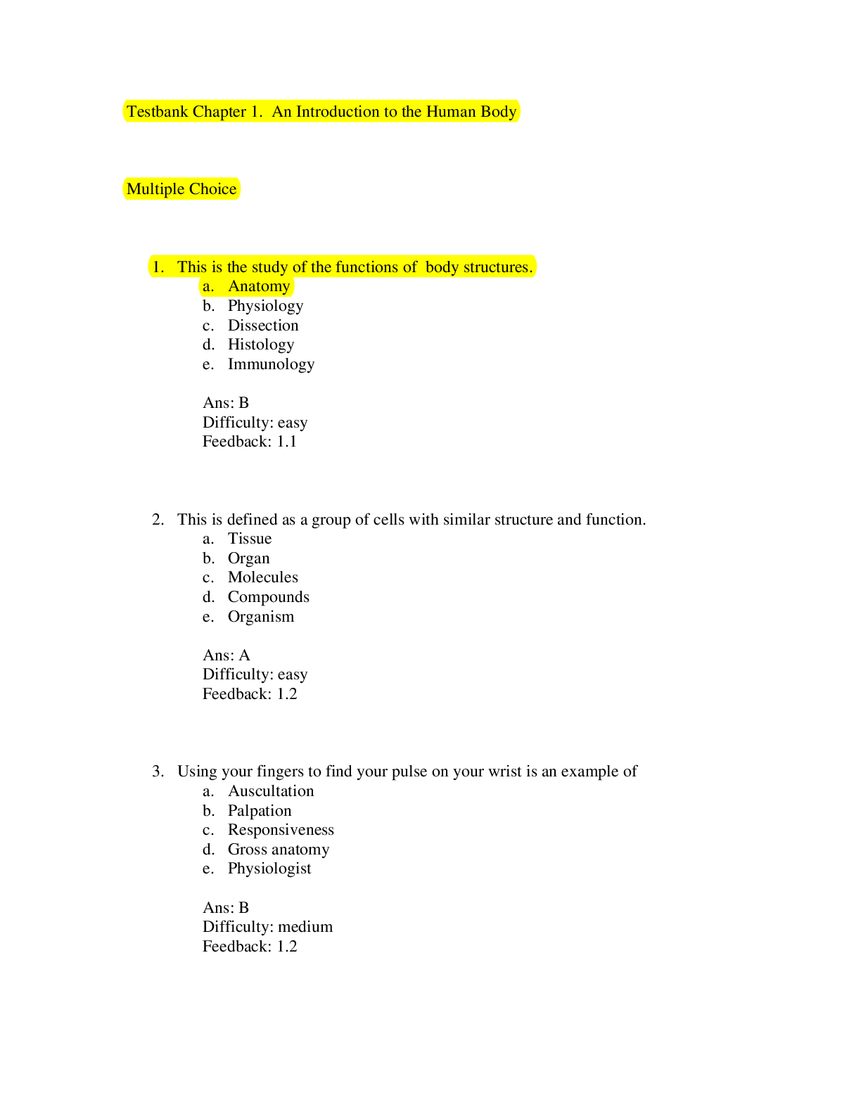
Reviews( 0 )
Document information
Connected school, study & course
About the document
Uploaded On
Mar 03, 2022
Number of pages
747
Written in

Additional information
This document has been written for:
Uploaded
Mar 03, 2022
Downloads
0
Views
38
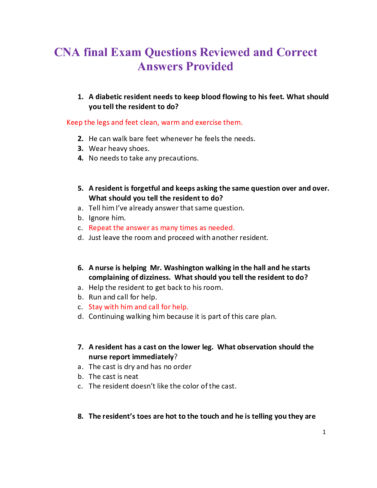
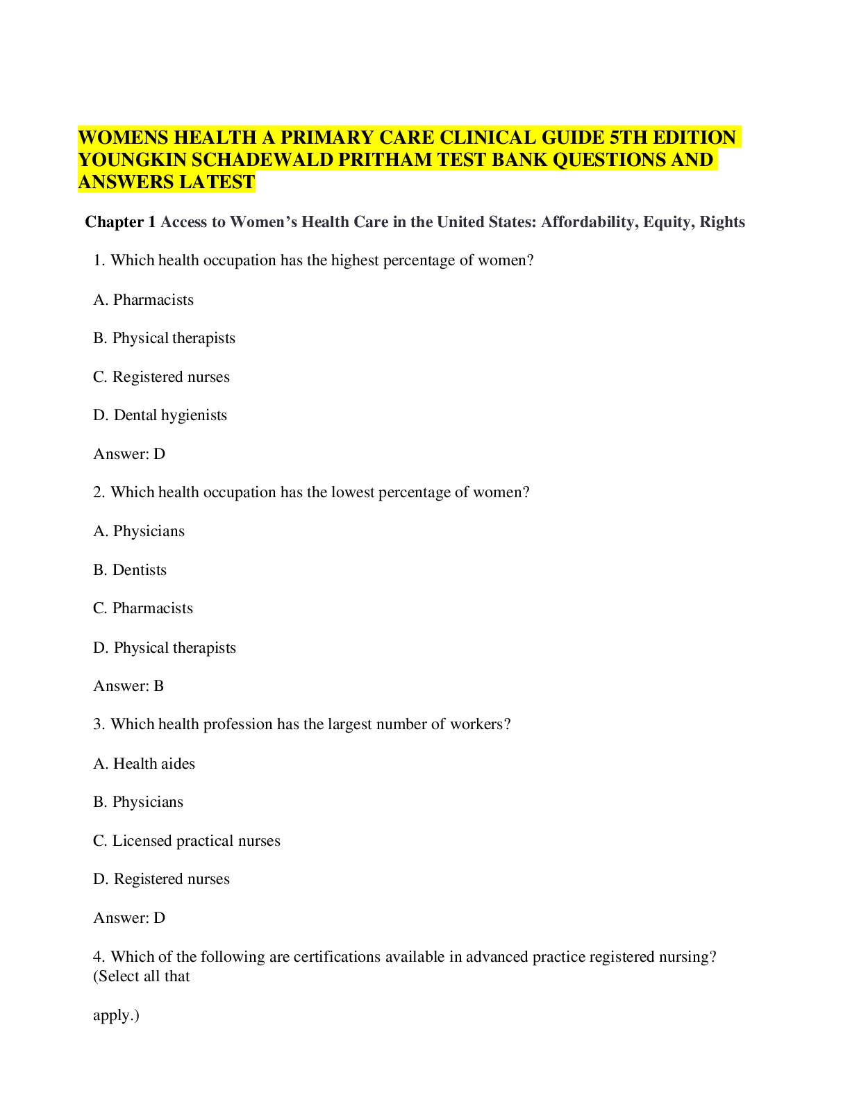


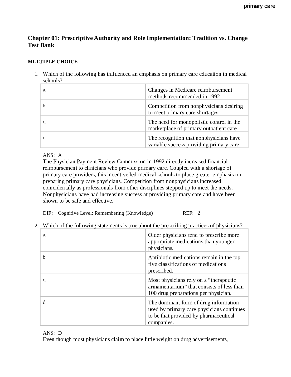


.png)