Health Care > EXAM > COC 2020 Final Exam Study Questions - Set 6 (Complete Solutions, Answered) (All)
COC 2020 Final Exam Study Questions - Set 6 (Complete Solutions, Answered)
Document Content and Description Below
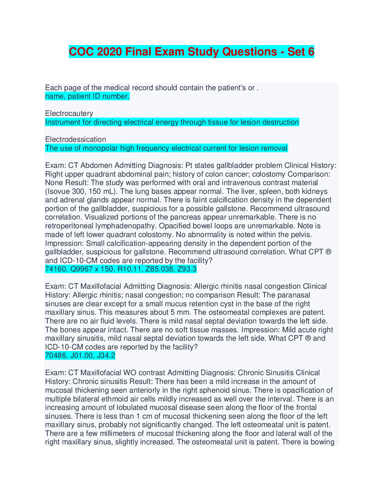
COC 2020 Final Exam Study Questions - Set 6 (Complete Solutions, Answered) Each page of the medical record should contain the patient's or . name, patient ID number. Electrocautery Instrument for di... recting electrical energy through tissue for lesion destruction Electrodessication The use of monopolar high frequency electrical current for lesion removal Exam: CT Abdomen Admitting Diagnosis: Pt states gallbladder problem Clinical History: Right upper quadrant abdominal pain; history of colon cancer; colostomy Comparison: None Result: The study was performed with oral and intravenous contrast material (Isovue 300, 150 mL). The lung bases appear normal. The liver, spleen, both kidneys and adrenal glands appear normal. There is faint calcification density in the dependent portion of the gallbladder, suspicious for a possible gallstone. Recommend ultrasound correlation. Visualized portions of the pancreas appear unremarkable. There is no retroperitoneal lymphadenopathy. Opacified bowel loops are unremarkable. Note is made of left lower quadrant colostomy. No abnormality is noted within the pelvis. Impression: Small calcification-appearing density in the dependent portion of the gallbladder, suspicious for gallstone. Recommend ultrasound correlation. What CPT ® and ICD-10-CM codes are reported by the facility? 74160, Q9967 x 150, R10.11, Z85.038, Z93.3 Exam: CT Maxillofacial Admitting Diagnosis: Allergic rhinitis nasal congestion Clinical History: Allergic rhinitis; nasal congestion; no comparison Result: The paranasal sinuses are clear except for a small mucus retention cyst in the base of the right maxillary sinus. This measures about 5 mm. The osteomeatal complexes are patent. There are no air fluid levels. There is mild nasal septal deviation towards the left side. The bones appear intact. There are no soft tissue masses. Impression: Mild acute right maxillary sinusitis, mild nasal septal deviation towards the left side. What CPT ® and ICD-10-CM codes are reported by the facility? 70486, J01.00, J34.2 Exam: CT Maxillofacial WO contrast Admitting Diagnosis: Chronic Sinusitis Clinical History: Chronic sinusitis Result: There has been a mild increase in the amount of mucosal thickening seen anteriorly in the right sphenoid sinus. There is opacification of multiple bilateral ethmoid air cells mildly increased as well over the interval. There is an increasing amount of lobulated mucosal disease seen along the floor of the frontal sinuses. There is less than 1 cm of mucosal thickening seen along the floor of the left maxillary sinus, probably not significantly changed. The left osteomeatal unit is patent. There are a few millimeters of mucosal thickening along the floor and lateral wall of the right maxillary sinus, slightly increased. The osteomeatal unit is patent. There is bowing of the nasal septum to the right. Impression: Sinusitis, mildly increased What CPT ® and ICD-10-CM codes are reported by the facility? 70486, J32.9 Exam: CT of the chest with intravenous contrast (Omnipaque 350, 150 mL) Admitting Diagnosis: Difficulty swallowing Result: The transverse aorta is prominent in size and measures approximately 3 centimeters in transverse dimension. There are a couple of small right peritracheal lymph nodes measuring a centimeter or less in size. No infiltrate or evidence of effusion is seen. There is evidence of old granulomatous exposure. Impression: Slight dilatation of the thoracic aorta. What CPT ®, HCPCS and ICD-10-CM codes are reported for the facility? 71260, Q9967 x 150, R13.10 Exam: MRI brain W/O and W contrast Admitting Diagnosis: Left facial weakness/Bell's palsy Clinical History: Comparison. Left facial weakness in a patient w/hx of Bell's Palsy and MS Result: MRI of the brain was obtained at 1.5 Tesla. Axial T1 weighted pre- and post-Gadolinium (1.6 mL) (iron-based contrast), T2 weighted FLAIR, and diffusion images were obtained. Sagittal T1 weighted and high resolution coronal and axial pre- and post-Gadolinium images of the internal auditory canals were obtained. The old study was available for comparison. The ventricles and sulci are within normal limits. There is no evidence of mass effect or midline shift. There are no extra-axial fluid collections. There are multiple punctuate areas of abnormal increased T2 weighted signal in the periventricular white matter and in the subcortical white matter of the centrum semiovale. Many of the foci are elongated and oriented toward the ventricles. Since the prior exam, there has been an increase in the number of hyperintense plaques. With contrast enhancement, none of the plaques appear to enhance at this time. The enhancement seen previously has resolved. The pattern and distribution is most characteristic for MS. The pituitary gland and cerebellum are unremarkable. There are multiple punctuate areas of hyperintense signal in the brain stem and brachium pontis. These do not enhance with gadolinium. There is no edema or mass effect from the lesions. There is a 7 mm focus of increased signal at the left CP angle. No abnormal signal, enhancement of discrete mass lesion is appreciated within the internal auditory canals. This would correlate with the patient's symptoms of a left facial palsy. Impression: Multiple hyperintense lesions predominately in the periventricular white matter with characteristic pattern for MS. None of the plaques currently enhance. There has been a fairly significant increase in the number of lesions since the last exam. Multiple lesions were also seen in the brain stem and the brachium pontis. Specifically, there is a 7 mm focus in the left CP angle which is probably the cause of the patient's left facial palsy. No discrete abnormality was seen in the internal auditory canals. What CPT ® and ICD-10-CM codes are reported by the facility? 70553, A9579 x 2, G51.0, G93.9, G35 Exam: MRI Lumbar Spine W/O Contrast Admitting Diagnosis: MRI-L spine, low back pain Clinical History: No comparison; low back pain which radiates to the right lower extremity Result: Vertebral body height is normal. The bone marrow signal appears normal. The visualized portions of the spinal cord appear normal. The foramina are patent in the sagittal images. There is normal disc space height and hydration. The intervertebral discs appear normal. There is specifically no evidence of disc herniation, spinal or foraminal stenosis. Impression: Negative What CPT ® and ICD-10-CM codes are reported for the facility? 72148, M54.5 Exam: MRI right upper extremity joint W/O contrast Admitting Diagnosis: RT shoulder pain Indication: Right shoulder pain and numbness; no comparison study Result: There are a few small subchondral cysts in the humeral head adjacent to the greater tuberosity. These lie subjacent to the supraspinatus insertion. No other marrow space abnormality is identified. There are mild degenerative changes in the right acromioclavicular joint with small osseocartilaginous spur inferiorly. Undersurface of the acromion is flat. No joint effusion is seen. No abnormal periarticular fluid collections are identified. Glenoid labrum is intact. Long head of the biceps is intact and is normally positioned. Supraspinatus tendon shows no evidence of full thickness tear and there is no evidence of fluid in the subacromial/ subdeltoid bursa. I cannot exclude a small partial thickness tear along the distal most aspect of the right supraspinatus tendon in its inferior border. Subscapularis and infraspinatus tendons are intact. Impression: Minor localized degenerative diseases of right shoulder. Possible small partial thickness tear along inferior border of right supraspinatus tendon at tendon insertion, otherwise negative. What CPT ® and ICD-10-CM codes are reported by the facility? 73221, M19.011 Examples of procedures considered "inpatient only" include: Surgical thoracoscopy, laminectomies, vertebral corpectomy, enterostomies, risky invasive procedures Facial Controls facial muscles around the eyes, forehead, external ear, and mouth; sensa-tion of taste; and certain salivary and lacrimal (tear) glands Facilities report Medicare outpatient clinic (E/M) visits with code(s): G0463 Rationale: Clinic E/M visits (99201-99215) are reported to Medicare by the facility with HCPCS Level II code G0463. Field: Geometric area defined by a collimator at the skin surface Flow cytometry is performed for DNA analysis. What CPT ® code is reported? 88182 Food moves through the digestive tract by what means? Peristalsis For Medicare how is each claim paid for outpatient facility reimbursement? Each claim is paid based on the determined interim outpatient reimbursement rate. Rationale: During the year-end cost report settlement, the prior year's entire outpatient claims are analyzed via a computer system. This method is to determine an interim reimbursement rate on which to pay the following year's Medicare claims. For OPPS, critical care is paid at two levels. What is the distinguishing factor for payment? One level for critical care services, another level when trauma activation occurs in addition to critical care For surgical procedures involving a primary surgeon and an assistant surgeon, who is the person responsible for the information in the procedural note? Primary surgeon Rationale: For surgical procedures with more than one surgeon, the primary surgeon is responsible for the procedural note. A resident, intern, or assistant can dictate the note, but the primary surgeon must indicate agreement by reading and signing it. For the UB-04 Form, which provider type qualifier is used to report the rendering provider? 82 Fractionation: Division of total planned dose into number of smaller doses given over time Friends brought a young male with type 1 diabetes to the emergency department, in a comatose state. He was admitted with ketoacidosis and was resuscitated with saline hydration via insulin drip. After regaining consciousness, the patient reported that the morning of admission he was experiencing nausea and vomiting and decided not to take his insulin because he had not eaten. He was treated with intravenous hydration and insulin drip. By the following morning, his laboratory work was within normal range and he was experiencing no symptoms. What ICD-10-CM code(s) are reported? E10.11 Ganglion cyst: are noncancerous lumps that most commonly develop along the tendons or joints of your wrists or hands. They also may occur in the ankles and feet. Ganglion cysts are typically round or oval and are filled with a jellylike fluid. Glossopharyngeal Responsible for swallowing, secretion of saliva, sensations of the throat and taste sensations for the back of the tongue Hospitals are required to provide a list of standard charges on the internet and update the list at least annually. True [Show More]
Last updated: 1 week ago
Preview 1 out of 12 pages
Reviews( 0 )
Document information
Connected school, study & course
About the document
Uploaded On
Apr 24, 2024
Number of pages
12
Written in

Additional information
This document has been written for:
Uploaded
Apr 24, 2024
Downloads
0
Views
4