Biology > STUDY GUIDE > NSG 331 Final Exam study guide 2019/2020 – Marian University | NSG331 Final Exam study guide 2019/ (All)
NSG 331 Final Exam study guide 2019/2020 – Marian University | NSG331 Final Exam study guide 2019/2020
Document Content and Description Below
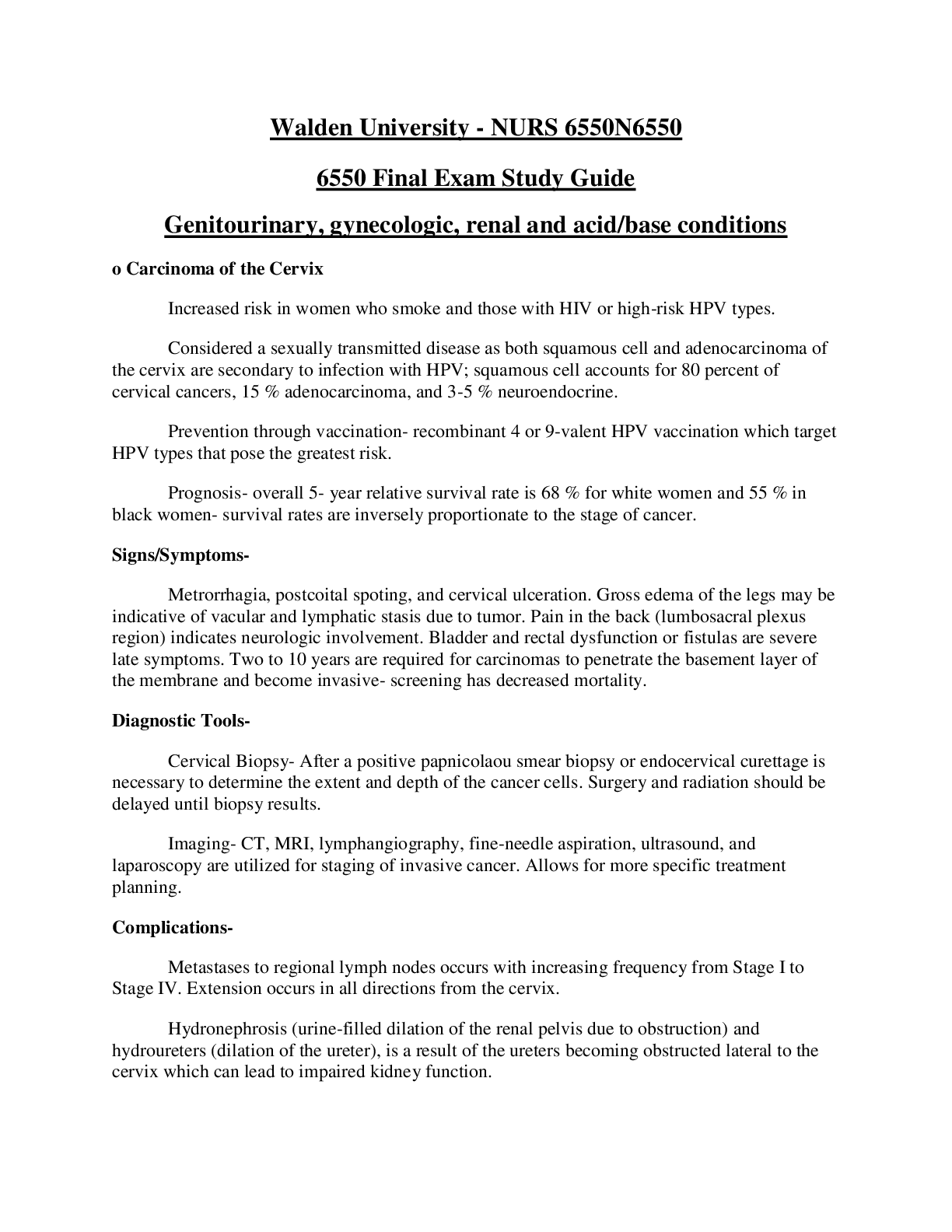
NSG 331 Final Exam study guide 2019/2020 – Marian University NSG 331 Final Exam Test Plan Questions evenly distributed between modules. Dosage: 2 questions Review the case studies done by cla... ssmates before the final exam Topics Disorders Head and neck cancer Head and Neck Cancer • Incidence: o 2-3% of all malignancies o Men>women o May involve • Nasal cavity • Para-nasal sinuses • Nasopharynx • Oropharynx • Larynx • Oral cavity • Salivary glands o Most people have advance disease at time of diagnosis • Risk Factors: o Cigarette smoking (85% of cancers) o Alcohol o Occupational exposure to asbestos, wood dust, mustard gas, petroleum products o Chronic laryngitis o Voice abuse o Genetics o HPV infection o Poor oral hygiene • Manifestations o Early – vary with location of the tumor • Oral cavity – white (leukoplakia)/red (erythroplakia) patch in mouth, ulcer that does not heal, change in the fit of dentures • Lump in throat, change in quality of voice • Laryngeal - Hoarseness that lasts for more than 2 weeks • Sore throat (unilateral), otalgia (ear pain), swelling or lumps in the neck • Interprofessional Care o Diagnostic Assessment • Hx & Physical exam • Indirect pharyngoscopy and laryngoscopy • Endoscopy • Biopsy • Chest x-ray • Barium swallow • CT / MRI / PET scan o Management • Surgery • Vocal cord stripping – removal of outer layer of tissue on vocal cords (early stage) – does not change speech • Laser surgery – inserted to vaporize / remove tumor • Cordectomy – part/all vocal cords are removed (changes speech – hoarse voice(partial); loss of voice (full removal)) • Partial or total laryngectomy – removal (full or partial) of larynx • Pharyngectomy – part/all of throat is removed • Lymph node removal with neck dissection • Tracheostomy – stoma / alternate pathway • Reconstructive procedures • Radiation therapy • Chemotherapy • Targeted therapy • Physical therapy • Occupational therapy • Speech therapy Laryngectomy Laryngeal Cancer • Manifestations o Hoarseness o Pain in throat o Dysphagia o Neck masses • Diagnosis o Visual exam of larynx o Biopsy o CT/MRI o Chest x-ray o Barium swallow study • Treatment o Early: partial laryngectomy, chemo,radiation, temp. trach., soft voice o Advanced cancers: total laryngectomy, radical neck, permanent trach. Stoma. No voice, unable to smell, decr. taste Nursing management for laryngectomy • Watch for complications: o Airway obstruction o Hemorrhage – monitor VS o Carotid artery rupture o Fistula formation • Elevate HOB – decreases edema and reduces pressure on esophagus o NO FLAT BEDS • Flex neck forward • Trach/stoma care • Wound assessment/care • NG feedings – d/t location of surgery and complications of chemo and radiation Nursing diagnosis for laryngectomy o Risk for Aspiration o Ineffective Airway Clearance o Risk for Impaired Gas Exchange o Impaired Nutrition: Less than Body Requirements o Risk for Infection Artificial Larynx • Discharge teaching o Stoma care o Self tube feedings o Fluids o Humidification o Suction prn o No swimming o Shower with guard o Carry ID o Cover stoma when outside o Continue speech therapy o No smoking Video of speech after laryngectomy https://www.youtube.com/watch?v=R4azcU6i2IE Pneumonia Pneumonia Etiology • Most likely to occur when defense become incompetence or overwhelmed by the virulence or quantity of infectious agents • Organisms that cause pneumonia reach the lung by three ways: o Aspirations of normal flora from the nasopharynx or oropharynx. Many organisms that cause pneumonia are normal inhabitants of the pharynx in healthy adults o Inhalation of microbes present in the air. Examples include Mycoplasma pneumonia and fungal pneumonias o Hematogenous spread from a primary infection elsewhere in the body. Examples are streptococci and staphylococcus aureus from infective endocarditis. Pathophysiology of Pneumonia • Slightly different depending on organisms, but they all cause inflammatory response • Consolidation occurs when the normally air-filled alveoli become filled with fluid and debris • Mucus production increases Clinical Manifestations • Cough, fever, chills, dyspnea, tachypnea, and pleuritic chest pain • Cough may or may not be productive • Sputum: green, yellow, or bloody • Older adult may not have classic symptoms: o Confusion or stupor o Hypothermia rather than fever o Nonspecific manifestations: diaphoresis, anorexia, fatigue, myalgias, and HA. o Fine or coarse crackles o If consolidation occurs: ♣ Bronchial breath sounds ♣ Egophony (a change in the sounds of the voice) ♣ Increases fremitus o Patient with pleural effusion may exhibit dullness to percussion over the affected areas Classifications of Pneumonia • Causative Agents o Bacteria o Viruses o Mycoplasma organisms o Fungi o Parasites o Some chemicals • Clinical Classification o Community-acquired pneumonia (CAP) ♣ Acute infection of the lung occurring in patients who have not been hospitalized or resided in a long-term care facility within 14 days of the onset of symptoms ♣ Treatment: • At home or hospitalization depending on severity • Empiric antibiotic therapy – the initiation of treatment before definitive diagnosis or causative agent is confirmed. • Should be started as soon as CAP is suspected o Hospital-acquired pneumonia (HAP) also known as nosocomial pneumonia ♣ Ex. Ventilator-associated pneumonia (VAP) – a type of HAP, refers to pneumonia that occurs more than 48 hours after endotracheal intubation ♣ Treatment • Initiated based on risk factors, early verses late onset, and probable organism • Antibiotic therapy is adjusted after sputum culture results are back if needed • HAP and VAP are associated with longer hospital stays, increased associated costs, sicker patients, and increased risk of morbidity and mortality ♣ Major problems in treatment is multi-drug resistance organisms • Ex. Primary culprits include methicillin-resistant staphylococcus aureus and gram-negative bacilli • Other Types o Aspiration Pneumonia ♣ Conditions that increase risk • Decreased LOC (decreases gag and cough reflexes) • Difficulty swallowing • Insertion of a NG tube with or without feeding ♣ Typically more than one organism is identified on sputum culture, including aerobes and anaerobes ♣ Usually a bacterial infection ♣ Aspiration of gastric acid content causes chemical (noninfectious) pneumonitis, which may not require antibiotic therapy but secondary bacterial infections can occur 48 to 72 hours later. o Necrotizing Pneumonia ♣ Rare complication of bacterial lung infection ♣ Characterized by liquefaction and sometimes cavitation of lung tissue ♣ Causative organisms include: staphylococcus, klebsiella, and streptococcus ♣ Lung abscesses typically occur ♣ S&S: immediate respiratory insufficiency and/or failure, leukopenia, and bleeding in airways ♣ Treatment: long term antibiotic therapy and possible surgery o Opportunistic Pneumonia ♣ Inflammation and infection of the lower respiratory tract in immunocompromised patients ♣ At risk for bacterial and viral pneumonia ♣ The person may also develop an infection from micro-organisms that do not normally cause disease, such as pneumocystis jiroveci and cytomegalovirus • P. jiroveci rarely occurs in the healthy individual but is the most common from of pneumonia in people with HIV • Slow and subtle onset with symptoms of fever, tachycardia, dyspnea, nonproductive cough and hypoxemia • Chest xray shows diffuse bilateral infiltrates • Treatment consist of Bactrim, Septra either IV or orally depending on severity • CMV, a herpes virus, can cause viral pneumonia • Most are asymptomatic or mild, but severe can occur in people with impaired immune response. • Most common life threatening infectious complications after hematopoietic stem cell transplant • Treatment: anti-viral medications and high dose immunoglobulin Types of Pneumonia • Pneumocystis jiroveci pneumonia (PJP) Complications of Pneumonia • Atelectasis • Pleurisy • Pleural effusion • Bacteremia • Pneumothorax • Meningitis • Acute Respiratory Failure • Sepsis/Septic Shock • Lung abscess – not a common complication Diagnostic Studies • Chest x-ray • Sputum specimen for culture and gram stain • Blood cultures are done for the severely ill patient • ABGs • C-reactive proteins (CRP) and pro-calcitonin are being explored as possible ways to help physicians distinguish between pneumonia from cardiac and respiratory failure Interprofessional Care for Pneumonia • Pneumococcal vaccine • Prompt treatment with antibiotics is essential • Supportive care • No definitive treatment for majority of viral pneumonias • Antivirals for influenza pneumonia • Drug Therapy • Nutrition Nursing Assessment • Subjective Data o Past health history: lung cancer, COPD, diabetes, malnutrition, chronic debilitating disease o Use of antibiotics, corticosteroids, chemotherapy, or immunosuppressants o Recent abdominal or thoracic surgery o Recent intubation o Tube feedings o Smoking o Alcoholism o Respiratory infections o Nutritional intake o Activity o Dyspnea o Cough o Pain • Objective Data o Vital Signs o Oxygen saturation o Fever o Restlessness or lethargy o Splinting affected area o Tachypnea o Asymmetric chest movements o Use of accessory muscles o Crackles o Friction rub o Dullness on percussion o Increased tactile fremitus o Sputum amount and color o Tachycardia o Changes in mental status Nursing Management • Nursing diagnosis o Impaired gas exchange o Ineffective breathing pattern o Acute pain (chest) o Activity intolerance • Outcomes o Clear breath sounds o Normal breathing patterns o No signs of hypoxia o Normal chest x-ray o Normal WBC count o Absence of complications related to pneumonia Nursing Implementation • Health Promotion • Prevent pneumonia in at risk patients • Acute Care • Acute Intervention Review Questions A 56-year-old normally healthy patient at the clinic is diagnosed with bacterial community-acquired pneumonia. Before treatment is prescribed, the nurse asks the patient about an allergy to a. amoxicillin b. erythromycin c. sulfonamides d. cephalosporins The nurse is caring for a patient with pneumonia. If a pleural effusion is developing, the nurse would expect which finding? a. Barrel-shaped chest b. Paradoxical respirations c. Hyperresonance on percussion Localized decreased breath sounds Tuberculosis Tuberculosis • Infectious disease caused by Mycobacterium tuberculosis • Lungs most commonly infected • but any organ can be infected • 1/3 of world’s population has TB • Leading cause of death in patients with HIV/AIDs • Prevalence is decreasing in the United States Risk Factors for TB • Homeless • Residents of inner-city neighborhoods • Foreign-born persons • Living or working in institutions (includes health care workers) • IV injecting drug users • Poverty, poor access to health care • Immunosuppression Multi-drug resistant tuberculosis (MDR-TB) • Resistance to 2 of the most potent first-line anti-TB drugs • Extensively drug-resistant TB (XDR-TB) resistant to any fluoroquinolone plus any injectable antibiotic • Several causes for resistance occur o Incorrect prescribing o Lack of case management o Nonadherence Etiology and Pathophysiology • Spread via airborne particles • Can be suspended in air for minutes to hours • Transmission requires close, frequent, or prolonged exposure • NOT spread by touching, sharing food utensils, kissing, or other physical contact • Factors that influence the likelihood of transmission • number of organisms expelled into the air • concentration of organisms • length of time of exposure • immune system of the exposed person • https://www.youtube.com/watch?v=yR51KVF4OX0 • Once inhaled, particles lodge in bronchioles and alveoli • Local inflammatory reaction occurs • Ghon lesion or focus – represents a calcified TB granuloma (the hallmark of a primary TB infection) • Infection walled off and further spread stopped • The formation of a granuloma is a defensive mechanism aimed at walling off the infection and preventing further spread • Only 5% to 10% will develop active TB • Aerophilic (oxygen-loving) – causes affinity for lungs • Infection can spread via lymphatics and grow in other organs as well • Cerebral cortex • Spine • Epiphyses of the bone • Adrenal glands Classification • Classes - TABLE 27-8 o 0 = No TB exposure o 1 = Exposure, no infection o 2 = Latent TB, no disease o 3 = TB, clinically active o 4 = TB, not clinically active o 5 = TB suspected • Primary infection o When bacteria are inhaled and initiate an inflammatory reaction o most people’s immune system will keep them from actually developing the disease • Latent TB infection (LTBI) o Infected but no active disease o positive skin test but are asymptomatic o cannot transmit to others but can development active TB o immunosuppression, DM, poor nutrition, aging, pregnancy, stress, and chronic disease can precipitated the reactivation of LTBI • Active TB disease o Primary TB - if it develops within the first two years o Reactivation TB (post-primary) - TB disease occuring 2 years after the initial infection o if the disease is laryngeal or pulmonary, the patient is considered infectious and can transmit the disease to others. Clinical Manifestations • LTBI – asymptomatic • Pulmonary TB o Takes 2-3 weeks to develop symptoms o Initial dry cough that becomes productive o Constitutional symptoms (fatigue, malaise, anorexia, weight loss, low-grade fever, night sweats) o Dyspnea and hemoptysis late symptoms • Can also present more acutely o High fever o Chills, generalized flu-like symptoms o Pleuritic pain o Productive cough o Crackles and/or adventitious breath sounds • Extrapulmonary TB manifestations dependent on organs infected o ex. renal TB can cause dysuria and hematuria o ex. bone and joint TB may cause severe pain o ex. TB meningitis causes HA, vomiting, and lymphadenopathy • Immunosuppressed people and older adults are less likely to have fever and other signs of an infection o Carefully investigate respiratory problems in HIV patients • Rule out opportunistic diseases o A change in cognitive function may be the only initial sign of TB in an older person Complications • Appropriately treated pulmonary TB heals without complications, except for scarring and residual cavitation within the lung • Miliary TB o Large numbers of organisms spread via the bloodstream to distant organs o Fatal if untreated o Manifestations progress slowly and vary depending on which organs are infected o Fever, cough, and lymphadenopathy occur o Can include hepatomegaly and splenomegaly • Pleural TB - specific type of extrapulmonary TB o Chest pain, fever, cough, and a unilateral pleural effusion are common o Pleural effusion • Bacteria in pleural space cause inflammation. • Pleural exudates of protein-rich fluid o Empyema • Large numbers of tubercular organisms in pleural space o Diagnosis is confirmed by AFB cultures and pleural biopsy • TB pneumonia o Large amounts of bacilli discharged from granulomas into lung or lymph nodes o Manifests as bacterial pneumonia • Other organ development o Spinal destruction o Bacterial meningitis - affects central nervous system o Peritonitis Diagnostic Studies • Tuberculin skin test (TST) o AKA: Mantoux test o Uses purified protein derivative (PPD) injected intradermally o Assess for induration in 48 – 72 hours o Presence of induration (not redness) at injection site indicates development of antibodies secondary to exposure to TB • Tuberculin skin test (TST) o Positive if ≥15 mm induration in low-risk individuals o Response ↓ in immunocompromised patients • Reactions ≥5 mm considered positive o two step skin test is used to prevent misinterpretation • recommended for health care workers and for individuals who have a decreased response to allergens • Interferon-γ gamma release assays (IGRAs) o Blood tests that detects T-cells in response to Mycobacterium tuberculosis o Includes QuantiFERON-TB and T-SPOT.TB tests o Rapid results - few hours o Several advantages over TST but more expensive o one patient visit o not subject to reader bias o have no booster phenomenon o are not affected by priot bacillus Calmette-Guerin (BCG) vaccination o Chest x-ray o Cannot make diagnosis solely on x-ray o because other diseases, such as sarcoidosis, can mimic the appearance of TB o May appear normal in a patient with TB o Upper lobe infiltrates, cavitary infiltrates, lymph node involvement, and pleural and/or pericardial effusion suggest TB o Bacteriologic studies o Required for diagnosis o Consecutive sputum samples obtained on 3 different days o Stained sputum smears examined for AFB o Culture results can take up to 8 weeks o Can also examine samples from other suspected TB sites o gastric washings o CSF o fluid from effusion or abscess - - - -- - - - - - - - - - - - - - - - - - - - - - - - - - Glipizide (Glucotrol) • Sulfonylureas • Stimulate release of insulin from pancreatic islets • Decrease glycogenolysis and gluconeogenesis • Enhance cellular sensitivity to insulin • Side effects: o Weight gain, ***HYPOGLYCEMIA Metformin (Glucophage) • Biguanides • Decreases rate of hepatic glucose production • Augments glucose uptake by tissues, especially muscles • Most effective first line treatment for type 2 DM • Side effects: o Diarrhea, lactic acidosis • Nursing considerations: o MUST BE HELD 1-2 DAYS BEFORE IV CONTRAST MEDIA GIVEN AND FOR 48 HOURS AFTER • Drug Alert: o Do not use in patients with kidney disease, liver disease, or heart failure. Lactic acidosis is rare complication of metformin accumulation. o IV contrast media that contain iodine pose a risk of acute kidney injury, which could exacerbate metformin-induced lactic acidosis o To reduce risk of kidney injury, discontinue metformin a day or two before the procedure o May be resumes 48 hours after the procedure, assuming kidney function is normal o Do not use in people who drink excessive amounts of alcohol Take with food to minimize GI side effectsDiagnostics Urinalysis: pH, specific gravity, protein, glucose, nitrites, leukocyte esterase • Urinalysis PP 1024-1031 o First morning void (more concentrated/likely to contain abnormal constituents) o Examine urine within 1 hour – otherwise keep refrigerated ♣ Bacteria multiply ♣ RBC hemolyze ♣ Casts disintegrate ♣ Urine becomes alkaline (d/t urea splitting bacteria) • Creatinine clearance – 70-135 o Collect 24-hour urine specimen ♣ First specimen in morning discarded then every void collected o Must be refrigerated, iced, or some kind of preservative o Creatinine clearance closely approximates GFR o Also, good to do a blood serum creatinine test during that time period o Most accurate indicator of renal function o Measure of amount of active muscle tissue – more muscle = higher value o After age 40 – decreases every year by 1 mL/min/year • Normal urinalysis – MEMORIZE THESE! o Clear, amber o pH: acidic (4.0-8.0) o Specific gravity: 1.03-1.030 o BUN: 8-20 o Creatinine: 0.5-1.5 o GFR-Glomerular Filtration Rate: >60 o Protein: random protein (dipstick)- 0-trace, 24-hour protein (quantitative)- <150 mg/day o Glucose: none o Nitrites: none, presence indicates bacteriuria o Leukocyte: 0-5/hpf o Esterase: none, it is an enzyme present in WBCs, indicating pyuria • Dipstick urinalysis o Identify presence of ♣ Nitrites – indicates bacteria ♣ WBC’s ♣ Leukocyte esterase - enzyme present in WBC’s that indicate pyuria – pus in urine Hemoglobin A1c – PP 1115-1118, 1659 • indicates the amount of glucose linked to hemoglobin, also called glycosylated hemoglobin • Assesses long term glycemic control during the previous 3 months • Goal is below 7% • Nursing responsibility is to inform the patient that fasting is not necessary and that blood sample will be done Cystoscopy – pp1025-1030 • Inspects interior of bladder with a tubular lighted scope • UseS: insert ureteral catheters, remove calculi, obtain biopsies of bladder lesions, treat bleeding lesions • Lithotomy position is used • Procedure may be done using local or general anesthesia, depending on patient’s needs and condition • Complications include urinary retention, urinary tract hemorrhage, bladder infection and perforation of bladder • Nursing responsibility o Before: force fluids or give IV fluids if general anesthesia is to be used, ensure consent is signed, explain procedure, give preoperative medication. o After: explain that burning on urination, pink-tinged urine, and urinary frequency are expected effects. • Observe for bright red bleeding, which is not normal. Assist with ambulation because orthostatic hypotension may occur. Offer warm sitz baths, heat, & mild analgesics to relieve discomfort. • Endourologic procedures for stones • Flexible ureteroscope inserted to remove stones from renal pelvis/UUT • Endoscopic procedure – inspects interior side of the bladder – inserted through urethra • Can remove calculi, obtain biopsy specimens, treat bleeding lesions • Fluids usually given before the procedure – often give meds before as well • Burning during urination can occur after the procedure, urine can have a pink tinge, or inc frequency • Also, could have orthostatic hypotension o • Trousseau’s sign – p 284 • Positive Trousseau’s (B & C) or Chvostek’s (A) sign = Tetany o • Carpal spasm induced by inflating BP cuff above systolic BP for a few min • Tests for HYPOCALCEMIA, also for hypomagnesemia Chvostek’s sign – P 284 • Rxn of facial muscle to light touch to facial nerve in front of the ear – facial muscles contract • Tests for HYPOCALCEMIA, also for hypomagnesemia CBC: WBC, Hgb, Htc, platelets, Red blood cell (RBC) – PP 599-600 • WBC: 4000-1100, elevations aver 1100 indicate infection, inflammation, tissue injury, death and malignancies, count less than 4000 is associated w/ bone marrow depression, severe or chronic illness • Hgb: female 11.7-15.5 g/dL, male 13.2-17.3 g/dL, measurement of gas-carrying capacity of RBC, reduced in cases of anemia, hemorrhage, and hemodilution (fluid excess), increased in polycythemia, hemoconcentration (fluid deficit/dehydration) • Hct: female 35-47%, male 39-50 %, measurement of packed cell volume of RBCs expressed as a percentage of the total blood volume • Platelets: 150,000-400,000 (150-400x10^9) • RBC: female 3.8-5.1x10^6, male 4.3-5.7x10^6 Prostate needle biopsy – PP1275-1281 • Needed to confirm the diagnosis of prostate cancer • Typically done using a transrectal approach • US probe enables urologist to visualize abnormalities where biopsy needles are to be placed into the prostate • Suspicious area is located, biopsy needles inserted through rectum wall into prostate to obtain tissue samples Fasting blood glucose pp1115-1118 • 70-99 mg/dL • Measures circulating glucose levels • Before: patient should fast 8-12 hours, water intake is permitted • Many medications may influence results Postprandial blood glucose • Variant of dumping syndrome o Uncontrolled gastric emptying of a bolus of fluid high in carbs into the small intestine o Results in hyperglycemia and release of excess insulin which leads to reflex hypoglycemia o 2 Hours after eating – symptoms similar to any hypoglycemic reaction Computed Tomography (CT)- p 603 • Noninvasive radiologic examination using computer assisted x-ray • Contrast medium often is used in abdominal studies of liver or spleen • Before: investigate iodine sensitivity if contrast medium is used (shellfish allergy), IV and or oral contrast may be given prior to procedure depending on area being studied. o Patient may need to be NPO 4 hours prior to study, assess renal function before test. • After: encourage patient to drink fluids to avoid renal problems with contrast, if ordered. Blood urea nitrogen (BUN) – p 1026 • 6-20 mg/dL, 2.1-7.1 mmol/L • Used to detect renal problems • Increased BUN indicates impaired kidney function • Concentration of urea in the blood is regulated by rate at which kidney excretes urea • Non-renal factors may increase BUN o rapid cell destruction from infections o Fever o GI bleeding, trauma o Athletic activity o Excessive muscle breakdown. • Explain test and watch for post puncture bleeding. Creatinine – P 1026 • 0.6-1.3 mg/Dl • More reliable than BUN as a determinant of renal function • Increased levels indicate impaired renal function • End product of muscle and protein metabolism and is released at a constant rate • Explain test and watch for post puncture bleeding. Prothrombin time (PT) / International normalized ratio (INR) • Prothrombin Time – P 601: 11-16 sec, assessment of extrinsic coagulation • INR – P 601: 2-3 is desired therapeutic level with warfarin • Blood Lab Tests o Diagnostic Test o Normal Range o Activated clotting time (ACT) o 70-120 sec o Activated partial thromboplastin time (aPTT) o 25-35 sec o International normalized ratio (INR) o 2-3 o Hemoglobin o F: 11.7-15.5 g/dL; M: 13.2-17.3 g/dL o Hematocrit o F: 35-47%; M: 39-50% o Platelet count o 150,000-400,000 µL o D-dimer o <250 mcg/L o Fibrin monomer complex o <6.1 mg/L • Potassium 3.5-5 mEq Sodium – 135-145 mEq Arterial Blood Gas Values (ABGs) • Normal: MEMORIZE THESE!!! o pH: 7.35-7.45 o pCO2: 35-45 o pO2: 80-100 o HCO3: 22-26 • Diagnose in six steps: o Evaluate pH o Analyze PaCO2 o Analyze HCO3 o Determine if CO2 or HCO3 matches the alteration o Decide if the body is attempting to compensate o Evaluate PaO2 = If abnormal – hypoxemia is present • Acid/Base Mnemonic – ROME o Respiratory - Opposite ♣ Alkalosis: Incr. pH, Decr. PaC02 ♣ Acidosis: Decr. pH, Incr. PaCO2 o Metabolic - Equal ♣ Alkalosis: Incr. pH, Incr. HCO3 ♣ Acidosis: Decr. pH, Decr. HCO3 • ABG’s with Compensation o Resp. Acidosis: Incr. HCO3 o Respiratory Alkalosis: Decr. HCO3 o Metabolic Acidosis: Decr. CO2 o Metabolic Alkalosis: Incr. CO2 o Compensatory change is always in the same direction as the pathologic (primary) change. ♣ Partial compensation - pH will not be wi/in normal range ♣ Complete compensation - pH will be back to normal Albumin – p 273 • 3.5-5.0 g/dL • If albumin level is low, likely to see edema in patient (oncotic pressure) • Indicates malnourishment – impairs ability to bind/distribute drugs, bind calcium Alanine aminotransferase (ALT) – p 852 • 10-40 U/L, elevated in liver damage and inflammation TNM classification • Anatomic extent of disease involvement – solid tumors (i.e. not leukemia) o T = tumor size and invasiveness o N = Presence/absence of spread to lymph nodes o M = Metastasis to distant organ sites o o Tis = Tumor in situ – no tendency to invade or metastasize o Done at the completion of diagnostic workup to guide effective treatment o Surgical staging – staging done at surgical excision, exploration, and/or lymph node sampling [Show More]
Last updated: 1 year ago
Preview 1 out of 171 pages
Instant download

Buy this document to get the full access instantly
Instant Download Access after purchase
Add to cartInstant download
Reviews( 0 )
Document information
Connected school, study & course
About the document
Uploaded On
Jul 04, 2020
Number of pages
171
Written in
Additional information
This document has been written for:
Uploaded
Jul 04, 2020
Downloads
0
Views
40

