*NURSING > EXAM > AGNP BOARD EXAM QUESTIONS Orthopedics Assessment.[DETAILED ANSWERS WELL EXPLAINED MOST TESTED PART I (All)
AGNP BOARD EXAM QUESTIONS Orthopedics Assessment.[DETAILED ANSWERS WELL EXPLAINED MOST TESTED PART IN AGNP BOARD EXAM QUESTIONS .GRADE A+
Document Content and Description Below
AGNP BOARD EXAM QUESTIONS Orthopedics Assessment.[DAGNP BOARD EXAM QUESTIONS Orthopedics Assessment (317 Questions). Question: The axioscapular group of muscles include which one of the following?... Supraspinatus Trapezius Correct Subscapularis Pectoralis major Explanation: The axioscapular group attaches the trunk to the scapula and includes the trapezius, rhomboids, serratus anterior, and levator scapulae. The scapulohumeral group of muscles extends from the scapula to the humerus and includes the muscles inserting directly on the humerus. This group includes the supraspinatus, infraspinatus, teres minor, and subscapularis. The axiohumeral muscle group attaches the trunk to the humerus and includes the pectoralis major and minor, and the latissimus dorsi. Question: An example of a cartilaginous joint would be the: vertebral bodies of the spine. Correct skull. shoulder. knee. Explanation: Vertebral bodies of the spine and the pubic symphysis of the pelvis are examples of cartilaginous joints. Examples of synovial joints include the shoulder, knee, hip, wrist, distal radioulnar, elbow, and carpals. The skull is an example of the fibrous joint. Question: The part of the ulna that forms the outer prominence of the elbow is referred to as the: olecranon bursa. olecranon fossa. olecranon process. Correct olecranon. Explanation: The part of the ulna that forms the outer prominence of the elbow is referred to as the olecranon process. This process fits into the fossa of the humerus when the arm is extended. Question: To assess muscle tone in the legs, support the patient's thigh with one hand, grasp the foot with the other, and: extend the patient's feet. flex and extend the patient's knee and ankle on each side. Correct have the patient try to lift the foot. feel for jerkiness in the calf. Explanation: To assess muscle tone in the legs, support the patient's thigh with one hand, grasp the foot with the other, and flex and extend the patient's knee and ankle on each side noting for any resistance to the movements. Question: When grading muscle strength, a grade of three would indicate: no muscular contraction detected. barely detectable trace of contraction. active movement of the body part with gravity eliminated. active movement against gravity. Correct Explanation: A grade of three would indicate active movement against gravity. Zero muscular strength would indicate no muscular contraction was noted on exam. A grade of one indicates a barely detectable trace of contraction noted on exam. For active movement of the body part with gravity eliminated, a grade of two would be noted. Question: Joints in which bones have intervening layers of fibrous tissue or cartilage holding the bones together are referred to as: cartilaginous joints. synovial joints. fibrous joints. Correct extra-articular joints. Explanation: Fibrous joints, such as the sutures of the skull, have intervening layers of fibrous tissue or cartilage holding the bones together. The bones are almost in direct contact and do not allow movement. Cartilaginous joints, such as those between vertebrae and the symphysis pubis, are slightly movable. In these joints, fibrocartilaginous discs separate the bony surfaces. Joints in which bones do not touch each other, and the joint articulations are freely moveable (within the limits surrounding ligaments) are called synovial joints. Extra-articular refers to the structures of selected regions of the joint and types of movement. Question: Passive flexion, varus stress, and external rotation of the lower leg evaluates the: medial meniscus. Correct lateral meniscus. lateral collateral ligament (LCL). posterior cruciate ligament (PCL). Explanation: Passive flexion, varus stress, and external rotation of the lower leg evaluates the medial meniscus. Question: When examining the knee, the presence of a palpable fluid wave with the returning fluid wave into the suprapatellar pouch is noted. This positive sign for effusion of the knee is known as the: balloon sign. Correct bulge sign. balloting sign. McMurray's sign. Explanation: A positive balloon sign for effusion in the knee is the presence of a palpable fluid wave with a returning fluid wave into suprapatellar pouch. When examining the knee, a fluid wave on the medial side between the patella and the femur is noted. This positive sign for effusion is known as the bulge sign. Balloting of the patella is tested by compressing the suprapatellar pouch and pushing the patella sharply against the femur. If fluid returns to the suprapatellar pouch, then an effusion of the knee is diagnosed. McMurray's test checks for tears in the medial meniscus. Question: The Abduction (or Valgus) Stress Test is a maneuver used to assess the function of the: Achilles tendon. medial meniscus. medial collateral ligament (MCL). Correct lateral collateral ligament (LCL). Explanation: The Abduction (or Valgus) Stress Test is a maneuver that evaluates the function of the medial collateral ligament. To perform this test, place the knee in thirty degrees of flexion. While stabilizing the knee, abduct the ankle. If the knee joint abducts greater than the uninjured knee, the test is positive. This is suggestive of a medical collateral ligament tear. Question: The dorsiflexors muscles in the foot include the: posterior tibial muscle. gastrocnemius. toe flexors. toe extensors. Correct Explanation: The dorsiflexors in the foot include the anterior tibial muscles and the toe extensors. Question: Thenar atrophy suggests: an ulnar nerve disorder. a median nerve disorder. Correct a radial nerve disorder. a superficial branch of the radial nerve. Explanation: Thenar atrophy suggests a median nerve disorder such as carpal tunnel syndrome. This is evidenced by muscle wasting in the palm of the hand. Question: Pouches of synovial fluid that cushion the movement of tendons and muscles over bone or other joint structures are referred to as: synovial joints. bursae. Correct joint capsule. synovial membrane. Explanation: Pouches of synovial fluid that cushion the movement of tendons and muscles over bone or other joint structures are referred to as bursae. (Bursae is plural. Bursa is singular). Question: A patient experienced a neck injury yesterday and presents to the nurse practitioner with aching paracervical pain and stiffness. Other complaints include dizziness, malaise, and fatigue. These findings may be associated with: mechanical neck pain. mechanical neck pain with whiplash. Correct cervical radiculopathy. cervical myelopathy. Explanation: In patients with mechanical neck pain with whiplash, the paracervical pain and stiffness begins the day after injury and may be accompanied by occipital headaches, dizziness, and malaise. Mechanical neck pain is described as aching pain in the cervical paraspinal muscles and ligaments with associated muscle spasm, stiffness, and tightness in the upper back and shoulder, lasting up to 6 weeks. With cervical radiculopathy, nerve root compression is the etiology. Symptoms may include sharp burning or tingling pain in the neck and one arm with associated paresthesias. In cervical myelopathy, cervical cord compression, the neck pain is associated with bilateral weakness and paresthesias in both upper and lower extremities. Question: Women who wear high-heeled shoes with narrow toe boxes are at risk of developing all of the following forefoot abnormalities except: hallux valgus. metatarsalgia. Achilles tendinitis. Correct Morton's neuroma. Explanation: Women who wear high-heeled shoes with narrow toe boxes are at risk of developing hallux valgus, metatarsalgia, and Morton's neuroma. Achilles tendinitis more commonly occurs in runners and affects the posterior foot as opposed to the forefoot. Question: Anserine bursitis arises from: excessive running. Correct excessive kneeling. arthritis. trauma Explanation: Anserine bursitis arises from excessive running, valgus knee deformity, fibromyalgias, and osteoarthritis. Prepatellar bursitis (“housemaid’s knee”) arises from excessive kneeling. A popliteal or “baker’s” cyst arises from distention of the gastrocnemius semimembranous bursa from underlying arthritis or trauma. Question: A 64-year-old man complains of worsening pain that radiates from the right buttock to the posterior upper thigh. This is a common complaint associated with: osteoporosis. degenerative disc disease (DDD). sciatica. Correct cauda equina. Explanation: Sciatica is characterized as a constant pain in one side of the buttock that radiates to the leg. The pain worsens while sitting. Patients with osteoporosis have no symptoms until bone fracture occurs. Degenerative disc disease (DDD) involves chronic lower back or neck pain and spasms. Cauda equina involves lower back pain, weakness, numbness of lower extremities, and possible loss of bladder control. Question: The ankle is a hinge formed by the tibia, fibula, and the: Achilles tendon. talus. Correct deltoid ligament. calcaneus. Explanation: The ankle is a hinge formed by the tibia, fibula, and the talus. The tibia and fibula act as a mortise, stabilizing the joint while bracing the talus like an inverted cup. Question: The group of muscles that lies medial and swings the thigh toward the body is known as the: abductor group. extensor group. flexor group. adductor group. Correct Explanation: The group of muscles that lies medial and swings the thigh toward the body is known as the adductor group. The group of muscles that lies laterally and swings the thigh away from the body is known as the abductor group. The group of muscles that lies posteriorly and extends the thigh is known as the extensor group. The group of muscles that lies anteriorly and flexes the thigh is known as the flexor group. Question: When examining the elbow for range of motion, the nurse practitioner instructs the patient to turn his palm upward. This motion is an example of: extension. flexion. supination. Correct pronation. Explanation: Instructing the patient to turn his palm upward is supination. Extension occurs with straightening the elbow. Flexion occurs with bending the elbow. Turning the palms downward demonstrates pronation. Question: The adductor tubbercle of the knee is located: lateral surface. medial surface. Correct anterior surface. posterior surface. Explanation: The adductor tubercle of the knee is located on the medial surface of the knee. Question: A patient complains of low back pain when he walks, but improvement with rest or lumbar flexion. This type of low back pain is referred to as: radicular low back pain. mechanical low back pain. sciatica. pseudoclaudication. Correct Explanation: Lumbar spinal stenosis or "pseudoclaudication" refers to pain in the back or legs where the patient walks but improves with rest, lumbar flexion, or both. Radicular pain, or sciatica, presents with shooting pains below the knee, into the lateral leg or posterior calf. It may be accompanied by paresthesias and/or weakness in the affected leg. Mechanical low back pain often arises from muscle and ligament injuries (~70%) or age- related intervertebral disc or facet disease. Common symptoms include aching pain in the lumbosacral area that radiates to the upper leg. Common risk factors include heavy lifting, poor conditioning, and obesity. Question: When examining the foot of a patient, the nurse practitioner notes tenderness of the posterior medial malleolus. This could be suggestive of: a bone spur. plantar fasciitis. a ligamentous injury. tibial tendinitis. Correct Explanation: Tenderness along the posterior medial malleolus suggests posterior tibial tendinitis. Bone spurs may be present on the calcaneus as bony projections and may cause numbness, tenderness, or pain. Localized tenderness on examination of the ankle joint could be suggestive of arthritis, infection of the ankle, or ligamentous injury. Focal heel tenderness on palpation of the plantar fascia suggests plantar fasciitis. Question: The posterior drawer sign is used to assess instability of the: anterior cruciate ligament (ACL). posterior cruciate ligament (PCL). Correct lateral collateral ligament (LCL). medial collateral ligament (MCL). Explanation: The posterior drawer sign is used to test instability of the posterior cruciate ligament. A positive posterior drawer sign occurs when the proximal tibia falls backward when force is applies to the PCL. This suggests PCL injury. Question: The nurse practitioner instructs the patient to place one hand behind his back and touch his shoulder blade. This shoulder movement elicits: extension. flexion. internal rotation. Correct external rotation. Explanation: The nurse practitioner instructs the patient to place one hand behind his back and touch his shoulder blade. This shoulder movement elicits internal rotation. Extension is tested by asking the patient to move his arm behind himself. Flexion is tested by asking the patient to move his arm in front of his body. External rotation is tested by asking the patient to raise his arm to his shoulder, bend his elbow, and rotate his forearm toward the ceiling. Question: The nurse practitioner instructs the patient to move his ear to his shoulder. This maneuver assesses: cervical flexion. cervical extension. rotation. lateral bending. Correct Explanation: Looking over one shoulder and then the other would be assessing rotation of the neck and having the patient bring his ear to his shoulder would be assessing lateral bending of the neck. Assessing neck extension would be having the patient look upward at the ceiling. Bringing the chin to the chest would be assessing flexion of the neck. Question: The lateral end of the clavicle that articulates with the acromion process of the scapula is referred to as the: glenohumeral joint. sternoclavicular joint. acromioclavicular joint. Correct manubrium joint. Explanation: The acromioclavicular joint lies at the lateral end of the clavicle and articulates with the acromion process of the scapula. The convex medial end of the clavicle articulates with the concave hollow in the upper sternum to form the sternoclavicular joint. The glenohumeral joint is where the head of the humerus articulates with the shallow glenoid fossa of the scapula. This joint is deeply situated and not normally palpable. There is no manubrium joint; it is the broad upper part of the sternum. Question: The synovial cavity occupies the: prepatellar bursa. negative infrapatellar space. Correct anserine bursa. suprapatellar pouch. Explanation: The concavities noted on each side and above the patella are known as the negative infrapatellar space. The synovial cavity of the knee occupies these areas. Question: When performing a musculoskeletal examination, the nurse practitioner instructs the patient to move his arm in front of himself and overhead. This motion of the shoulder girdle would be an example of: adduction. abduction. flexion. Correct extension. Explanation: When performing a musculoskeletal examination, the nurse practitioner instructs the patient to move his arm in front and overhead. This motion of the shoulder girdle would be an example of flexion. Extension occurs when the patient moves his arm behind himself. Abduction occurs when the patient moves his arms away from the body laterally and overhead. Adduction occurs when the patient crosses his arm in front of his body. Question: The structure that projects from the spinal column posteriorly in the midline is referred to as the: articular process. spinous process. Correct articular facets. vertebral foramen. Explanation: The structure that projects from the spinal column posteriorly in the midline is referred to as the spinous process. The articular processes are located on each side of the vertebra at the junction of the pedicles and the laminae, also referred to as the articular facets. The vertebral foramen encloses the spinal cord. Question: The nerve that provides sensation to the palm and palmar surface of most of the thumb, second and third fingers, and half of the fourth digit is the: ulnar nerve. radial nerve. median nerve. Correct flexor retinaculum. Explanation: The nerve that provides sensation to the palm and palmar surface of most of the thumb, second and third fingers, and half of the fourth digit is the median nerve. The ulnar nerve innervates half of the fourth digit and the fifth digit. The radial nerve innervates the dorsal web space of thumb and index finger. The flexor retinaculum is a ligament. Question: Tenderness over the sacroiliac joint is commonly noted in patients with: arthritis. spondylolisthesis. ankylosing spondylitis. Correct thoracic kyphosis. Explanation: Tenderness over the sacroiliac joint is commonly noted in patients with sacroiliitis or ankylosing spondylitis. Question: Which one of the following ligaments of the foot is most at risk for injury from inversion? Anterior talofibular ligament Correct Deltoid ligament Calcaneofibular ligament Posterior talofibular ligament Explanation: The three ligaments of the foot with higher risk for injury are the anterior talofibular ligament, the calcaneofibular ligament and the posterior talofibular ligament. The anterior talofibular ligament is most at risk in injury from inversion (heel bows inward) injuries. Question: A patient complains of a sharp burning pain in the neck and right arm with associated paresthesias and weakness. These symptoms may be associated with: mechanical neck pain. mechanical neck pain with whiplash. cervical radiculopathy. Correct cervical myelopathy. Explanation: With cervical radiculopathy, nerve root compression is the etiology. Symptoms may include sharp burning or tingling pain in the neck and one arm with associated paresthesias. Mechanical neck pain is described as aching pain in the cervical paraspinal muscles and ligaments with associated muscle spasm and stiffness and tightness in the upper back and shoulder, lasting up to 6 weeks. In patients with mechanical neck pain with whiplash, the paracervical pain and stiffness begins the day after injury and may be accompanied by occipital headaches, dizziness, and malaise. In cervical myelopathy, cervical cord compression, the neck pain is associated with bilateral weakness and paresthesias in both upper and lower extremities. Question: When examining the patient for wrist flexion, the nurse practitioner instructs the patient with his palms down to: point his fingers toward the ceiling. move his fingers toward the midline. move his fingers away from the midline. point his fingers toward the floor. Correct Explanation: When examining the patient for wrist flexion, the nurse practitioner instructs the patient to place his palms down and to point his fingers toward the floor. Extension occurs with pointing fingers toward the ceiling. Adduction occurs with bringing the fingers toward the midline. Abduction occurs with having the patient move his fingers away from his midline. Question: Which of the following substances is essential for bone health and muscle function? Vitamin B12 Calcium Correct Magnesium Phosphorus Explanation: Calcium is the most abundant mineral in the body and is essential for bone health, muscle function, nerve transmission, vascular function, intracellular signaling, and hormonal secretion. Vitamin B12 is responsible for normal cognitive function and peripheral nerve health. Vitamin B12 is essential for absorption of dietary calcium. Magnesium contributes to the structural development of bone and is required for the synthesis of DNA, RNA, and the antioxidant, glutathione. The main function of phosphorus is in the formation of bones and teeth. Question: When examining the ankle and foot, the nurse practitioner instructs the patient to dorsiflex and plantar flex the foot at the ankle. This maneuver assesses the: talocalcaneal joint. tibiotalar joint. transverse tarsal joint. metatarsophalangeal joint. Explanation: Dorsiflexion and plantar flexion of the foot at the ankle assesses the ankle joint, also known as the tibiotalar joint. Question: The longest part of the sternum extending from the end of the manubrium to the beginning of the xiphoid process is referred to as the: body of the sternum. manubrium. xiphoid process. acromion process. Explanation: The body of the sternum is the longest part and extends from the end of the manubrium to the beginning of the xiphoid process. The manubrium, (Latin for handle), or manubrium sterni, is the broad upper part of the sternum. It has a quadrangular shape, narrowing from the top, which gives it four borders. The xiphoid process is the pointed end of the sternum located at the inferior end. The acromion process is an extension of the spine of the scapula and located at the highest point of the shoulder. Question: Physical signs associated with cervical radiculopathy from nerve root compression include: weakness in the triceps and finger flexors and extensors. Correct decreased cervical range of motion. neck flexion with resulting sensation of electrical shock radiating down the spine. local neck muscle tenderness. Explanation: Weakness in the triceps and finger flexors and extensors is associated with cervical radiculopathy from nerve root compression. Mechanical neck pain with whiplash results in decreased neck range of motion, perceived weakness of the upper extremities, and paracervical tenderness. Physical signs associated with cervical myelopathy from cervical cord compression may include hyperreflexia, clonus at the wrists, knee or ankle, gait disturbances and positive Lhermitte's sign: neck flexion with resulting sensation of electrical shock radiating down the spine. Local neck muscle tenderness is associated with mechanical neck pain. Question: The axiohumeral group of muscles include which one of the following? Supraspinatus Trapezius Subscapularis Pectoralis major Correct Explanation: The axiohumeral muscle group attaches the trunk to the humerus and includes the pectoralis major and minor, and the latissimus dorsi. The axioscapular group attaches the trunk to the scapula and includes the trapezius, rhomboids, serratus anterior, and levator scapulae. The scapulohumeral group of muscles extends from the scapula to the humerus and includes the muscles inserting directly on the humerus. This group includes the supraspinatus, infraspinatus, teres minor, and subscapularis. Question: A normal finding in the musculoskeletal assessment of a 3-year-old child would be the presence of: a C-shaped spine. genu varum. genu-valgum. Correct a club foot. Explanation: Infants start out with bowlegs because of their folded position while in the mother's womb. The legs begin to straighten once the child starts to walk (at about 12 to 18 months). By age 3, the child becomes "knock-kneed". When the child stands, the knees touch but the ankles do not. "Knock knees" are usually not treated. If the problem continues after age 7, the child may use a night brace. Club foot is considered abnormal and may require surgical correction. The presence of a C-spine would be considered abnormal and need further evaluation. Question: The group of muscles that lies lateral and moves the thigh away from the body is known as the: abductor group. Correct extensor group. flexor group. adductor group. Explanation: The group of muscles that lies laterally and swings the thigh away from the body is known as the abductor group. The group of muscles that lies medial and swings the thigh toward the body is known as the adductor group. The group of muscles that lies posteriorly and extends the thigh is known as the extensor group. The group of muscles that lies anteriorly and flexes the thigh is known as the flexor group. Question: The patella rests on the: lateral condyle of the femur. lateral epicondyle of the tibia. head of the fibula. articulating surface of the femur. Correct Explanation: The patella rests on the anterior articulating surface of the femur midway between the epicondyles, embedded in the tendon of the quadriceps muscle. Question: Located on the anterior aspect of the distal femur, the patella slides on this grove during flexion and extension of the knee. The name of this groove is the: patellar groove. trochlear groove. Correct femoral groove. distal tuberosity. Explanation: Located on the anterior aspect of the distal femur, the patella slides on the trochlear groove during flexion and extension of the knee. The other structures are not grooves. Question: The anterior drawer sign is used to assess instability of the: anterior cruciate ligament (ACL). Correct posterior cruciate ligament (PCL). lateral collateral ligament (LCL). medial collateral ligament (MCL). Explanation: The anterior drawer sign is used to evaluate the anterior cruciate ligament (ACL) for instability. A forward jerk showing the contours of the upper tibia is a positive anterior drawer sign suggestive of an ACL tear. Question: Flexion contracture of the knee suggests hamstring tightness or: quadriceps tightness. limb paralysis. Correct prepatellar bursitis. quadriceps weakness. Explanation: Flexion contracture (inability to extend fully) is seen in hamstring tightness or limb paralysis. Swelling over the patella suggests prepatellar bursitis. Stumbling or "giving way" of the knee during the heel strike phase of gait suggests quadriceps weakness or abnormal patellar tracking. Question: The nurse practitioner instructs the patient to bend at the waist moving from side to side from a standing position. This maneuver would elicit: flexion of the lumbar spine. extension of the lumbar spine. lateral movement of the lumbar spine. rotation of the lumbar spine. Correct Explanation: Instructing the patient to bend at the waist and to rotate from side to side from a standing position creates rotation of the lumbar spine. Extension is noted by having the patient bend backwards as far as possible. Bending forward while trying to touch the toes is an example of flexion of the lumbar spine. Bending to the left and right sides from the waist is an example of lateral bending. Question: The axiohumeral group of muscles: pulls the shoulder backward. rotates the shoulder laterally. produce internal rotation of the shoulder. Correct draws the shoulder blade forward. Explanation: The axiohumeral group produces internal rotation of the shoulder. The axioscapular group pulls the shoulder backward and rotates the scapula. The scapulohumeral group of muscles rotates the shoulder laterally, including the rotator cuff, and depresses and rotates the the head of the humerus. The serratus anterior draws the shoulder blade forward. Question: When examining the ankle and foot of a patient, the nurse practitioner instructs the patient to point the foot toward the floor. This motion assesses: ankle extension. ankle flexion. Correct ankle inversion. ankle eversion. Explanation: Having the patient point the foot toward the floor assesses ankle flexion. Having the patient point the foot toward the ceiling assesses ankle extension. Having the patient move the heel inward assesses ankle inversion. Having the patient move the heel outward assesses eversion. Question: Genu varum refers to: toeing inward or outward of the feet. bow leggedness. Correct knock knees. clubfoot. Explanation: The term genu valgum refers to knock knees and genu varum refers to bow leggedness. Talipes equinovarus is the term that describes a clubfoot. Toeing inward or outward of the feet is known as tibial torsion. Question: A patient presents with midline, lumbar back pain. The nurse practitioner should assess for: muscle strain. sacroiliitis. bursitis. disc herniation. Correct Explanation: For midline back pain, assess for musculoligamentous injury, disc herniation, vertebral collapse, spinal cord metastases, and, rarely, epidural abscess. Muscle strain, sacroiliitis, bursitis, and sciatica is suggestive of non-midline back pain. Question: Acromioclavicular arthritis usually arises from prior direct injury to the: glenohumeral joint. shoulder girdle. Correct rotator cuff. subacromial bursa. Explanation: Acromioclavicular arthritis usually arises from prior direct injury to the shoulder girdle resulting in degenerative changes. Question: The patellar tendon continues below the knee joint and inserts distally on the: head of the tibia. tibial tuberosity. Correct medial condyle of the tibia. lateral condyle of the tibia. Explanation: The patellar tendon continues below the knee joint and inserts distally on the tibial tuberosity. Question: The quadriceps femoris muscle of the thigh: flexes the knee. extends the knee. Correct abducts the knee. adducts the knee. Explanation: The quadriceps femoris muscle of the thigh extends the knee. The hamstring muscles flex the kne Question: A thickened nodule overlying the flexor tendon of the 4th finger and possibly the 5th finger near the distal palmar crease is suggestive of: trigger finger. Dupuytren's contracture. Correct thenar atrophy. a ganglion. Explanation: The first sign of a Dupuytren’s contracture is a thickened nodule overlying a flexor tendon of the finger especially near the distal palmar crease. Subsequently, the skin in this area puckers, and a thickened fibrotic cord develops between palm and finger. Finger extension is limited, but flexion is usually normal. Question: Upon examination of the left shoulder, the nurse practitioner notes an inflamed right subacromial bursa. An X-ray of the left shoulder demonstrates a calcium deposit in the AC joint. The patient holds her arm close to her side and experiences extreme pain when attempting any movement of the arm. These symptoms are most likely indicative of: a complete rotator cuff tear. adhesive capsulitis. rotator cuff tendinitis. calcific tendinitis. Correct Explanation: Calcific tendinitis involves the supraspinatus tendon and an inflamed subacromial bursa. It is associated with deposition of calcium salts resulting in disabling attacks of shoulder pain severely limiting motions due to the pain. Adhesive capsulitis, or frozen shoulder, refers to fibrosis of the glenohumeral joint capsule resulting in a dull aching pain in the shoulder. It progresses to restriction of active and passive range of motion and tenderness with external rotation. With complete rotator cuff tears, active abduction and forward flexion at the glenohumeral joint are severely impaired. A characteristic shrug of the shoulder is noted with a positive arm drop on the affected side. Reports of sharp "catches" of pain, grating, and weakness in the shoulder when lifting the arm overhead are symptoms suggestive of rotator cuff tendinitis or impingement syndrome. Question: Pain and crepitus over the patella suggests: roughening of the patellar undersurface. Correct a degenerative patella. a partial or complete patellar tendon tear. synovial thickening over the knee joint. Explanation: Pain and crepitus over the patella suggests roughening of the patellar undersurface that articulates with the femur. Tenderness over the patellar tendon or inability to extend the knee suggests a partial or complete tear of the patellar tendon. A degenerative patella produces pain with compression and patellar movement during quadriceps contraction. Swelling above and adjacent to the patella suggest synovial thickening or effusion of the knee joint. Question: A popliteal or "baker's" cyst arises from: excessive running. fibromyalgia. excessive kneeling. trauma. Correct Explanation: A popliteal or “baker’s” cyst arises from distention of the gastrocnemius semimembranous bursa from underlying arthritis or trauma. Anserine bursitis arises from running, valgus knee deformity, fibromyalgias, and osteoarthritis. Prepatellar bursitis (“housemaid’s knee”) arises from excessive kneeling. Question: When examining the elbow for range of motion, the nurse practitioner instructs the patient to bend his elbow. This motion is an example of: extension. flexion. Correct supination. pronation. Explanation: Instructing the patient to bend his elbow is an example of flexion. Extension occurs with straightening the elbow. Turning the palms upward demonstrate supination. Turning the palms downward demonstrates pronation. Question: The muscle of the scapulohumeral group that runs above the glenohumeral joint and inserts on the greater tubercle is known as the: infraspinatus muscle. teres minor muscle. subscapularis muscle. supraspinatus muscle. Correct Explanation: The muscle of the scapulohumeral group that runs above the glenohumeral joint and inserts on the greater tubercle is known as the supraspinatus. The infraspinatus and teres minor muscles cross the glenohumeral joint posteriorly and insert on the greater tubercle. The subscapularis muscle originates on the anterior surface of the scapula and crosses the joint anteriorly and inserts on the lesser tubercle. Question: The subtalar joint is located: between the medial malleolus and the talus. where the talus and calcaneus meet. Correct between the tibia and the talus. at the distal end of the tibia. Explanation: The subtalar or talocalcaneal joint is located where the talus and calcaneus meet. This joint allows for inversion and eversion of the foot. Question: When performing an examination of a painful left hip on a adult, there is a palpable bogginess over the area. This finding is referred to as: edema. tissue tenderness. crepitus. synovitis. Correct Explanation: Synovitis is a term used to describe a palpable bogginess or doughiness over a joint area. Crepitus is an audible or palpable "crunching" during movement of tendons or ligaments over bone or areas of cartilage loss. This may occur painlessly in joints but is more significant when associated with symptoms or signs. Edema or swelling is the build-up of excess fluid in surrounding tissues. Tissue tenderness is a term used to describe an area that is sensitive to touch, usually related to trauma. Synovitis is a term used to describe a palpable bogginess or doughiness over a joint area. Question: The primary hip flexor is the: gluteus maximus. gluteus minimus. abductor group. iliopsoas. Correct Explanation: The primary hip flexor is the iliopsoas. It extends from above the iliac crest to the lesser trochanter. The extensor group lies posteriorly and extends the thigh. The adductor group is medial and swings the thigh toward the body. The abductor group is lateral, extends from the iliac crest to the greater trochanter and moves the thigh away from the body. Question: To palpate the lateral meniscus, place the knee in flexion and palpate along the: lateral joint line of the knee. Correct on either side of the patella. upper edge of the tibial plateau. top of the patella. Explanation: The lateral meniscus is palpated on the lateral joint line by placing the patient's knee in slight flexion. To palpate the medial meniscus, slightly internally rotate the tibia and press on the medial soft tissue along the upper edge of the tibial plateau. To palpate the tibiofemoral joint, face the patient's knee and place the thumbs in the soft-tissue depressions on either side of the patellar tendon. Question: To test the thumb for extension, ask the patient to: move his thumb across his palm and touch the base of the fifth finger. move his thumb from the base of the fifth finger and then as far away from the palm as possible. Correct touch the thumb to each of the other fingertips. place the fingers and thumbs in the neutral position with the palm up and then move the thumb anteriorly away from the palm. Explanation: To test extension, ask the patient to move his thumb from the base of the fifth finger, across the palm, and then as far away from the palm as possible. To test the thumb for flexion, ask the patient to move his thumb to touch the base of the fifth finger. Touching the thumb to each of the other fingers tests opposition. Placing the fingers and thumbs in the neutral position with the palm up and moving the thumb anteriorly away from the palm assesses abduction. Moving the thumb back to its neutral position assesses adduction. Question: Blue sclera, weak muscles, and increased joint flexibility during a newborn assessment may be suggestive of: Marfan's syndrome. muscular dystrophy. osteogenesis imperfecta. Correct retinopathy of the newborn. Explanation: Clinical manifestations of osteogenesis imperfecta include, poor alignment of bones, brittle bones, increased joint flexibility, weak muscles, short statue, blue sclera, and possible cataracts. The other choices are not consistent with these signs and symptoms. Question: The group of muscles that lies anteriorly and flexes the thigh is known as the: abductor group. extensor group. flexor group. Correct adductor group. Explanation: The group of muscles that lies anteriorly and flexes the thigh is known as the flexor group. The gluteus medius and minimus muscles are part of the abductor group of muscles that move the thigh away from the body and help to stabilize the pelvis during the stance phase of gait. The gluteus maximus is the primary extensor of the hip. The muscles in the adductor group arise from the rami of pubis and ischium. Question: The tibiotalar joint is located between the: tibia and the calcaneus. the tibia and the talus. Correct medial malleolus and the talus. talus and the calcaneus. Explanation: The tibiotalar joint is located between the tibia and the talus. This joint allows dorsiflexion and plantarflexion of the ankle. Question: A structural channel beneath the palmar surface of the wrist and proximal hand is known as the: median nerve plexus. carpal tunnel. Correct carpal sheath. flexor retinaculum. Explanation: A structural channel beneath the palmar surface of the wrist and proximal hand is known as the carpal tunnel. The carpal sheath covers the tendons. The flexor retinaculum is the transverse ligament that holds the sheath in place. Question: The metatarsophalangeal joints are located: in the ball of the foot. proximal to the web of the toes. Correct under the talus. in the arch of the foot. Explanation: The metatarsophalangeal joints are located proximal to the web of the toes. These joints allow for flexion, extension, abduction, adduction and circumduction of the foot. Question: The structure that supports weight during sitting is the: intervertebral disc. coccyx. Correct sacrum. sacroiliac junction. Explanation: The coccyx is the final segment of the vertebral column and serves as an attachment for various tendons, muscles, and ligaments. It also supports weight while sitting. The vertebral column angles sharply posteriorly and becomes immovable at the lumbosacral junction. The intervertebral discs cushion movement between vertebrae and allow the vertebral column to curve, flex, and bend. The sacroiliac joint overlays the posterior superior iliac spine. Question: Which finger of the dominant hand is usually the "pleximeter"? Thumb Forefinger Middle Finger Correct Ring finger Explanation: The middle finger on the examiner's hand is termed the pleximeter. It is used to tap on the middle finger that is placed on the patient. This finger is used against the skin to displace any air between it and the body part being percussed. Question: When examining the foot of a patient, the nurse practitioner notes localized tenderness over the ankle joint. This could be suggestive of: a bone spur. plantar fasciitis. a ligamentous injury. Correct tibial tendinitis. Explanation: Localized tenderness on examination of the ankle joint could be suggestive of arthritis, infection of the ankle, or ligamentous injury. Bone spurs may be present on the calcaneus as bony projections and may cause numbness, tenderness, or pain. Focal heel tenderness on palpation of the plantar fascia suggests plantar fasciitis. Tenderness along the posterior medial malleolus suggests posterior tibial tendinitis. Question: The medial and lateral menisci: connects the medial femoral epicondyle to the medial condyle of the tibia. flex the knee. cushion the action of the femur on the tibia. Correct extend the knee. Explanation: The medial and lateral menisci are fibrocartilaginous discs that cushion the action of the femur on the tibia. The medial collateral ligament connects the medial femoral epicondyle to the medial condyle of the tibia. The hamstring muscles flex the knee. The quadriceps femoris extends the knee. Question: A patient complains of aching pain in the lumbosacral area that radiates into the upper leg. This type of low back pain is referred to as: radicular low back pain. mechanical low back pain. Correct sciatica. pseudoclaudication pain. Explanation: Mechanical low back pain often arises from muscle and ligament injuries (~70%), or age-related intervertebral disc or facet disease. Common symptoms include aching pain in the lumbosacral area that radiates to the upper leg. Common risk factors include heavy lifting, poor conditioning, and obesity. Radicular low back pain or sciatica presents with shooting pains below the knee, into the lateral leg or posterior calf. It may be accompanied by paresthesias and/or weakness in the affected leg. Pseudoclaudication pain refers to lumbar spinal stenosis and presents with pain in the back or legs with walking that improves with rest, lumbar flexion, or both. Question: When examining the foot of a patient, the nurse practitioner notes focal heel tenderness on palpation of the plantar fascia. This could be suggestive of: a bone spur. plantar fasciitis. Correct a ligamentous injury. tibial tendinitis. Explanation: Focal heel tenderness on palpation of the plantar fascia suggests plantar fasciitis. Bone spurs may be present on the calcaneus as bony projections and may cause numbness, tenderness, or pain. Localized tenderness on examination of the ankle joint could be suggestive of arthritis, infection of the ankle, or ligamentous injury. Tenderness along the posterior medial malleolus suggests posterior tibial tendinitis. Question: A small, tuberculated eminence, curved a little forward, and giving attachment to the radial collateral ligament of the elbow-joint is referred to as the: dorsal epicondyle of the humerus. ventral epicondyle of the humerus. lateral epicondyle of the humerus. Correct anterior epicondyle of the humerus. Explanation: A small, tuberculated eminence, curved a little forward, and giving attachment to the radial collateral ligament of the elbow-joint is referred to as the lateral epicondyle of the humerus. Question: Physical signs associated with cervical myelopathy from cervical cord compression include: weakness in the triceps and finger flexors and extensors. decreased cervical range of motion. neck flexion with resulting sensation of electrical shock radiating down the spine. Correct local neck muscle tenderness. Explanation: Physical signs associated with cervical myelopathy from cervical cord compression may include hyperreflexia, clonus at the wrists, knee or ankle, gait disturbances and positive Lhermitte's sign: neck flexion with resulting sensation of electrical shock radiating down the spine. Weakness in the triceps and finger flexors and extensors is associated with cervical radiculopathy from nerve root compression. Mechanical neck pain with whiplash results in decreased neck range of motion, perceived weakness of the upper extremities, and paracervical tenderness. Local neck muscle tenderness is associated with mechanical neck pain. Question: When examining the knee, a fluid wave on the medial side between the patella and the femur is noted. This positive sign for effusion of the knee is known as the: balloon sign. bulge sign. Correct balloting sign. McMurray's sign. Explanation: When examining the knee, a fluid wave on the medial side between the patella and the femur is noted. This positive sign for effusion is known as the bulge sign. A positive balloon sign in the knee is the presence of a palpable fluid wave with a returning fluid wave into suprapatellar pouch. Balloting the patella occurs by compressing the suprapatellar pouch and pushing the patella sharply against the femur, causing fluid to return to the suprapatellar pouch. McMurray's test checks for tears in the medial meniscus. Question: When screening for scoliosis, assessment should include: measuring and comparing the length of the child's legs. obtaining x-rays the spine of the child. observing the back while the child is bending forward. Correct asking the child to touch the thigh to the abdomen. Explanation: The screening test used to reveal curvature of the spine is having the child bend forward. Structural scoliosis will demonstrate rib humps and the curves of functional scoliosis will disappear. Discrepancy in leg length is not the purpose of screening for scoliosis. X-rays of the spine are only taken after an abnormality is found. Having the child put the thigh to the abdomen is a test for slipped capital femoral epiphysis not scoliosis. Question: A long curved bone along the uppermost part of the ilium is known as the: anterior superior iliac spine. iliac crest. Correct superior ramus of pubis. pubic symphysis. Explanation: A long curved bone along the uppermost part of the ilium is known as the iliac crest. The anterior superior iliac spine is the area where the iliac crest terminates anteriorly. The superior ramus of pubis are the pubic bones that help form the obturator foramen. The pubic symphysis is a cartilage-like articulation between the pubic bones. Question: Pain that radiates along the dermatome of a nerve due to inflammation or irritation of a nerve root is referred to as: polyarticular joint pain. extra-articular joint pain. monoarticular joint pain. radicular pain. Correct Explanation: Radicular pain refers to pain that radiates along the dermatome of a nerve due to inflammation or irritation of a nerve root (at its connection to the spinal column), as with sciatica pain. If joint pain is localized and involves only one joint, it is monoarticular. Extra-articular is a term used to describe joint structures and would include periarticular ligaments, tendons, bursae, muscle, fascia, bones, nerves, and overlying skin. Joint pain that is polyarticular involves several joints. Question: The scapulohumeral group of muscles include which one of the following? Supraspinatus Correct Trapezius Serratus anterior Pectoralis major Explanation: The scapulohumeral group of muscles extends from the scapula to the humerus and includes the muscles inserting directly on the humerus. This group includes the supraspinatus, infraspinatus, teres minor, and subscapularis. The axioscapular group attaches the trunk to the scapula and includes the trapezius, rhomboids, serratus anterior, and levator scapulae. The axiohumeral muscle group attaches the trunk to the humerus and includes the pectoralis major and minor, and the latissimus dorsi. Question: Where the head of the humerus articulates with the shallow glenoid fossa of the scapula is known as the: glenohumeral joint. Correct sternoclavicular joint. acromioclavicular joint. manubrium joint. Explanation: The glenohumeral joint is where the head of the humerus articulates with the shallow glenoid fossa of the scapula. This joint is deeply situated and not normally palpable. The acromioclavicular joint lies at the lateral end of the clavicle and articulates with the acromion process of the scapula. The convex medial end of the clavicle articulates with the concave hollow in the upper sternum to form the sternoclavicular joint. There is no manubrium joint; it is the broad upper part of the sternum. Question: The nurse practitioner instructs the patient to bend forward and try to touch his toes. This maneuver would elicit: flexion of the lumbar spine. Correct extension of the lumbar spine. lateral movement of the lumbar spine. rotation of the lumbar spine. Explanation: Bending forward while trying to touch the toes is an example of flexion of the lumbar spine. Extension would be noted by having the patient bend backward as far as possible. Instructing the patient to rotate from side to side in a standing position would create rotation of the back. Bending to the side from the waist would be an example of lateral bending. Question: When examining the elbow, swelling over the olecranon process is noted. This finding could be suggestive of: synovitis. bursitis. Correct osteomyelitis. arthritis. Explanation: Swelling over the olecranon process is found in olecranon bursitis. Inflammation or synovial fluid noted in the olecranon process occurs in arthritis. Question: Plantar flexion of the foot is powered by the posterior muscle, toe flexors, and the: tibiotalar joint. posterior talofibular ligament. anterior talofibular ligament. gastrocnemius muscle. Correct Explanation: Plantar flexion of the foot is powered by the posterior muscle, toe flexors, and the gastrocnemius muscle. Question: Upon examination of the foot and ankle, the nurse practitioner notes point tenderness over the posterior aspects of the right malleolus. Additionally, the patient is unable to bear weight after 4 steps. This finding is most consistent with: Achilles tendinitis. an ankle fracture. Correct a ligamentous injury. rheumatoid arthritis. Explanation: Point tenderness over the posterior aspects of the right malleolus with an inability to bear weight after 4 steps could be consistent with an ankle fracture. Rheumatoid nodules and tenderness may be associated with Achilles tendinitis. Localized tenderness on examination of the ankle joint could be suggestive of arthritis, infection of the ankle, or ligamentous injury. Tenderness on compression of the foot is an early sign of rheumatoid arthritis. Question: The lateral collateral ligament of the knee: crosses obliquely from the anterior medial tibia to the lateral femoral condyle. connects the lateral femoral epicondyle and the head of the fibula. Correct crosses from the posterior tibia and lateral meniscus to the medial femoral condyle. cushions the action of the femur on the tibia. Explanation: The lateral collateral ligament (LCL) connects the lateral femoral epicondyle and the head of the fibula. The anterior cruciate ligament (ACL) crosses obliquely from the anterior medial tibia to the lateral femoral condyle, preventing the tibia from sliding forward on the femur. The posterior cruciate ligament (PCL) crosses from the posterior tibia and lateral meniscus to the medial femoral condyle, preventing the tibia from slipping backward on the femur. The medial and lateral menisci are fibrocartilaginous discs that cushion the action of the femur on the tibia. Question: On examination of the feet, the nurse practitioner observes a painful thickening of the skin on the bony prominence of the left fifth toe. This lesion is most likely a: plantar wart. corn. Correct callus. neuropathic ulcer. Explanation: A corn is a painful conical thickening of skin that results from recurrent pressure on normally thin skin and characteristically occur over bony prominences such as the fifth toe. A plantar wart is a hyperkeratotic lesion caused by human papillomavirus and is usually seen on the sole of the foot. Characteristically, small dark spots give a stippled appearance to a wart and normal skin lines stop at the wart's edge. Tenderness occurs if pinched side to side. A callus usually develops on the soles of the feet where the skin is thick and is exposed to constant pressure. It is tender to direct pressure. A neuropathic ulcer appears as a deep, infected, and indolent lesion on the sole of the foot. The tip off is that it is painless due to the diminished or absent pain sensation as seen in diabetic neuropathy. Question: The deep intrinsic muscles of the back assist with: extension. Correct flexion. rotation. lateral bending. Explanation: The deep intrinsic muscles of the back assist with extension. The psoas muscle group and muscles of the abdominal wall assist with flexion. The abdominal muscles and intrinsic muscles of the back assist with rotation. Lateral bending uses the abdominal muscles and intrinsic muscles of the back. Question: The medial collateral ligament of the knee: connects the medial femoral epicondyle to the medial condyle of the tibia. Correct flex the knee. cushion the action of the femur on the tibia. extend the knee. Explanation: The medial collateral ligament connects the medial femoral epicondyle to the medial condyle of the tibia and is crucial to stabilizing the knee. The medial and lateral menisci are fibrocartilaginous discs that cushion the action of the femur on the tibia. The hamstring muscles flex the knee. The quadriceps femoris extends the knee. Question: The groove of the metacarpophalangeal joint can be palpated by having the patient: extend his hand. flex his hand. Correct supinate his hand. pronate his hand. Explanation: The groove of the metacarpophalangeal joints can be palpated by having the patient flex his hand. Question: The posterior cruciate ligament of the knee: crosses obliquely from the anterior medial tibia to the lateral femoral condyle. connects the lateral femoral epicondyle and the head of the fibula. crosses from the posterior tibia and lateral meniscus to the medial femoral condyle. Correct cushions the action of the femur on the tibia. Explanation: The posterior cruciate ligament (PCL) crosses from the posterior tibia and lateral meniscus to the medial femoral condyle, preventing the tibia from slipping backward on the femur. The anterior cruciate ligament (ACL) crosses obliquely from the anterior medial tibia to the lateral femoral condyle, preventing the tibia from sliding forward on the femur. The lateral collateral ligament (LCL) connects the lateral femoral epicondyle and the head of the fibula. The medial and lateral menisci are fibrocartilaginous discs that cushion the action of the femur on the tibia. Question: Focal tenderness over the trochanter confirms: tendinitis. bursitis. Correct muscle spasm. sciatica. Explanation: Focal tenderness over the trochanter confirms trochanter bursitis. Tenderness over the posterolateral surface of the greater trochanter occurs in localized tendinitis or muscle spasm form referred hip pain and iliotibial band tendinitis. Pain that radiates along the dermatome of the sciatic nerve due to inflammation or irritation of the nerve root at it connection to the spinal column would be associated with sciatic pain. Question: The thick curved extension of the superior border of the scapula is referred to as the: xiphoid process. acromion process. coracoid process. Correct manubrium. Explanation: The thick curved extension of the superior border of the scapula is referred to as the coracoid process. This structure overhangs the glenoid cavity and gives attachment to the short head of the biceps, the coracobrachial muscle, the smaller pectoral muscle, and the coracoacromial ligament. The xiphoid process is the pointed part of the sternum located at the inferior end. The acromion process is an extension of the spine of the scapula and located at the highest point of the shoulder. The manubrium, (Latin for handle), or manubrium sterni, is the broad upper part of the sternum. Question: When performing a musculoskeletal examination, the nurse practitioner instructs the patient to move his arm out to the side, away from his body, and overhead. This motion of the shoulder girdle would be an example of: adduction. abduction. Correct flexion. extension. Explanation: When performing a musculoskeletal examination, the nurse practitioner instructs the patient to move his arm away from the body laterally and overhead. This motion of the shoulder girdle would be an example of abduction. An example of adduction occurs when the patient moves his arm across his body. Flexion occurs when the patient raise his arm in front and overhead. Extension occurs when the patient to moves his arm behind himself. Question: Tenderness over the scapulohumeral muscle group with the inability to abduct the arm above the shoulder level would be consistent will all of the following conditions except: sprains. tears. tendon rupture of the rotator cuff. synovitis of the glenohumeral joint. Correct Explanation: Tenderness over the SITS (Supraspinatus, Infraspinatus, Teres minor Subscapularis) muscle insertions and inability to abduct the arm above shoulder level are seen in sprains, tears, and tendon rupture of the rotator cuff. Tenderness and effusion suggest synovitis of the glenohumeral joint. Question: The semimembranous bursa of the knee lies: between the patella and the overlying skin. 1-2 inches below the knee joint on the medial surface. upward and deep to the quadriceps muscle. on the posterior and medial surface of the knee. Correct Explanation: The large semimembranosus bursa lies on the posterior and medial surfaces of the knee. The prepatellar bursa lies between the patella and the overlying skin. The suprapatellar pouch of the knee lies upward and deep to the quadriceps muscle. The anserine bursa lies 1-2 inches below the knee joint on the medial surface. Question: When performing a musculoskeletal exam on a patient with mechanical low back pain, osteoporosis is suspected. Positive findings would include all of the following except: calf wasting. Correct thoracic kyphosis. percussion tenderness over the spinous process. fracture in the hip. Explanation: When performing a musculoskeletal exam on a patient with mechanical low back pain, osteoporosis is suspected. Positive findings would include all of the following except calf wasting. Calf wasting is generally observed with radicular low back pain. An example would be sciatica from disc herniation. Question: A 35 year old female patient presents to the nurse practitioner with complaints of pain in the joints of both hands accompanied by stiffness, especially in the morning with tenderness and warmth to touch. Examination reveals swelling of the synovial tissue and limitation of motion. These findings are consistent with: osteoarthritis. rheumatoid arthritis. Correct gouty arthritis. polymyalgia rheumatica. Explanation: Complaints of pain in the joints of the hands accompanied by stiffness, especially in the morning with tenderness and warmth to touch, swelling of the synovial tissue or tendons, and limitation of motion are consistent with rheumatoid arthritis. Osteoarthritis usually involves a single joint with tenderness to touch, and brief stiffness in the early morning and after a period of inactivity. Gouty arthritis often affects the big toe and less often, the dorsa of the foot, ankles, knees, and elbows. The swelling is usually around the joint with extreme tenderness, erythema, and very warm to touch. Polymyalgia rheumatica affects the muscles rather than the joints. Question: The joint in the knee formed by the patella and the femur is known as the: patella joint. tibiofemoral joint. patellofemoral joint. Correct trochlear joint. Explanation: The joint in the knee formed by the patella and the femur is known as the patella femoral joint. Question: When examining the ankle and foot of a patient, the nurse practitioner instructs the patient to move the heel outward. This motion assesses: ankle extension. ankle flexion. ankle inversion. ankle eversion. Correct Explanation: Having the patient move the heel outward assesses eversion. Having the patient point the foot toward the ceiling assesses ankle extension. Having the patient point the foot toward the floor assesses ankle flexion. Having the patient move the heel inward assesses inversion. Question: On examination of the foot, the nurse practitioner notes acute inflammation of the first metatarsophalangeal joint. This finding could be consistent with: Achilles tendinitis. gout. Correct a ligamentous injury. rheumatoid arthritis. Explanation: Acute inflammation of the first metatarsophalangeal joint suggests gout. Question: What test is performed when the nurse practitioner instructs the patient to hold his wrists in flexion for 60 seconds while pressing the backs of his hands together to form right angles? Finkelstein's test Tinel's test Phalen's test Correct Thumb abduction Explanation: To test Phalen's sign the patient would hold his wrists in flexion for 60 seconds while pressing the backs of his hands together to form right angles. Numbness and tingling would constitute a positive Phalen's sign. Asking the patient to point his thumb upward while the examiner applies downward resistance tests thumb abduction. To test thumb movement, instruct the patient to grasp his thumb against his palm and then move the wrist toward the midline in ulnar deviation. This maneuver is commonly known as Finkelstein's test. The examiner would tap lightly on the median nerve in the carpal tunnel to assess Tinel's test. Question: The nurse practitioner is examining the elbow of a 16-year-old male athlete. Increased pain is noted when he tries to extend his wrist against resistance. This finding is most consistent with: lateral epicondylitis. Correct medial epicondylitis. olecranon bursitis. arthritis of the elbow. Explanation: Lateral epicondylitis (tennis elbow) follows repetitive extension of the wrist or pronation–supination of the forearm. Pain and tenderness develop 1 cm distal to the lateral epicondyle and possibly in the extensor muscles close to it. When the patient tries to extend the wrist against resistance, pain increases. Question: A patient reports pain of the left shoulder when attempting to lift his arm away from his body or when he tries to flex the arm forward. A characteristic shrug of the left shoulder with a positive arm drop is noted. These symptoms could be suggestive of: a complete rotator cuff tear. Correct adhesive capsulitis. rotator cuff tendinitis. calcific tendinitis. Explanation: With complete rotator cuff tears, active abduction and forward flexion at the glenohumeral joint are severely impaired. A characteristic shrug of the shoulder is noted with a positive arm drop on the affected side. Reports of sharp "catches" of pain, grating, and weakness in the shoulder when lifting the arm overhead are symptoms suggestive of rotator cuff tendinitis or impingement syndrome. With complete rotator cuff tears, active abduction and forward flexion at the glenohumeral joint are severely impaired. A characteristic shrug of the shoulder is noted with a positive arm drop on the affected side. Adhesive capsulitis or frozen shoulder, refers to fibrosis of the glenohumeral joint capsule resulting in a dull aching pain in the shoulder. It progresses to restriction of active and passive range of motion. Calcific tendinitis involves the supraspinatus tendon and is associated with deposition of calcium salts. This results in disabling attacks of shoulder pain severely limiting motion due to the pain. Question: The quadrangular shape of the broad upper part of the sternum is referred to as the: body of the sternum. manubrium. Correct xiphoid process. acromion process. Explanation: The manubrium, (Latin for handle), or manubrium sterni, is the broad upper part of the sternum. It has a quadrangular shape, narrowing from the top, which gives it four borders. The body of the sternum is the longest part and extends from the end of the manubrium to the beginning of the xiphoid process. The xiphoid process is the pointed end of the sternum located at the inferior end. The acromion process is an extension of the spine of the scapula and located at the highest point of the shoulder. Question: When grading muscle strength, a grade of four would indicate: no muscular contraction detected. barely detectable trace of contraction. active movement of the body part with gravity eliminated. active movement against gravity with some resistance. Correct Explanation: A grade of four would indicate active movement against gravity with some resistance. Zero muscular strength would indicate no muscular contraction was noted on exam. A grade of one indicates a barely detectable trace of contraction noted on exam. For active movement of the body part with gravity eliminated, a grade of two would be noted. Question: Which of the following symptoms would be suggestive of lumbar spinal stenosis? Calf wasting Thigh pain after 30 seconds of lumbar extension Correct Absent ankle jerk Loss of normal lumbar lordosis Explanation: Lumbar spinal stenosis arises from hypertrophic degenerative disease of one or more vertebral facets and thickening of the ligamentum flavum, causing narrowing of the spinal canal. Symptoms include posture flexed forward, lower extremity weakness, hyporeflexia, and thigh pain after 30 seconds of lumbar extension. Calf wasting and absent ankle jerk are typically noted in patients with sciatica or disc disease. Loss of the normal lumbar lordosis is a common cause of low back pain. Question: The abdominal muscles and intrinsic muscles of the back assist with: extension. flexion. rotation. Correct supination. Explanation: Lateral bending and rotation use the abdominal muscles and intrinsic muscles of the back. The psoas muscle group and muscles of the abdominal wall assist with flexion. The deep intrinsic muscles of the back assist with extension. Question: Swelling noted over the tibial tubercle suggests: anserine bursitis. infrapatellar bursitis. Correct prepatellar bursitis. semimembranous bursitis. Explanation: Swelling over the patella suggests prepatellar bursitis. Swelling over the tibial tubercle suggests infrapatellar bursitis. Swelling 1-2 inches below the knee joint and on the medial surface are suggestive of anserine bursitis. Semimembranous bursitis would be suggested by swelling on the posterior and medial surface of the knee. Question: When performing a spinal exam, the nurse practitioner noted the appearance of poor posture and a "hump" appearance of the upper back. This finding could be suggestive of: a spinal compression. scoliosis a vertebral fracture. thoracic kyphosis. Correct Explanation: Thoracic kyphosis usually presents with back pain and stiffness as well as a curvature of the thoracic spine. Scoliosis would present with a lateral curvature of the spine. Vertebral tenderness could be suggestive of a vertebral fracture. Symptoms of spinal compression include back pain, muscle weakness, bowel or bladder impairment. Thoracic kyphosis presents with back pain and stiffness as well as a curvature of the thoracic spine. Question: The structure that creates a channel for the spinal nerve roots is known as the: intervertebral foramen. Correct transverse foramen. articular facets. vertebral foramen. Explanation: The intervertebral foramen creates a channel for the spinal nerve roots. The vertebral foramen encloses the spinal cord. The transverse foramen creates a channel for the vertebral artery. The articular processes are located on each side of the vertebra at the junction of the pedicles and the laminae, also referred to as the articular facets. Question: To test the latissimus dorsi, teres major, posterior deltoid, and triceps brachii muscles, the nurse practitioner would have the patient perform which shoulder movement? Adduction abduction. flexion. extension. Correct Explanation: To test the latissimus dorsi, teres major, posterior deltoid, and triceps brachii muscles, the nurse practitioner would have the patient extend his arm. Question: The area at the posterior aspect of the spine lateral to the sacroiliac joint is known as the: posterior superior iliac spine. Correct ischial tuberosity. superior ramus of pubis. pubic symphysis. Explanation: The posterior superior aspect of the spine lateral to the sacroiliac joint is known as the posterior superior iliac spine. The ischial tuberosity is a large swelling posteriorly on the superior ramus of the ischium. It marks the lateral boundary of the pelvic outlet and bares most of the weight when sitting. The superior ramus of pubis are the pubic bones that help form the obturator foramen. The pubic symphysis is a cartilage-like articulation between the pubic bones. Question: The lateral malleolus is located at the: lateral end surface of the tibia. distal end of the tibia. distal end of the fibula. Correct lateral surface of the fibula. Explanation: The lateral malleolus is located at the distal end of the fibula. Question: When performing a spinal exam, the patient complains of vertebral tenderness. This finding could be suggestive of: a spinal compression. sacroiliitis. a vertebral fracture. Correct thoracic kyphosis. Explanation: Complaints of vertebral tenderness during a spinal exam could be suggestive of a vertebral fracture. Symptoms of spinal compression include back pain, muscle weakness, bowel or bladder impairment. Tenderness over the sacroiliac joint is commonly noted in patients with sacroiliitis or ankylosing spondylitis. Thoracic kyphosis usually presents with back pain and stiffness as well as a curvature of the thoracic spine. Question: The medial epicondyle of the femur is located on the: lateral surface. medial surface. Correct anterior surface. posterior surface. Explanation: The medial epicondyle of the femur is located on the medial surface of the knee along with the adductor tubercle, medial epicondyle of the femur, and the medial condyle of the tibia. Question: The area located between the olecranon process and the skin is known as the: olecranon fossa. olecranon bursa. Correct acromion process. olecranon. Explanation: The area located between the olecranon process and the skin is known as the olecranon bursa. Question: When performing a musculoskeletal examination, the nurse practitioner instructs the patient to move his arm behind himself. This motion of the shoulder girdle would be an example of: adduction. abduction. flexion. extension. Correct Explanation: When performing a musculoskeletal examination, the nurse practitioner instructs the patient to move his arm behind himself. This motion of the shoulder girdle would be an example of extension. Flexion occurs when the patient moves his arm in front and overhead. Abduction occurs when the patient moves his arms away from the body laterally and overhead. An example of adduction occurs when the patient crosses his arm in front of his body. Question: The groove of the radiocarpal joint is located: on the ventral surface of the wrist. between the metacarpophalangeal joint and the carpal bones. on the dorsum of the wrist. Correct between the distal interphalangeal joint and the proximal interphalangeal joint. Explanation: The groove of the radiocarpal joint is located on the dorsum of the wrist. Question: Which nerve in the arm originates in the axilla and travels down the arm in a shallow depression on the surface of the humerus? Median nerve Ulnar nerve Radial nerve Correct Brachial plexus Explanation: The radial nerve originates in the axilla and travels down the arm in a shallow depression (radial groove) on the surface of the humerus. The median nerve is located on the ventral forearm and is just medial to the brachial artery in the antecubital fossa. The ulnar nerve runs posteriorly in the ulnar groove between the medial epicondyle and the olecranon process. The brachial plexus runs from the spine through the neck, the axilla, and into the arm. Question: The extension of the spine of the scapula located at the highest point of the shoulder is referred to as the: body of the sternum. manubrium. xiphoid process. acromion process. Correct Explanation: The acromion process is an extension of the spine of the scapula and located at the highest point of the shoulder. The manubrium, (Latin for handle), or manubrium sterni, is the broad upper part of the sternum. It has a quadrangular shape, narrowing from the top, which gives it four borders, the body of the sternum is the longest part and extends from the end of the manubrium to the beginning of the xiphoid process. The xiphoid process is the pointed end of the sternum located at the inferior end. Question: When grading muscle strength, a grade of two would indicate: no muscular contraction detected. barely detectable trace of contraction. active movement of the body part with gravity eliminated. Correct active movement against gravity with some resistance. Explanation: For active movement of the body part with gravity eliminated, a grade of two would be noted. Zero muscular strength would indicate no muscular contraction was noted on exam. A grade of one would indicate a barely a detectable trace of contraction noted on exam. A grade of four would indicate active movement against gravity with some resistance. Question: The anterior cruciate ligament of the knee: crosses obliquely from the anterior medial tibia to the lateral femoral condyle. Correct connects the lateral femoral epicondyle and the head of the fibula. crosses from the posterior tibia and lateral meniscus to the medial femoral condyle. cushions the action of the femur on the tibia. Explanation: The anterior cruciate ligament (ACL) crosses obliquely from the anterior medial tibia to the lateral femoral condyle, preventing the tibia from sliding forward on the femur. The lateral collateral ligament (LCL) connects the lateral femoral epicondyle and the head of the fibula. The posterior cruciate ligament (PCL) crosses from the posterior tibia and lateral meniscus to the medial femoral condyle, preventing the tibia from slipping backward on the femur. The medial and lateral menisci are fibrocartilaginous discs that cushion the action of the femur on the tibia. Question: When assessing the knee, the examiner instructs the patient to sit and swing his lower leg toward midline. This motion assesses knee: flexion. extension. internal rotation. Correct external rotation. Explanation: Internal rotation of the knee is elicited by having the patient swing his lower leg toward the midline while sitting. Instructing the patient to bend his knee assesses knee flexion. Having the patient straighten his leg assesses extension of the knee. Instructing the patient to swing his leg away from his midline while sitting would be a maneuver to assess external rotation of the knee. Question: When examining the elbow for range of motion, the nurse practitioner instructs the patient to straighten his elbow. This motion is an example of: extension. Correct flexion. supination. pronation. Explanation: Instructing the patient to straighten his elbow is an example of extension. Flexion occurs with bending the elbow. Turning the palms upward demonstrates supination. Turning the palms downward demonstrates pronation. Question: The nurse practitioner suspects a rotator cuff tear in a patient who is unable to: squeeze the examiner's left hand with his right hand. flex his elbow. touch his left scapula with his right hand. Correct supinate his left wrist. Explanation: The nurse practitioner suspects a rotator cuff tear on a patient who is unable to touch his left scapula with his right hand. Several other maneuvers that could suggest rotator cuff tear or injury include Neer's and Hawkin's tests, and a drop arm test. Question: When performing a musculoskeletal examination, the nurse practitioner instructs the patient to move his arm in front of his body. This motion of the shoulder girdle would be an example of: adduction. abduction. flexion. Correct extension. Explanation: When performing a musculoskeletal examination, the nurse practitioner instructs the patient to move his arm in front of his body. This motion of the shoulder girdle would be an example of flexion. Extension occurs when the patient moves his arm behind himself. Abduction occurs when the patient moves his arm away from the body laterally and overhead. Adduction occurs when the patient moves his arm across his body. Question: When assessing the knee, the examiner instructs the patient to sit and swing his lower leg away from midline. This motion would assess knee: flexion. extension. internal rotation. external rotation. Correct Explanation: Instructing the patient to swing his leg away from his midline while sitting assesses external rotation of the knee. Having the patient straighten his leg assesses extension of the knee. The examiner instructs the patient to bend his knee. This maneuver assesses knee flexion. Internal rotation of the knee is elicited by having the patient swing his lower leg toward the midline while sitting. Question: The nerve that provides sensation to the dorsal web of the thumb and index finger is the: ulnar nerve. radial nerve. Correct median nerve. flexor retinaculum. Explanation: The radial nerve innervates the dorsal web space of the thumb and index finger. The ulnar nerve innervates half of the fourth digit and the entire fifth digit. The nerve that provides sensation to the palm and palmar surface of most of the thumb, second and third fingers, and half of the fourth digit is the median nerve. The flexor retinaculum is a ligament. Question: When describing muscle strength, the term quadriplegia means: impaired strength. absence of strength. weakness of one half of the body. inability to move all four extremities. Correct Explanation: Quadriplegia means inability to move or paralysis of all four limbs. Hemiparesis refers to weakness of one half of the body. Impaired strength is called weakness, or paresis. Absence of strength is called paralysis, or plegia. Question: To test the thumb for flexion, ask the patient to: move his thumb across his palm and touch the base of the fifth finger. Correct move his thumb from the base of the fifth finger and then as far away from the palm as possible. touch the thumb to each of the other fingertips. place the fingers and thumbs in the neutral position with the palm up and then move the thumb anteriorly away from the palm. Explanation: To test the thumb for flexion, ask the patient to move his thumb to touch the base of the fifth finger. To test extension, ask the patient to move his thumb from the base of the fifth finger, across the palm, and then as far away from the palm as possible. Touching the thumb to each of the other fingers tests opposition. Placing the fingers and thumbs in the neutral position with the palm up and moving the thumb anteriorly away from the palm assesses abduction. Moving the thumb back to its neutral position assesses adduction. Question: The joints that are distal to the knuckle and best felt on either side of the extensor tendon are the: proximal interphalangeal joints. distal interphalangeal joints. metacarpophalangeal joints. Correct radiocarpal joints. Explanation: The joints that is distal to the knuckle and best felt on either side of the extensor tendon are the metacarpophalangeal joints. Question: The structure that encloses the spinal cord is known as the: articular process. spinous process. articular facets. vertebral foramen. Correct Explanation: The vertebral foramen encloses the spinal cord. The structure that projects from the spinal column posteriorly in the midline is referred to as the spinous process. The articular processes are located on each side of the vertebra at the junction of the pedicles and the laminae, also referred to as the articular facets. Question: Physical signs associated with mechanical neck pain include: weakness in the triceps and finger flexors and extensors. decreased cervical range of motion. neck flexion with resulting sensation of electrical shock radiating down the spine. local neck muscle tenderness. Correct Explanation: Local neck muscle tenderness is associated with mechanical neck pain. Weakness in the triceps and finger flexors and extensors is associated with cervical radiculopathy from nerve root compression. Mechanical neck pain with whiplash results in decreased neck range of motion, perceived weakness of the upper extremities, and paracervical tenderness. Physical signs associated with cervical myelopathy from cervical cord compression may include hyperreflexia, clonus at the wrists, knee or ankle, gait disturbances and positive Lhermitte's sign: neck flexion with resulting sensation of electrical shock radiating down the spine. Question: The paravertebral muscles are located: at the anterior surface of the vertebrae. on both sides of the midline of the vertebrae. Correct between the vertebrae. in front of the cervical vertebrae. Explanation: The paravertebral muscles are located on both sides of the vertebrae and extend downward to the entire spine. Muscles that attach to the anterior surface of the vertebrae include the psoas muscles and the muscles of the abdominal wall. The small intrinsic muscles are located between the vertebrae. Prevertebral muscles run in front of the cervical vertebrae, and they contract generally to flex the neck and bow the head. Question: Contracture of the sternocleidomastoid muscle could result in lateral deviation and rotation of the head. This condition is suggestive of: arthritis. torticollis. Correct spondylolisthesis. thoracic kyphosis. Explanation: Contracture of the sternocleidomastoid muscle could result in lateral deviation and rotation of the head. This condition is suggestive of torticollis. Question: When examining the knee, swelling above and adjacent to the patella was noted. This finding could be suggestive of: roughening of the patellar undersurface. a degenerative patella. a partial or complete patellar tendon tear. synovial thickening over the knee joint. Correct Explanation: Swelling above and adjacent to the patella would suggest synovial thickening or effusion of the knee joint. Pain and crepitus over the patella suggest roughening of the patellar undersurface that articulates with the femur. A degenerative patella may be noted by pain with compression and patellar movement during quadriceps contraction. Tenderness over the patellar tendon or inability to extend the knee suggests a partial or complete tear of the patellar tendon. Question: The lateral bone that serves as a strut between the scapula and the sternum is known as the: humerus. acromion process. clavicle. Correct coracoid process. Explanation: The lateral bone that serves as a strut between the scapula and the sternum is known as the clavicle. The humerus is the long bone of the upper arm. The acromion process is an extension of the spine of the scapula and located at the highest point of the shoulder. The thick curved extension of the superior border of the scapula is referred to as the coracoid process. Question: Dynamic stabilizers of the shoulder are referred to as those structures that are: incapable of movement. capable of movement. Correct bony structures of the shoulder girdle. responsible for joint stability. Explanation: Dynamic stabilizers are capable of movement and include the SITS muscles of the rotator cuff (supraspinatus, infraspinatus, teres minor, and subscapularis). These muscles move the humerus and compress and stabilize the humeral head within the glenoid cavity. Static stabilizers are incapable of movement and include the bony structures of the shoulder girdle, the labrum, the articular capsule, and the glenohumeral ligaments that add to joint stability. Question: To test the thumb for opposition, ask the patient to: move his thumb across his palm and touch the base of the fifth finger. move his thumb from the base of the fifth finger and then as far away from the palm as possible. touch the thumb to each of the other fingertips. Correct place the fingers and thumbs in the neutral position with the palm up and then move the thumb anteriorly away from the palm. Explanation: Touching the thumb to each of the other fingers tests opposition. To test the thumb for flexion, ask the patient to move his thumb to touch the base of the fifth finger. To test extension, ask the patient to move his thumb from the base of the fifth finger, across the palm, and then as far away from the palm as possible. Placing the fingers and thumbs in the neutral position with the palm up and moving the thumb anteriorly away from the palm assesses abduction. Moving the thumb back to its neutral position assesses adduction. Question: The anserine bursa of the knee lies: between the patella and the overlying skin. 1-2 inches below the knee joint on the medial surface. Correct upward and deep to the quadriceps muscle. on the posterior and medial surface of the knee. Explanation: The prepatellar bursa lies between the patella and the overlying skin. The suprapatellar pouch of the knee lies upward and deep to the quadriceps muscle. The anserine bursa lies 1-2 inches below the knee joint on the medial surface. The large semimembranosus bursa lies on the posterior and medial surfaces of the knee. Question: Flexion and extension of the neck occurs primarily between the skull and: cervical vertebra 1 (C1). Correct cervical vertebra 2 (C2). cervical vertebrae 1 and 2 (C1 and C2). cervical vertebrae 2-7 (C2-7) Explanation: Flexion and extension of the neck occurs primarily between the skull and cervical vertebra 1, the atlas. Rotation of the neck occurs between the skull and C1- C2, the axis. Lateral bending of the neck occurs at cervical vertebrae 2-7 (C2-7). Question: When examining the ankle and foot, the nurse practitioner stabilizes the heel and inverts and everts the forefoot. This maneuver assesses the: talocalcaneal joint. tibiotalar joint. transverse tarsal joint. Correct metatarsophalangeal joint. Explanation: To assess the transverse tarsal joint, stabilize the heel and invert and evert the forefoot. Question: To test the thumb for adduction, ask the patient to place the fingers and thumbs in the neutral position with the palm up and move the thumb anteriorly away from the palm and then: move his thumb across his palm and touch the base of the fifth finger. move his thumb from the base of the fifth finger and then as far away from the palm as possible. touch the thumb to each of the other fingertips. back to its neutral position. Correct Explanation: Placing the fingers and thumbs in the neutral position with the palm up and moving the thumb anteriorly away from the palm and then back to its neutral position assesses adduction. To test the thumb for flexion, ask the patient to move his thumb to touch the base of the fifth finger. To test extension, ask the patient to move his thumb from the base of the fifth finger, across the palm, and then as far away from the palm as possible. Touching the thumb to each of the other fingers tests opposition. Placing the fingers and thumbs in the neutral position with the palm up and moving the thumb anteriorly away from the palm assesses abduction. Question: When examining the ankle and foot, the nurse practitioner stabilizes the ankle with one hand, grasps the heel with the other and inverts and everts the foot by turning the heel inward and outward. This maneuver assesses the: talocalcaneal joint. Correct tibiotalar joint. transverse tarsal joint. metatarsophalangeal joint. Explanation: To assess the talocalcaneal (subtalar) joint, the nurse practitioner would stabilize the ankle with one hand, grasp the heel with the other and invert and evert the foot by turning the heel inward and outward. An arthritic joint is painful when the ankle is moved in any direction. However, a ligamentous sprain produces maximal pain when the ligament is stretched. Question: When examining the knee, the patient complains of pain with compression and patellar movement during quadriceps contraction. This finding could be suggestive of: roughening of the patellar undersurface. a degenerative patella. Correct a partial or complete patellar tendon tear. synovial thickening over the knee joint. Explanation: A degenerative patella may be noted by pain with compression and patellar movement during quadriceps contraction. Pain and crepitus over the patella suggests roughening of the patellar undersurface that articulates with the femur. Tenderness over the patellar tendon or inability to extend the knee suggests a partial or complete tear of the patellar tendon. Swelling above and adjacent to the patella would suggest synovial thickening or effusion of the knee joint. Question: The lesser tubercle of the humerus is located: in front of the head of the humerus, and is directed medially and anteriorly. Correct lateral to the head of the humerus. on the lower half of the humerus. on the posterior surface of the humerus. Explanation: The lesser tubercle of the humerus, although smaller, is more prominent than the greater tubercle. It is situated in front, and is directed medially and anteriorly. The greater tubercle is situated lateral to the humeral head and lesser tubercle. Question: On examination of the left wrist, the nurse practitioner notes a slightly tender .75 cm swelling along the joint capsule during flexion of the wrist. This finding could be suggestive of: a rheumatoid nodule. a ganglion cyst. Correct chronic tophaceous gout. osteoarthritis. Explanation: Ganglia are cystic, round, usually nontender swellings along tendon sheaths or joint capsules, frequently at the dorsum of the wrist. Flexion of the wrist makes ganglia more prominent; extension tends to obscure them. Question: Stumbling or "giving way" of the knee during the heel strike phase of gait suggests: hamstring tightness. limb paralysis. prepatellar bursitis. quadriceps weakness. Correct Explanation: Stumbling or "giving way" of the knee during the heel strike phase of gait suggests quadriceps weakness or abnormal patellar tracking. Flexion contracture (inability to extend fully) is seen in limb paralysis or hamstring tightness. Swelling over the patella suggests prepatellar bursitis. Question: The greater tubercle of the humerus is located: in front of the head of the humerus, and is directed medially and anteriorly. lateral to the head of the humerus. Correct on the lower half of the humerus. on the posterior surface of the humerus. Explanation: The greater tubercle is situated lateral to the humeral head and lesser tubercle. The lesser tubercle of the humerus, although smaller, is more prominent than the greater tubercle: it is situated in front, and is directed medially and anteriorly. Question: To test the supraspinatus, anterior and lateral deltoid, and pectoralis major, the nurse practitioner would have the patient perform which shoulder movement? Adduction Abduction Correct Flexion Extension Explanation: To test the supraspinatus, anterior and lateral deltoid, and pectoralis major, the nurse practitioner would have the patient abduct his arm. Question: Stiffness in a joint and limited motion after inactivity is commonly referred to as: pseudoclaudication. gelling. Correct diffuse idiopathic hyperostosis. sciatica. Explanation: Stiffness in a joint and limited motion after inactivity is commonly referred to as "gelling". It is typically seen in patients with degenerative joint disease. Lumbar spinal stenosis or "pseudoclaudication" refers to pain in the back or legs when walking, and improves with rest and lumbar flexion. Diffuse idiopathic hyperostosis (DISH) is an inflammatory polyarthritic condition causing chronic back stiffness and affecting men older than 50 years. Sciatica symptoms usually include a shooting pain below the knee, commonly in the lateral leg or posterior calf and accompanied by low back pain. Question: Lateral epicondylitis is also referred to as: golfer's elbow. tennis elbow. Correct pitcher's elbow. quarterback elbow. Explanation: Tenderness with or without effusion over the olecranon process and the lateral epicondyle is referred to as lateral epicondylitis or "tennis elbow". Medial epicondylitis or "golfer's" and "pitcher's elbow" is tenderness over the median epicondyle. "Quarterback elbow" is not a term generally used in the medical profession. However, quarterbacks are known to experience lateral epicondylitis or "tennis elbow" from throwing the football. Question: When describing muscle strength, the term paresis means: impaired strength. Correct absence of strength. weakness of one half of the body. inability to move all four extremities. Explanation: Impaired strength is called weakness, or paresis. Absence of strength is called paralysis, or plegia. Hemiparesis refers to weakness of one half of the body. Quadriplegia means paralysis of all four limbs. Question: When discussing the musculoskeletal system, which of the following statements is consistent with extra-articular disease? Extra-articular disease involves swelling and tenderness of the entire joint. Extra-articular disease limits both active and passive range of motion. Extra-articular disease is usually due to stiffness or pain. Extra-articular disease typically involves selected regions of the joint and types of movement. Correct Explanation: Extra-articular disease typically involves selected regions of the joint and types of movement. Articular disease typically involves swelling and tenderness of the entire joint and limits both active and passive range of motion due either to stiffness or to pain. Question: The psoas muscle group and muscles of the abdominal wall assist with: extension. flexion. Correct rotation. lateral bending. Explanation: The psoas muscle group and muscles of the abdominal wall assist with flexion. The deep intrinsic muscles of the back assist with extension. The abdominal muscles and intrinsic muscles of the back assist with rotation. Lateral bending uses the abdominal muscles and intrinsic muscles of the back. Question: The joint that articulates with the concave condyles of the tibia is the: patella. tibiofemoral joint. Correct patellofemoral joint. trochlear joint. Explanation: Two condylar tibiofemoral joints are formed by the convex curves of the medial and lateral condyles of the femur as they articulate with the concave condyles of the tibia. Question: When grading muscle strength, a grade of one would indicate: no muscular contraction detected. barely detectable trace of contraction. Correct active movement of the body part with gravity eliminated. active movement against gravity with some resistance. Explanation: A grade of one would indicate a barely detectable trace of contraction noted on exam. Zero muscular strength would indicate no muscular contraction noted on exam. For active movement of the body part with gravity eliminated, a grade of two would be noted. A grade of four would indicate active movement against gravity with some resistance. Question: When assessing the knee, the examiner instructs the patient to bend his knee. This motion would assess knee: flexion. Correct extension. internal rotation. external rotation. Explanation: When assessing the knee, the examiner instructs the patient to bend his knee. This maneuver would assess knee flexion. Straightening of the leg assesses extension of the knee. Internal rotation of the knee could be elicited by having the patient swing his lower leg toward the midline while sitting. Instructing the patient to swing his leg away from his midline while sitting would assess external rotation of the knee. Question: The structure that appears as a large swelling posteriorly on the superior ramus of the ischium and bears most of the weight in sitting, is known as the: anterior superior iliac spine. ischial tuberosity. Correct acetabulofemoral joint. ilium. Explanation: The ischial tuberosity is a large swelling posteriorly on the superior ramus of the ischium. It marks the lateral boundary of the pelvic outlet and bears most of the weight when sitting. Question: To palpate the trochanteric bursa, position the patient: on one side, hip flexed, and externally rotated. on one side, hip flexed, and internally rotated. Correct on one side, hip extended, and externally rotated. on one side, hip extended, and internally rotated. Explanation: To palpate the trochanteric bursa, position the patient on one side, hip flexed, and internally rotated. Question: Inspection of the hip begins with careful observation of a patient's gait. A patient's foot moves forward without bearing weight. This is known as the: swing phase of gait. Correct stance phase of gait. push off phase of gait. heel strike phase of gait. Explanation: Inspection of the hip begins with careful observation of a patient's gait. There are 2 phases of gait: stance and swing. The swing phase occurs when the foot moves forward and does not bear weight. The stance phase occurs when the foot is on the ground bearing weight. Question: When describing muscle strength, the term hemiplegia means: impaired strength. absence of strength. paralysis of one half of the body. Correct weakness of one half of the body Explanation: Hemiplegia refers to paralysis of one half of the body. Hemiparesis refers to weakness of one half of the body. Impaired strength is called weakness, or paresis. Absence of strength is called paralysis, or plegia. Question: A patient reports sharp "catches" of pain, grating, and weakness in the right shoulder when lifting the arm overhead. These symptoms could be suggestive of: a complete rotator cuff tear. adhesive capsulitis. rotator cuff tendinitis. Correct calcific tendinitis. Explanation: Reports of sharp "catches" of pain, grating, and weakness in the shoulder when lifting the arm overhead are symptoms suggestive of rotator cuff tendinitis or impingement syndrome. With complete rotator cuff tears, active abduction and forward flexion at the glenohumeral joint are severely impaired. A characteristic shrug of the shoulder is noted with a positive arm drop on the affected side. Adhesive capsulitis, or frozen shoulder, refers to fibrosis of the glenohumeral joint capsule resulting in a dull aching pain in the shoulder. It progresses to restriction of active and passive range of motion. Calcific tendinitis involves the supraspinatus tendon and is associated with deposition of calcium salts. This results in disabling attacks of shoulder pain severely limiting motions due to the pain. Question: The nurse practitioner instructs the patient to raise his arm to his shoulder, bend his elbow, and rotate his forearm toward the ceiling. This shoulder movement elicits: extension. flexion. internal rotation. external rotation. Correct Explanation: The nurse practitioner instructs the patient to raise his arm to his shoulder, bend his elbow, and rotate his forearm toward the ceiling. Placing one hand behind his back and touching his shoulder blade tests internal rotation. Extension is tested by asking the patient to move his arm behind himself. Flexion is tested by asking the patient to move his arm in front of his body. Question: Crepitus with flexion and extension of the knee suggests: tendinitis. osteoarthritis. Correct rheumatoid arthritis. fibromyalgia. Explanation: Crepitus with flexion and extension of the knee most likely suggests osteoarthritis. Question: To palpate the medial meniscus, slightly internally rotate the tibia and palpate the medial soft tissue along the: lateral joint line of the knee. on either side of the patella. upper edge of the tibial plateau. Correct top of the patella. Explanation: To palpate the medial meniscus, slightly internally rotate the tibia and palpate the medial soft tissue along the upper edge of the tibial plateau. The lateral meniscus is palpated on the lateral joint line by placing the patient's knee in slight flexion. To palpate the tibiofemoral joint, face the patient's knee and place the thumbs in the soft- tissue depressions on either side of the patellar tendon. Question: The small intrinsic muscles are located: at the anterior surface of the vertebrae. on either side of the midline of the vertebrae. between the vertebrae. Correct in front of the cervical vertebrae. Explanation: The small intrinsic muscles are located between the vertebrae. Prevertebral muscles run in front of the cervical vertebrae, and they contract generally to flex the neck and bow the head. The paravertebral muscles are located on both sides of the vertebrae and extend downward to the entire spine. Muscles that attach to the anterior surface of the vertebrae include the psoas muscles and the muscles of the abdominal wall. Question: Collagen fibers connecting muscle to bone are known as: tendons. Correct fibrous connective tissue. ligaments. cartilage. Explanation: Tendons are collagen fibers connecting muscle to bone. Fibrous connective tissue provides support and shock absorption to surrounding organs and bones. Ligaments are ropelike bundles of collagen fibrils that connect bone to bone. Cartilage is a fibrous connective tissue that provides the model in which bone develops. It is a smooth covering over the ends of bones in a joint and is more flexible than bone. Question: When assessing a 3-month-old for developmental dysplasia of the hips (DDH), which one of the following symptoms would be suspicious of dysplasia? Limitation of adduction of the affected extremity, shortening of the femur and negative Ortolani's sign Limitation of abduction of the affected extremity, shortening of the femur and positive Ortolani's sign Correct Limitation of adduction of the affected extremity, Trendelenburg's sign and symmetry of gluteal folds Limitation of abduction of the affected extremity, asymmetry of gluteal folds, and lengthening of the femur on the affected side Explanation: Developmental dysplasia of the hips (DDH) presents with limitation of abduction, shortening of the extremity, as the head of the femur does not fit into the acetabulum, and a positive Ortolani's (it clicks when maneuvered). DDH presents with asymmetry of the gluteal folds. Question: The muscle of the scapulohumeral group that originates on the anterior surface of the scapula and crosses the joint anteriorly and inserts on the lesser tubercle is the: infraspinatus muscle. teres minor muscle. subscapularis muscle. Correct supraspinatus muscle. Explanation: The subscapularis muscle originates on the anterior surface of the scapula and crosses the joint anteriorly and inserts on the lesser tubercle. One of the muscles of the scapulohumeral group that crosses the glenohumeral joint posteriorly and inserts on the greater tubercle is the infraspinatus muscle. The other one is the teres minor muscle. The muscle that runs above the glenohumeral joint and inserts on the greater tubercle is known as the supraspinatus. Question: With the patient standing and the examiner sitting in the chair, the examiner should observe: alignment of the legs and feet. Correct the abdomen. the rectum. the axillary lymph nodes. Explanation: With the patient standing and the examiner sitting on a chair or stool, the examiner should observe the peripheral vascular system (by inspecting for varicose veins), examine the alignment of the spine and its range of motion, and examine the alignment of the legs and the feet. The gait can also be assessed with the examiner in the seated position. The other assessments cannot be performed effectively by a seated examiner. Question: Swelling on the posterior and medial surface of the knee would be suggestive of: anserine bursitis. infrapatellar bursitis. prepatellar bursitis. semimembranosus bursitis. Correct Explanation: Semimembranous bursitis would be suggested by swelling on the posterior and medial surface of the knee. Swelling 1-2 inches below the knee joint and on the medial surface would be suggestive of anserine bursitis. Swelling over the tibial tubercle suggests infrapatellar bursitis. Swelling over the patella suggests prepatellar bursitis. Question: When assessing a patient with complaints consistent with carpal tunnel syndrome, which one of the following symptoms is unlikely? Dropping objects Inability to twist lids off jars Tingling of the first three digits of the hand Numbness of the last two digits of the hand Correct Explanation: For complaints of dropping objects, inability to twist lids off jars, aching at the wrist or even the forearm, and numbness of the first three digits, consider carpal tunnel syndrome. Question: When discussing the musculoskeletal system, all of the following statements related to articular structure disease are true except which one? Articular disease typically involves swelling and tenderness of the entire joint. Articular disease typically limits both active and passive range of motion. Articular disease is usually due to stiffness or pain. Correct Extra articular disease typically involves selected regions of the joint and types of movement. Explanation: Articular disease typically involves swelling and tenderness of the entire joint and limits both active and passive range of motion due either to stiffness or to pain. Extra-articular disease typically involves selected regions of the joint and types of movement. Question: The area where the iliac crest terminates anteriorly on the ilium is known as the: anterior superior iliac spine. Correct iliac crest. superior ramus of pubis. pubic symphysis. Explanation: The anterior superior iliac spine is the area where the iliac crest terminates anteriorly along the ilium. A long curved bone along the uppermost part of the ilium is known as the iliac crest. The superior ramus of pubis are the pubic bones that help form the obturator foramen. The pubic symphysis is a cartilage-like articulation between the pubic bones. Question: A tool for assessing risk factors for osteoporotic fractures is the: DEXA. FRAX. Correct BRCA1. HAARM. Explanation: The FRAX calculator generates fracture risk based on age, body mass index, parental fracture history, use of glucocorticoids, presence of rheumatoid arthritis or secondary osteoporosis, and tobacco and alcohol use. It has been validated for black, Hispanic, and Asian women in the USA and has calculators that are country and continent specific. Duel energy x-ray absorptiometry, DEXA, is the optimal standard for measuring bone density. BRAC1 is a gene that can mutate and increase the risk of breast cancer. HAARM is the melanoma risk model. Question: With the patient in the dorsal decubitus position, have him slowly extend the knee while maintaining the varus stress and external rotation. If a snap on the medial joint line is palpated, this may indicate a positive test for a: lateral collateral ligament (LCL) tear. medial collateral ligament (MCL) tear. posterior cruciate ligament (PCL) tear. medial meniscal tear. Correct Explanation: With the patient in the dorsal decubitus position, have him slowly extend the knee while maintaining the varus stress and external rotation. If a snap on the medial joint line is palpated, this may indicate a medial meniscal tear or a positive McMurray test. The Adduction (Varus) Stress Test evaluates the function of the lateral collateral ligament, while the Abduction (Valgus) Stress Test evaluates the medial collateral ligament. To test the posterior cruciate ligament, the posterior drawer sign would be assessed. Question: The ligament of the foot that fans out from the inferior surface of the medial malleolus to the talus and proximal tarsal bones is the: anterior talofibular ligament. deltoid ligament. Correct calcaneofibular ligament. posterior talofibular ligament. Explanation: The ligament of the foot that fans out from the inferior surface of the medial malleolus to the talus and proximal tarsal bones is the deltoid ligament. This ligament protects against stress from eversion. Question: The structure that creates a channel for the vertebral artery is known as the: intervertebral foramen. transverse foramen. Correct articular facets. vertebral foramen. Explanation: The transverse foramen creates a channel for the vertebral artery. The intervertebral foramen creates a channel for the spinal nerve roots. The vertebral foramen encloses the spinal cord. The articular processes are located on each side of the vertebra at the junction of the pedicles and the laminae, also referred to as the articular facets. Question: The calcaneus is located: under the talus. Correct at the distal end of the tibia. at the distal end of the fibula. in the arch of the foot. Explanation: The calcaneus is located under the talus and is known as the heel bone. Question: A dislocation of the elbow joint caused by the sudden pull on the extended pronated forearm is referred to as: golfer's elbow. tennis elbow. nursemaid's elbow. Correct quarterback elbow. Explanation: "Nursemaid's elbow" refers to a dislocation of the elbow joint caused by a sudden pull on the extended pronated forearm. Medial epicondylitis or "golfer's" and "pitcher's elbow" is tenderness over the median epicondyle. "Quarterback elbow" is not a term generally used in the medical profession; however, quarterbacks are known to experience lateral epicondylitis or "tennis elbow" from throwing the football. Tenderness with or without effusion over the olecranon process and the lateral epicondyle is referred to as lateral epicondylitis or "tennis elbow". Question: The nurse practitioner instructs the patient to lie supine, bend his knee, and turn his lower leg and foot away from the midline. This maneuver would assess hip: abduction extension. external rotation. internal rotation. Correct Explanation: To assess for hip internal rotation, the patient would lie supine, bend his knee, and turn his lower leg and foot away from the midline. To assess hip abduction, the patient would lie supine and move his lower leg away from the midline. To assess hip extension, the patient would lie face up, bend his knees, place feet flat on the table, and lift his buttocks off the table. To assess for external rotation of the hip, the patient would lie supine, bend his knee, and turn the lower leg and foot toward the midline. Question: The concavities noted on each side and above the patella are known as the: prepatellar bursa. negative infrapatellar space. Correct anserine bursa. suprapatellar pouch. Explanation: The concavities noted on each side and above the patella are known as the negative infrapatellar space. The synovial cavity of the knee occupies these areas. Question: The nurse practitioner instructs the patient to lie supine, bend his knee, and move his lower leg toward the midline. This maneuver would assess hip: adduction Correct extension. external rotation. internal rotation. Explanation: To assess hip adduction, the patient would lie supine, bend his knee and move his lower leg toward the midline. To assess hip extension, the patient would lie face up, bend his knees, place feet flat on the table, and lift his buttocks off the table. To assess for external rotation of the hip, the patient would lie supine, bend his knee, and turn the lower leg and foot towards the midline. To assess for hip internal rotation, the patient would lie supine, bend his knee, and turn his lower leg and foot away from the midline. Question: The prevertebral muscles are located: at the anterior surface of the vertebrae. on either side of the midline of the vertebrae. between the vertebrae. in front of the cervical vertebrae. Correct Explanation: Prevertebral muscles run in front of the cervical vertebrae, and they contract generally to flex the neck and bow the head. The paravertebral muscles are located on both sides of the vertebrae and extend downward to the entire spine. Muscles that attach to the anterior surface of the vertebrae include the psoas muscles and the muscles of the abdominal wall. The small intrinsic muscles are located between the vertebrae. Question: On examination of the feet, the nurse practitioner observes a painless thickening of the skin under the ball of the foot. It is tender to direct pressure. This lesion is most likely a: plantar wart. corn. callus. Correct neuropathic ulcer. Explanation: A callus usually develops on the soles of the feet where the skin is thick and is exposed to constant pressure. It is tender to direct pressure. A plantar wart is a hyperkeratotic lesion caused by human papillomavirus and is usually seen in the sole of the foot. Characteristically, small dark spots give a stippled appearance to a wart and normal skin lines stop at the wart's edge. Tenderness occurs if pinched side to side. A corn is a painful conical thickening of skin that results from recurrent pressure on normally thin skin and characteristically occur over bony prominences such as the fifth toe. A neuropathic ulcer appears as a deep, infected, and indolent lesion on the sole of the foot. It is painless due to diminished or absent pain sensation as seen in diabetic neuropathy. Question: Lateral bending of the neck occurs primarily between the skull and: cervical vertebra 1 (C1). cervical vertebra 2 (C2). cervical vertebrae 1 and 2 (C1 and C2). cervical vertebrae 2-7 (C2-7) Correct Explanation: Lateral bending of the neck occurs at cervical vertebrae 2-7 (C2-7). Flexion and extension of the neck occurs primarily between the skull and cervical vertebra 1, the atlas. Rotation of the neck occurs between the skull and C1- C2, the axis. Question: Upon examination of the left shoulder, the left arm is at the patient's side. The elbow is flexed 90 degrees and the patient is instructed to supinate the forearm against the nurse practitioner's resistance; increased pain in the bicipital grove results. This finding confirms: supraspinatus tendinitis. bicipital tendinitis. Correct rotator cuff tendinitis. calcific tendinitis. Explanation: Increased pain in the bicipital grove when resistance is placed against a supinated forearm confirms bicipital tendinitis. Pain during resisted forward flexion of the shoulder with the elbow extended could also be a sign of bicipital tendinitis. Question: Olecranon bursitis may be caused by all of the following except: gout. trauma. frozen shoulder. Correct osteoarthritis. Explanation: Olecranon bursitis refers to swelling and inflammation of the olecranon bursa and may result from trauma, gout, or arthritis. Question: When examining the ankle and foot of a patient, the nurse practitioner instructs the patient to point the foot toward the ceiling. This motion assesses: ankle extension. Correct ankle flexion. ankle inversion. ankle eversion. Explanation: Having the patient point the foot toward the ceiling assesses ankle extension. Having the patient point the foot toward the floor assesses ankle flexion. Having the patient move the heel inward assesses inversion. Having the patient move the heel outward assesses eversion. Question: The muscle of the scapulohumeral group that crosses the glenohumeral joint posteriorly and inserts on the greater tubercle is known as the: infraspinatus muscle. Correct pectoralis major. subscapularis muscle. supraspinatus muscle. Explanation: One of the muscles of the scapulohumeral group that crosses the glenohumeral joint posteriorly and inserts on the greater tubercle is the infraspinatus muscle. The other one is the teres minor muscle. The pectoralis major muscle is situated on the anterior chest. The muscle that runs above the glenohumeral joint and inserts on the greater tubercle is known as the supraspinatus. The subscapularis muscle originates on the anterior surface of the scapula and crosses the joint anteriorly and inserts on the lesser tubercle. Question: A cartilage-like articulation between the pubic bones is known as the: anterior superior iliac spine. iliac crest. superior ramus of pubis. pubic symphysis. Explanation: The pubic symphysis is a cartilage-like articulation between the pubic bones. The anterior superior iliac spine is the area where the iliac crest terminates anteriorly on the ilium. A long curved bone along the uppermost part of the ilium is known as the iliac crest. The superior ramus of pubis are the pubic bones that help form the obturator foramen. Question: A patient complains of shooting pains below the knee radiating into the lateral leg and calf. This type of low back pain is referred to as: radicular low back pain. mechanical low back pain. lumbar spinal stenosis. pseudoclaudication pain. Explanation: Radicular low back pain, or sciatica, presents with shooting pains below the knee, into the lateral leg or posterior calf. It may be accompanied by paresthesias and/or weakness in the affected leg. Mechanical low back pain often arises from muscle and ligament injuries (~70%) or age-related intervertebral disc or facet disease. Common symptoms include aching pain in the lumbosacral area that radiates to the upper leg. Common risk factors include heavy lifting, poor conditioning, and obesity. Lumbar spinal stenosis or "pseudoclaudication" refers to pain in the back or legs with walking that improves with rest, lumbar flexion, or both. Question: Metatarsalgia is a term used to describe: inflammation of the metatarsophalangeal joint. pain in the metatarsals and the phalanges. pain and tenderness in the metatarsals. Correct pain at the junction of the tibia and the talus. Explanation: Metatarsalgia is a term used to describe pain and tenderness in the metatarsals. Activities such as running and jumping and wearing ill-fitting shoes can cause this condition. Question: On examination of a six-week-old infant, developmental hip dysplasia (DDH) is suspected. If DHH is present, it might be evidenced by: symmetrical gluteal folds. limited abduction of the affected leg. Correct limited adduction of the affected leg. lordosis. Explanation: In newborns and infants with developmental hip dysplasia (DHH), the gluteal skin folds are asymmetrical, there is limited abduction of the affected leg, and there is an audible hip click when pressure is applied while externally rotating the hip. Lordosis is not seen in this age group and is not associated with DHH. Question: When examining the ankle and foot of a patient, the nurse practitioner instructs the patient to move the heel inward. This motion assesses: ankle extension. ankle flexion. ankle inversion. Correct ankle eversion. Explanation: Having the patient move the heel inward would be assessing the motion of inversion. Having the patient point the foot toward the ceiling assesses ankle extension. Having the patient point the foot toward the floor assesses ankle flexion. Having the patient move the heel outward assesses eversion. Question: When examining the knee, which of the following symptoms would be indicative of a positive Abduction (Valgus) Stress Test? Pain in the lateral joint line Pain in the medial joint line Correct Pain in the anterior joint line A click along the medial joint line Explanation: The Abduction (or Valgus) Stress Test is a maneuver that evaluates the function of the medial collateral ligament. To perform this test, place the knee in thirty degrees of flexion. While stabilizing the knee, abduct the ankle. If the knee joint abducts greater than the uninjured knee, the test is positive. This is suggestive of a medical collateral ligament tear. When tenderness extends more to the proximal or distal joint line, the collateral ligament can be the cause of pain instead of the meniscus. Question: When examining the medial and lateral meniscus, a click along the medial joint with valgus stress, external rotation, and leg extension suggests a probable tear of the: anterior portion of the medial meniscus. anterior portion of the lateral meniscus. posterior portion of the lateral meniscus. posterior portion of the medial meniscus. Correct Explanation: When examining the medial and lateral meniscus, a click along the medial joint with valgus stress, external rotation, and leg extension suggests a probable tear of the posterior portion of the medial meniscus. Question: The prepatellar bursa of the knee lies: between the patella and the overlying skin. Correct 1-2 inches below the knee joint on the medial surface. upward and deep to the quadriceps muscle. on the posterior and medial surface of the knee. Explanation: The prepatellar bursa lies between the patella and the overlying skin. The suprapatellar pouch of the knee lies upward and deep to the quadriceps muscle. The anserine bursa lies 1-2 inches below the knee joint on the medial surface. The large semimembranosus bursa lies on the posterior and medial surfaces of the knee. Question: The gluteus medius and minimus muscles are part which group of muscles in the thigh? Abductor group Correct Extensor group Flexor group Adductor group Explanation: The gluteus medius and minimus muscles are part of the abductor group of muscles that move the thigh away from the body and help to stabilize the pelvis during the stance phase of gait. The gluteus maximus is primary extensor of the hip. The iliopsoas is the primary hip flexor. The muscles in the adductor group arise from the rami of pubis and ischium. Question: When examining the ankle and foot, the nurse practitioner moves the proximal phalanx of each toe up and down. This maneuver assesses the: talocalcaneal joint. tibiotalar joint. transverse tarsal joint. metatarsophalangeal joint. Correct Explanation: To assess the metatarsophalangeal joint, move the proximal phalanx of each toe up and down. Pain during this maneuver could suggest synovitis. Question: An example of a fibrous joint would be the: vertebral bodies of the spine. skull. Correct shoulder. pubic symphysis of the pelvis. Explanation: The skull is an example of the fibrous joint. Examples of synovial joints include the shoulder, knee, hip, wrist, distal radioulnar, elbow, and carpals. Vertebral bodies of the spine and the pubic symphysis of the pelvis are examples of cartilaginous joints. Question: The axioscapular group of muscles: pulls the shoulder backward. Correct rotates the shoulder laterally. produce internal rotation of the shoulder. draws the shoulder blade forward. Explanation: The axioscapular group pulls the shoulder backward and rotates the scapula. The scapulohumeral group of muscles rotates the shoulder laterally, including the rotator cuff, and depresses and rotates the head of the humerus. The axiohumeral group produces internal rotation of the shoulder. The serratus anterior draws the shoulder blade forward. Question: A condition resulting from a forceful throwing motion and causing the shoulder to "slip out of the joint" when the arm is abducted and externally rotated is known as: acromioclavicular arthritis. adhesive capsulitis. anterior dislocation of the humerus. Correct frozen shoulder. Explanation: A condition resulting from a forceful throwing motion and causing the shoulder to "slip out of the joint" when the arm is abducted and externally rotated is known as anterior dislocation or subluxation of the humerus. Question: When describing muscle strength, the term paralysis means: impaired strength. absence of strength. Correct weakness of one half of the body. inability to move all four extremities. Explanation: Absence of strength is called paralysis, or plegia. Impaired strength is called weakness, or paresis. Hemiparesis refers to weakness of one half of the body. Quadriplegia means paralysis of all four limbs. Question: Another term for medial epicondylitis is: golfer's elbow. Correct tennis elbow. nursemaid's elbow. quarterback elbow. Explanation: Medial epicondylitis or "golfer's" and "pitcher's elbow" is tenderness over the median epicondyle. "Quarterback elbow" is not a term generally used in the medical profession. However, quarterbacks are known to experience lateral epicondylitis or "tennis elbow" from throwing the football. Tenderness with or without effusion over the olecranon process and the lateral epicondyle is referred to as lateral epicondylitis or "tennis elbow". "Nursemaid's elbow" refers to a dislocation of the elbow joint caused by a sudden pull on the extended pronated forearm. Question: When assessing the knee, the examiner instructs the patient to straighten his knee. This motion would assess knee: flexion. extension. Correct internal rotation. external rotation. Explanation: Having the patient straighten his leg assesses extension of the knee. The examiner instructs the patient to bend his knee. This maneuver assesses knee flexion. Internal rotation of the knee could be elicited by having the patient swing his lower leg toward the midline while sitting. Instructing the patient to swing his leg away from his midline while sitting assesses external rotation of the knee. Question: When examining the knee, which of the following symptoms could be indicative of a positive Adduction (Varus) Stress Test? Pain in the lateral joint line Correct Pain in the medial joint line Pain in the anterior joint line A click along the medial joint line. Explanation: The Adduction (or Varus) Stress Test is a maneuver that evaluates the function of the lateral collateral ligament. To perform this test, the knee is held in 30 degrees of flexion. With one hand on the medial side of the knee and one hand on the ankle, an adduction force is gently applied. If pain is noted in the lateral joint line, this could be indicative of a lateral collateral ligament tear. When tenderness extends more to the proximal or distal joint line, the collateral ligament may be the cause of pain instead of the meniscus. Question: A sixty-five year-old patient is noted to have a positive drop arm test. This finding is consistent with: synovitis. a rotator cuff tear. Correct osteomyelitis. arthritis. Explanation: Age of 60 years or greater and a positive drop-arm test are the findings most likely to identify a degenerative rotator cuff tear. Question: The Lachman Test is used to assess instability of the: anterior cruciate ligament (ACL). Correct posterior cruciate ligament (PCL). lateral collateral ligament (LCL). medial collateral ligament (MCL). Explanation: The Lachman Test is used to assess instability of the anterior cruciate ligament. The knee is passively held in 30 degrees of flexion and the patient is asked to relax. With one hand, the distal femur is stabilized and with the other hand a gentle anterior force on the proximal tibia is applied. Comparison with the contralateral knee is essential. Question: The nerve that provides sensation to half of the fourth digit and fifth digit is the: ulnar nerve. Correct radial nerve. median nerve. flexor retinaculum. Explanation: The ulnar nerve innervates half of the fourth digit and the entire fifth digit. The nerve that provides sensation to the palm and palmar surface of most of the thumb, second and third fingers, and half of the fourth digit is the median nerve. The radial nerve innervates the dorsal web space of thumb and index finger. The flexor retinaculum is a ligament. Question: Which nerve in the arm is located in the ventral forearm and is just medial to the brachial artery in the antecubital fossa? Median nerve Correct Ulnar nerve Radial nerve Brachial plexus Explanation: The median nerve is located on the ventral forearm and is just medial to the brachial artery in the antecubital fossa. The ulnar nerve runs posteriorly in the ulnar groove between the medial epicondyle and the olecranon process. The radial nerve originates in the axilla and travels down the arm in a shallow depression (radial groove) on the surface of the humerus. The brachial plexus runs from the spine through the neck, the axilla, and into the arm. Question: The scapulohumeral group of muscles: pulls the shoulder backward. rotates the shoulder laterally. Correct produce internal rotation of the shoulder. draws the shoulder blade forward. Explanation: The scapulohumeral group of muscles rotates the shoulder laterally, including the rotator cuff, and depresses and rotates the head of the humerus. The axioscapular group pulls the shoulder backward and rotates the scapula. The axiohumeral group produces internal rotation of the shoulder. The serratus anterior draws the shoulder blade forward. Question: The nurse practitioner instructs the patient to look over one shoulder, then the other. This maneuver would assess cervical: flexion. extension. rotation. Correct lateral bending. Explanation: Looking over one shoulder and then the other assesses rotation of the neck. Asking the patient to move his ear to his shoulder assesses lateral bending of the neck. Assessing neck extension occurs by asking the patient to look upward at the ceiling. Moving the chin to the chest assesses flexion of the cervical spine. Question: On examination of the feet, the nurse practitioner observes a deep, infected lesion on the plantar surface of the left foot. The patient denies pain to touch. He has a history of diabetes. This lesion is most likely a: plantar wart. corn. callus. neuropathic ulcer. Correct Explanation: A neuropathic ulcer appears as a deep, infected, and indolent lesion on the sole or plantar surface of the foot. It is painless due to diminished or absent pain sensation as seen in diabetic neuropathy. A plantar wart is a hyperkeratotic lesion caused by human papillomavirus and is usually seen on the sole of the foot. Characteristically, small dark spots give a stippled appearance to a wart and normal skin lines stop at the wart's edge. Tenderness occurs if pinched side to side. A corn is a painful conical thickening of skin that results from recurrent pressure on normally thin skin and characteristically occur over bony prominences such as the fifth toe. A callus usually develops on the soles of the feet where the skin is thick and is exposed to constant pressure. It is tender to direct pressure. Question: The forward slippage of one vertebrae resulting in spinal cord compression is referred to as: arthritis. spondylolisthesis. Correct sacroiliitis. thoracic kyphosis. Explanation: The forward slippage of one vertebrae resulting in spinal cord compression is referred to as spondylolisthesis. Question: When examining the foot of a patient, the nurse practitioner notes a bony projection along the edge of the right calcaneus with minimal pain and tenderness to touch. This could be suggestive of: a bone spur. Correct plantar fasciitis. a ligamentous injury. tibial tendinitis. Explanation: Bone spurs may be present on the calcaneus as bony projections and may cause numbness, tenderness, or pain. Focal heel tenderness on palpation of the plantar fascia suggests plantar fasciitis. Localized tenderness on examination of the ankle joint could be suggestive of arthritis, infection of the ankle, or ligamentous injury. Tenderness along the posterior medial malleolus suggests posterior tibial tendinitis. Question: A six-year-old male complains of ankle pain and difficulty walking, but denies any recent injuries. Findings reveal ankle tenderness, decreased mobility and range of motion. The patients temperature of 102 degrees °F. These findings are consistent with: a fractured ankle. early onset of Paget's Disease. osteomyelitis. Correct osteoporosis. Explanation: Osteomyelitis is an acute or chronic infection of the bone usually caused by Staphylococcus. Typical symptoms include elevated temperature, chills, restlessness, severe bone pain unrelieved by analgesics or rest. There is usually swelling, redness and warmth at the site. This may be seen in children and older adults. Osteoporosis is usually seen in adults and is not known to produce bone pain. A fractured ankle would result from an injury. Paget's Disease is a chronic skeletal disease that affects adults in their forties. Question: A defect in the gastrocnemius and soleus muscles with tenderness and swelling could be suggestive of: osteoarthritis. Fibromyalgia Syndrome. Achilles tendinitis. a ruptured Achilles tendon. Correct Explanation: A defect in the gastrocnemius and soleus muscles with tenderness and swelling could be suggestive of a ruptured Achilles tendon. Tenderness and thickening of the tendon above the calcaneus, sometimes with a protuberant posterolateral bony process of the calcaneus, suggests Achilles tendinitis. Fibromyalgia and osteoarthritis do not usually exhibit tenderness and swelling in these muscles. Question: The nurse practitioner instructs the patient to extend and move his fingers as far apart from each other as possible. This maneuver assesses the fingers and thumbs for: adduction. abduction. Correct flexion. extension. Explanation: Instructing the patient to make a fist with the left hand and to place his left thumb on top of the distal fingers assesses flexion. Extension occurs when the patient is able to completely move the fingers away from the palm and fingers are most distal from the palm. Asking the patient to move the fingers as far apart from each other as possible demonstrates abduction. The fingers must be kept in the same plane. The ability to move the fingers so that each digit touches the finger next to it assesses adduction. The fingers must be kept in the same plane. Question: While evaluating the thumb, ask the patient to point his thumb upward as the nurse practitioner applies downward resistance. This maneuver tests: Finkelstein's test. Tinel's test. Phalen's test. thumb abduction. Correct Explanation: Asking the patient to point his thumb upward while the examiner applies downward resistance tests thumb abduction. To test thumb movement instruct the patient to grasp his thumb against his palm and then move the wrist toward the midline in ulnar deviation. This maneuver is commonly known as Finkelstein's test. The examiner taps lightly over the median nerve in the carpal tunnel to assess Tinel's sign. To assess Phalen's sign, the patient would hold his wrists in flexion for 60 seconds while pressing the backs of his hands together to form right angles. Question: A patient complains of lateral hip pain while pointing near the trochanter. This type of pain could be suggestive of: sciatica. radicular pain. polyarticular arthritis. bursitis. Correct Explanation: Lateral hip pain near the greater trochanter suggests trochanteric bursitis. Sciatica symptoms usually include a shooting pain below the knee, commonly in the lateral leg or posterior calf and accompanied by low back pain. Radicular pain refers to pain that radiates along the dermatome of a nerve due to inflammation or irritation of a nerve root, as with sciatica pain. Polyarticular arthritis refers to arthritis involving several joints. Question: The principal muscles involved when opening the mouth are the: masseter muscles. temporalis muscles. internal pterygoid muscles. external pterygoid muscles. Correct Explanation: The principal muscles opening the mouth are the external pterygoids. Closing the mouth are the muscles innervated by Cranial Nerve V, (trigeminal nerve) the masseter, the temporalis, and the internal pterygoids muscles. Question: Joint pain that is localized and involves one joint, would be documented as: polyarticular joint pain. extra-articular joint pain. monoarticular joint pain. Correct radicular pain. Explanation: If the joint pain is localized and involves only one joint, it is monoarticular. Extra- articular is a term used to describe joint structures and would include periarticular ligaments, tendons, bursae, muscle, fascia, bones, nerves, and overlying skin. Joint pain that is polyarticular involves several joints. Radicular pain refers to pain that radiates along the dermatome of a nerve due to inflammation or irritation of a nerve root at its connection to the spinal column, as with sciatica pain. Question: When performing an examination of a painful left elbow on a adult, inspection of the surrounding tissue reveals warmth. This finding could be suggestive of all of the following conditions except: arthritis. tendinitis. fibrosis. Correct osteomyelitis. Explanation: Warmth of an area upon palpation is usually suggestive of infection and can be noted in arthritis, tendinitis, bursitis, and osteomyelitis. Warmth is usually not noted in fibrosis. Question: Joints in which bones do not touch each other, and the joint articulations are freely moveable (within the limits surrounding ligaments) are called: cartilaginous joints. synovial joints. Correct fibrous joints. extra-articular joints. Explanation: Joints in which bones do not touch each other, and the joint articulations are freely moveable (within the limits surrounding ligaments) are called synovial joints. Some examples of synovial joints are the elbow, shoulder, and hip. Cartilaginous joints, such as those between vertebrae and the symphysis pubis, are slightly movable. Fibrocartilaginous discs separate the bony surfaces of cartilaginous joints. Fibrous joints, such as the sutures of the skull, have intervening layers of fibrous tissue or cartilage holding the bones together. The bones are almost in direct contact and do not allow movement. Extra-articular refers to the structures of selected regions of the joint and types of movement. Question: When examining the patient for wrist adduction, the nurse practitioner instructs the patient with his palms down to: point his fingers toward the ceiling. move his fingers toward the midline. Correct move his fingers away from the midline. point his fingers toward the floor. Explanation: Adduction occurs by moving fingers toward the midline. When examining the patient for wrist flexion, the nurse practitioner instructs the patient to position his palms down and to point his fingers toward the floor. Extension occurs with pointing fingers toward the ceiling. Abduction occurs by having the patient bring his fingers away from the midline. Question: Rotation of the neck occurs primarily between the skull and: cervical vertebra 1 (C1). cervical vertebra 2 (C2). cervical vertebrae 1 and 2 (C1 and C2). Correct cervical vertebrae 2-7 (C2-7) Explanation: Rotation of the neck occurs between the skull and C1- C2, the axis. Flexion and extension of the neck occurs primarily between the skull and cervical vertebra 1, the atlas. Lateral bending of the neck occurs at cervical vertebrae 2-7 (C2-7). Question: The gluteus maximus is known as the primary hip: abductor. extensor. Correct flexor. adductor. Explanation: The gluteus maximus is primary extensor of the hip. The gluteus medius and minimus muscles are part of the abductor group of muscles that move the thigh away from the body and help to stabilize the pelvis during the stance phase of gait. The iliopsoas is the primary hip flexor. The muscles in the adductor group arise from the rami of pubis and ischium. Question: On examination of the feet, the nurse practitioner notes a dusky red swelling extending beyond the margin of the metatarsophalangeal joint of the right great toe. It is hot on palpation and the patient states it is painful to touch. These findings are suggestive of: cellulitis. acute gouty arthritis. Correct acute tenosynovitis. a felon. Explanation: The metatarsophalangeal joint of the great toe is the initial site of attack in 50% of the episodes of acute gouty arthritis. It is characterized by a very painful and tender, hot, dusky red swelling that extends beyond the margin of the joint. It is easily mistaken for a cellulitis. However, cellulitis involves the dermis and subcutaneous tissue as opposed to the joint. Question: On examination of the shoulder, tenderness is noted just below the tip of the acromion with pain on abduction and rotation and loss of smooth movement. This finding could be suggestive of: septic arthritis. subacromial bursitis. Correct synovitis. rotator cuff tear. Explanation: Tenderness noted just below the tip of the acromion with pain on abduction and rotation and loss of smooth movement could be suggestive of subacromial bursitis. Septic arthritis, synovitis, and rotator cuff tear do not present with these symptoms. Question: The joint that provides most of the flexion and extension of the wrist is the: distal radioulnar joint. radiocarpal joint. Correct intercarpal joint. metacarpophalangeal joint. Explanation: The joint that provides most of the flexion and extension of the wrist is the radiocarpal or wrist joint. Question: Passive flexion, valgus stress, and internal rotation of the lower leg, evaluates the: medial meniscus. lateral meniscus. Correct lateral collateral ligament (LCL). posterior cruciate ligament (PCL). Explanation: Passive flexion, valgus stress, and internal rotation of the lower leg, evaluates the lateral meniscus. Question: When examining the patient for wrist abduction, the nurse practitioner instructs the patient to position his palms down and: point his fingers toward the ceiling. move his fingers toward the midline. move his fingers away from the midline. Correct point his fingers toward the floor. Explanation: Abduction occurs by having the patient bring his fingers away from the midline. When examining the patient for wrist flexion, the nurse practitioner instructs the patient to position his palms down and to point his fingers toward the floor. Extension occurs with pointing fingers toward the ceiling. Adduction occurs by moving fingers toward the midline. Question: The structure that cushions movement between vertebrae is referred to as the: vertebral column. intervertebral disc. Correct vertebral foramen. vertebral body. Explanation: The intervertebral discs cushion movement between vertebrae and allow the vertebral column to curve, flex, and bend. The vertebral column consists of 24 vertebrae stacked on the sacrum and coccyx. The vertebral foramen encloses the spinal cord. The vertebral body supports weight bearing. Question: Physical signs associated with mechanical neck pain with whiplash include: weakness in the triceps and finger flexors and extensors. decreased neck range of motion. Correct neck flexion with resulting sensation of electrical shock radiating down the spine. local neck muscle tenderness. Explanation: Mechanical neck pain with whiplash results in decreased neck range of motion, perceived weakness of the upper extremities, and paracervical tenderness. Weakness in the triceps and finger flexors and extensors is associated with cervical radiculopathy from nerve root compression. Physical signs associated with cervical myelopathy from cervical cord compression may include hyperreflexia, clonus at the wrists, knee or ankle, gait disturbances and positive Lhermitte's sign: neck flexion with resulting sensation of electrical shock radiating down the spine. Local neck muscle tenderness is associated with mechanical neck pain. Question: Swelling over the patella suggests: anserine bursitis. infrapatellar bursitis. prepatellar bursitis. Correct semimembranous bursitis. Explanation: Swelling over the patella suggests prepatellar bursitis. Swelling 1-2 inches below the knee joint and on the medial surface would be suggestive of anserine bursitis. Swelling over the tibial tubercle suggests infrapatellar bursitis. Semimembranous bursitis would be suggested by swelling on the posterior and medial surface of the knee. Question: When a patient complains of joint pain as progressing from one joint to another, the examiner should consider this pattern of involvement as migratory. This type of involvement would most likely be observed in a patient who has: gout. rheumatic fever. Correct bursitis. osteomyelitis. Explanation: A migratory pattern would involve migrating pain from joint to joint or steadily spreading from one joint to multiple joints. Examples of this type of joint pain migration is seen in patients who have rheumatic fever or gonococcal arthritis. Gout usually involves one joint and typically affects the first toe. Bursitis is consistent with extra- articular pain that occurs in inflammatory conditions. Osteomyelitis usually presents suddenly with a swollen joint and pain. This is more commonly seen in children. Question: The group of muscles that lies posteriorly and extends the thigh is known as the: abductor group. extensor group. Correct flexor group. adductor group. Explanation: The group of muscles that lies posteriorly and extends the thigh is known as the extensor group. The group of muscles that lies laterally and swings the thigh away from the body is known as the abductor group. The group of muscles that lies anteriorly and flexes the thigh is known as the flexor group. The group of muscles that lies medial and swings the thigh toward the body is known as the adductor group. Question: In the older adult, the test for leg mobility is known as the: 10-Minute Geriatric Scanner. timed "get up and go test". Correct ball rolling test. straight -leg test. Explanation: The test for leg mobility, also known as the timed “get up and go” test for gait and balance, is an excellent screen for risk of falling. Ask the patient to get up from a chair, walk 10 feet, turn, and return to the chair. Most older adults can complete this test in 10 seconds. Question: After attempting to elicit the Moro reflex in a newborn, the nurse practitioner identifies absence of movement of the left arm. The next assessment would be to: perform the Ortolani maneuver. elicit the Babinski reflex. examine the clavicle. Correct check the brachial pulses. Explanation: A positive Moro reflex occurs when both arms are extended. If this response is not elicited bilaterally, the nurse practitioner should assess the clavicle. If the clavicle is fractured, the Moro response will be demonstrated on the unaffected side only. Babinski reflex assesses for neurological abnormalities. Gallant reflex is checked while the infant is prone and assesses for trunk incurvation. Ortolani maneuver assesses for congenital hip dysplasia. Question: When examining the patient for wrist extension, the nurse practitioner instructs the patient to place his palms down and to: point his fingers toward the ceiling. Correct move his fingers toward the midline. move his fingers away from the midline. point his fingers toward the floor. Explanation: Extension occurs with pointing the fingers toward the ceiling. When examining the patient for wrist flexion, the nurse practitioner instructs the patient to place his palms down and to point his fingers toward the floor. Adduction occurs with bringing the fingers toward the midline. Abduction occurs with having the patient move his fingers away from his midline. Question: The shoulder derives its mobility from a complex interconnected structure which includes three large bones, three principal muscle groups, and: 1 joint. 2 joints. 3 joints. 4 joints. Correct Explanation: The shoulder derives its mobility from a complex interconnected structure of four joints, three large bones, and three principal muscle groups, often referred to as the shoulder girdle. Question: To test the thumb for abduction, ask the patient to: move his thumb across his palm and touch the base of the fifth finger. move his thumb from the base of the fifth finger and then as far away from the palm as possible. touch the thumb to each of the other fingertips. place the fingers and thumbs in the neutral position with the palm up and then move the thumb anteriorly away from the palm. Correct Explanation: Placing the fingers and thumbs in the neutral position with the palm up and moving the thumb anteriorly away from the palm assesses abduction. To test the thumb for flexion, ask the patient to move his thumb to touch the base of the fifth finger. To test extension, ask the patient to move his thumb from the base of the fifth finger, across the palm, and then as far away from the palm as possible. Touching the thumb to each of the other fingers tests opposition. Moving the thumb back to its neutral position assesses adduction. Question: If a patient presents with non-midline lumbar back pain, the nurse practitioner should assess for: vertebral collapse. spinal cord metastases. muscle strain. Correct disc herniation. Explanation: For midline back pain, assess for musculoligamentous injury, disc herniation, vertebral collapse, spinal cord metastases, and, rarely, epidural abscess. Muscle strain, sacroiliitis, trochanteric bursitis, sciatica, and hip arthritis is suggestive of non-midline back pain. Pyelonephritis and renal stones may also be suggestive of non-midline back pain. Question: When inspecting the shoulder and shoulder girdle, an elevation of the right shoulder was noted. This finding could be associated with: anterior shoulder dislocation. scoliosis. Correct atrophy of the supraspinatus muscle. rotator cuff tear. Explanation: Scoliosis may cause elevation of one shoulder. With anterior dislocation of the shoulder, the rounded lateral aspect of the shoulder appears flattened. Atrophy of the supraspinatus and infraspinatus with increased prominence of the scapular spine can appear within 2 to 3 weeks of a rotator cuff tear. Pain from a rotator cuff tear is usually located over the outside of the shoulder and upper arm. Question: Upon examination of the left shoulder, the patient complains of a dull, aching pain when attempting active or passive range of motion and localized tenderness with external rotation. These symptoms could be suggestive of: a complete rotator cuff tear. adhesive capsulitis. Correct rotator cuff tendinitis. calcific tendinitis. Explanation: Adhesive capsulitis, or frozen shoulder, refers to fibrosis of the glenohumeral joint capsule resulting in a dull aching pain in the shoulder. It progresses to restriction of active and passive range of motion and tenderness with external rotation. With complete rotator cuff tears, active abduction and forward flexion at the glenohumeral joint are severely impaired. A characteristic shrug of the shoulder is noted with a positive arm drop on the affected side. Reports of sharp "catches" of pain, grating, and weakness in the shoulder when lifting the arm overhead are symptoms suggestive of rotator cuff tendinitis or impingement syndrome. Calcific tendinitis involves the supraspinatus tendon and is associated with deposition of calcium salts. This results in disabling attacks of shoulder pain severely limiting motions due to the pain. Question: The nurse practitioner instructs the patient to lie supine, bend his knee, and turn his lower leg and foot across the midline. This maneuver would assess hip: flexion. extension. external rotation. Correct adduction Explanation: To assess for external rotation of the hip, the patient would lie supine, bend his knee, and turn the lower leg and foot towards the midline. The nurse practitioner instructs the patient to bend his knee to his chest and pull against his abdomen. This maneuver would assess hip flexion. To assess hip extension, the patient would lie face up, bend his knees, place feet flat on the table and lift his buttocks off the table. To assess hip adduction, the patient would lie supine, bend his knee and move his lower leg toward the midline. Question: When grading muscle strength, a five would indicate: no muscular contraction detected. barely detectable trace of contraction. active movement of the body part with gravity eliminated. active movement against full resistance without fatigue. Correct Explanation: A grade of five for muscle strength would indicate active movement against full resistance without fatigue. Zero muscular strength would indicate no muscular contraction was noted on exam. A grade of one indicates a barely detectable trace of contraction noted on exam. For active movement of the body part with gravity eliminated, a grade of two would be noted. Question: When evaluating a patient who complains of thumb pain, the nurse practitioner would test thumb movement by instructing the patient to place his thumb in the palm and then move the wrist toward the midline in ulnar deviation. This maneuver is commonly known as: de Quervain's test. Finkelstein's test. Correct Tinel's test. Phalen's test. Explanation: When evaluating a patient who complains of thumb pain, the nurse practitioner would test thumb movement by instructing the patient to place his thumb in his palm and then move the wrist toward the midline in ulnar deviation. This maneuver is commonly known as Finkelstein's test. Tinel's test assesses for median nerve compression. The examiner would tap lightly over the median nerve of the carpal tunnel. To test Phalen's sign, the patient would hold his wrists in flexion for 60 seconds while pressing the backs of his hands together to form right angles. Question: The nurse practitioner would tap lightly over the median nerve in the carpal tunnel to assess: Finkelstein's test. Tinel's test. Correct Phalen's test. thumb abduction. Explanation: The examiner taps lightly over the median nerve in the carpal tunnel to assess Tinel's sign. A positive Tinel's test may indicate carpal tunnel syndrome. Asking the patient to point his thumb upward while the examiner applies downward resistance tests thumb abduction. To test thumb movement, instruct the patient to grasp his thumb against his palm and then move his wrist toward the midline in ulnar deviation. This maneuver is commonly known as Finkelstein's test. To test Phalen's sign, the patient would hold his wrists in flexion for 60 seconds while pressing the backs of his hands together to form right angles. Question: Decreased spinal mobility in the lumbar region could be suggestive of: Dupuytren's contracture. torticollis. osteoarthritis. Correct kyphosis. Explanation: Decreased spinal mobility in the lumbar region could be suggestive of osteoarthritis or ankylosing spondylitis. Dupuytren's contracture affects the hands. Torticollis would present a lateral deviation and rotation of the head. Kyphosis affects the thoracic spine. Question: When performing an examination of a tender left finger on an adult, the surrounding tissue reveals warmth, edema, and redness. This finding could be suggestive of: carcinoma. muscular atrophy. synovitis. gouty arthritis. Correct Explanation: Redness, warmth, and edema over a tender joint suggest septic or gouty arthritis infection, or possibly rheumatoid arthritis. Question: When inspecting the face, asymmetry is noted. This finding could be suggestive of: trigeminal neuralgia. temporomandibular joint dysfunction syndrome. Correct temporal arthritis. a normal finding. Explanation: Facial asymmetry can be seen in temporomandibular joint dysfunction syndrome (TMJ) Question: Restrictions of internal and external rotation of the hip are sensitive indicators of: lumbar lordosis. arthritis. Correct scoliosis. kyphosis. Explanation: Restrictions of internal and external rotation of the hip are sensitive indicators of arthritis. Lumbar lordosis, scoliosis, and kyphosis do not generally affect internal or external rotation of the hip. Question: The hamstring muscles flex the knee and are located on the: anterior aspect of the thigh. posterior aspect of the thigh. Correct medial aspect of the thigh. lateral aspect of the thigh. Explanation: The set of muscles known as the hamstring muscles are located on the posterior aspect of the thigh. These flex the knee. Question: Another term used to describe rotator cuff tendinitis is: bicipital tendinitis. impingement syndrome. Correct frozen shoulder. acromioclavicular arthritis. Explanation: Another term used to describe rotator cuff tendinitis is impingement syndrome. Question: When performing an examination of a painful left wrist in an adult, there is a palpable crunching sound during flexion. This finding is referred to as: edema. tissue tenderness. crepitus. Correct synovitis. Explanation: Crepitus is an audible or palpable "crunching" during movement of tendons or ligaments over bone, or areas of cartilage loss. This may occur painlessly in joints but is more significant when associated with symptoms or signs. Edema or swelling is the build-up of excess fluid in surrounding tissues. Tissue tenderness is a term used to describe an area that is sensitive to touch, usually related to trauma. Synovitis is a term used to describe a palpable bogginess or doughiness over a joint's adjoining soft tissue. Question: The vertebral column angles sharply posteriorly and becomes immovable at the: intervertebral disc. coccyx. lumbosacral junction. Correct sacroiliac junction. Explanation: The vertebral column angles sharply posteriorly and becomes immovable at the lumbosacral junction. The intervertebral discs cushion movement between vertebrae and allow the vertebral column to curve, flex, and bend. The coccyx is the final segment of the vertebral column and serves as an attachment for various tendons, muscles, and ligaments. It also supports weight while sitting. The sacroiliac joint overlays the posterior superior iliac spine. Question: Risk factors related to osteoporosis include all of the following except: postmenopausal status in women. vitamin D deficiency. use of corticosteroids. male gender, age 35 and 45 years. Correct Explanation: Both men and women develop osteoporosis. Men are at risk of developing osteoporosis after the age of 50 years. One in four men over age 50 has an osteoporosis related fracture. Osteopenia affects 12 million men. Other risk factors for development of osteoporosis include prior fragility fracture, low dietary calcium intake, tobacco and alcohol use, chronic kidney disease, and organ transplantation. Question: Static stabilizers of the shoulder are referred to as those structures that are: muscular structures of the shoulder girdle. capable of movement. bony structures of the shoulder girdle. Correct responsible for stabilizing the humeral head in the glenoid cavity. Explanation: Static stabilizers are incapable of movement and include the bony structures of the shoulder girdle, the labrum, the articular capsule, and the glenohumeral ligaments that add to joint stability. Dynamic stabilizers are capable of movement and include the SITS muscles of the rotator cuff (supraspinatus, infraspinatus, teres minor, and subscapularis). These muscles move the humerus and compress and stabilize the humeral head within the glenoid cavity. Question: A 65 year old female patient presents to the nurse practitioner with complaints of pain in the right knee, occasional stiffness (especially in the morning) with minimal tenderness to touch. Examination reveals moderate swelling in the knee with a tender bony ridge along the joint margin. These findings are consistent with: osteoarthritis. Correct rheumatoid arthritis. gouty arthritis. polymyalgia rheumatica. Explanation: Osteoarthritis usually involves a single joint with tenderness to touch, and brief stiffness in the early morning and after a period of inactivity. Swelling in the joint is typical accompanied by tender bony ridges along the joint margins. Complaints of pain in the joints of the hands accompanied by stiffness, especially in the morning with tenderness and warmth to touch, swelling of the synovial tissue or tendons, and limitation of motion are consistent with rheumatoid arthritis. Gouty arthritis typically affects the base of the big toe, less often, the dorsa of the foot, ankles, knees, and elbows. The swelling is usually around the joint with extreme tenderness and erythema and sensitivity to touch. Polymyalgia rheumatica affects the muscles rather than the joints. Question: An irregular shaped bony feature at the lateral top of the femur is known as the: trochanteric bursa. greater trochanter. Correct acetabulofemoral joint. ilium. Explanation: An irregular shaped bony feature at the top of the femur is known as the greater trochanter. It is not to be confused with the head of the femur. Question: Peak bone mass is reached by age: 12 years. 18 years. 25 years. 30 years. Correct Explanation: Peak bone mass is reached by age 30. Question: Prepatellar bursitis arises from: excessive running. excessive kneeling. Correct arthritis. fibromyalgia. Explanation: Prepatellar bursitis (“housemaid’s knee”) arises from excessive kneeling. Anserine bursitis arises from running, valgus knee deformity, fibromyalgias, and osteoarthritis. A popliteal or “baker’s” cyst arises from distention of the gastrocnemius semimembranous bursa from underlying arthritis or trauma. Question: The nurse practitioner instructs the patient bend backward as far as possible. This maneuver would elicit: flexion of the lumbar spine. extension of the lumbar spine. Correct lateral movement of the lumbar spine. rotation of the lumbar spine. Explanation: Extension would be noted by having the patient bend backward as far as possible. Bending forward while trying to touch the toes is an example of flexion of the lumbar spine. Instructing the patient to rotate from side to side in a standing position produces rotation of the back. Bending to the side from the waist is an example of lateral bending. Question: When examining the elbow for range of motion, the nurse practitioner instructs the patient to turn his palms downward. This motion is an example of: extension. flexion. supination. pronation. Correct Explanation: Turning the palms downward demonstrates pronation. Instructing the patient to bend his elbow demonstrates flexion. Extension occurs with straightening the elbow. Turning the palms upward demonstrates supination. Question: Vasculitis on the extremities and an erythematous to salmon-colored rash over the trunk are skin lesions that may be attributed to: scleroderma. rheumatoid arthritis. Correct systemic lupus erythematosus (SLE). thrombocytopenic purpura. Explanation: Erythematous to salmon-colored rashes are skin conditions associated with rheumatoid arthritis. Scleroderma presents as a thickened, taut, and shiny skin appearance. Systemic lupus erythematosus associated skin lesions include: malar (butterfly) rash, discoid rash, alopecia, vasculitis, oral ulcers, and Raynaud’s phenomenon. Thrombocytopenia presents with generalized petechiae and ecchymosis. Question: Joints in which bones are slightly moveable and fibrocartilaginous discs separate the bony surfaces are referred to as: cartilaginous joints. Correct synovial joints. fibrous joints. extra-articular joints. Explanation: Cartilaginous joints, such as those between vertebrae and the symphysis pubis, are slightly movable. In these joints, fibrocartilaginous discs separate the bony surfaces. Joints in which bones do not touch each other, and the joint articulations are freely moveable (within the limits surrounding ligaments) are called synovial joints. Some examples of synovial joints are the elbow, shoulder, and hip. Fibrous joints, such as the sutures of the skull, have intervening layers of fibrous tissue or cartilage holding the bones together. The bones are almost in direct contact and do not allow movement. Extra-articular refers to the structures of selected regions of the joint and types of movement. Question: Which nerve runs from the spine through the neck, the axilla, and into the arm? Median nerve Ulnar nerve Radial nerve Brachial plexus Correct Explanation: The brachial plexus is a network of nerve fibers that runs from the spine through the neck, the axilla, and into the arm. This network of nerves passes through the cervico- axillary canal to reach the axilla and innervates brachium (upper arm), antebrachium (forearm), and hand. The radial nerve originates in the axilla and travels down the arm in a shallow depression (radial groove) on the surface of the humerus. The median nerve is located on the ventral forearm and is just medial to the brachial artery in the antecubital fossa. The ulnar nerve runs posteriorly in the ulnar groove between the medial epicondyle and the olecranon process. Question: Which nerve in the arm runs posteriorly in the ulnar groove between the medial epicondyle and the olecranon process? Median nerve Ulnar nerve Correct Radial nerve Brachial plexus Explanation: The ulnar nerve runs posteriorly in the ulnar groove between the medial epicondyle and the olecranon process. The median nerve is located on the ventral forearm and is just medial to the brachial artery in the antecubital fossa. The radial nerve originates in the axilla and travels down the arm in a shallow depression (radial groove) on the surface of the humerus. The brachial plexus runs from the spine through the neck, the axilla, and into the arm. Question: The pointed part of the sternum located at the inferior end is known as the: body of the sternum. manubrium. xiphoid process. Correct acromion process. Explanation: The xiphoid process is the pointed part of the sternum located at the inferior end. The manubrium, (Latin for handle), or manubrium sterni, is the broad upper part of the sternum. It has a quadrangular shape, narrowing from the top, which gives it four borders. The body of the sternum is the longest part and extends from the end of the manubrium to the beginning of the xiphoid process. The acromion process is an extension of the spine of the scapula and located at the highest point of the shoulder. Question: Swelling noted 1-2 inches below the knee joint and on the medial surface of the knee would be suggestive of: anserine bursitis. Correct infrapatellar bursitis. prepatellar bursitis. semimembranosus bursitis. Explanation: Swelling 1-2 inches below the knee joint and on the medial surface would be suggestive of anserine bursitis. Swelling over the tibial tubercle suggests infrapatellar bursitis. Swelling over the patella suggests prepatellar bursitis. Semimembranous bursitis would be suggested by swelling on the posterior and medial surface of the knee. Question: When grading muscle strength, a zero would indicate: no muscular contraction detected. Correct barely detectable trace of contraction. active movement of the body part with gravity eliminated. active movement against gravity with some resistance. Explanation: Zero muscular strength would indicate that no muscular contraction was noted on exam. A grade of one would indicate that there was barely a detectable trace of contraction noted on exam. For active movement of the body part with gravity eliminated, a grade of two would be noted. A grade of four would indicate active movement against gravity with some resistance. Question: The suprapatellar pouch of the knee lies: between the patella and the overlying skin. 1-2 inches below the knee joint on the medial surface. upward and deep to the quadriceps muscle. Correct on the posterior and medial surface of the knee. Explanation: The suprapatellar pouch of the knee lies upward and deep to the quadriceps muscle. The prepatellar bursa lies between the patella and the overlying skin. The anserine bursa lies 1-2 inches below the knee joint on the medial surface. The large semimembranosus bursa lies on the posterior and medial surfaces of the knee. Question: The nurse practitioner instructs the patient look upward at the ceiling. This maneuver assesses cervical: flexion. extension. Correct rotation. lateral bending. Explanation: Assessing neck extension occurs by asking the patient to look upward at the ceiling. Bringing the chin to the chest assesses flexion of the cervical spine. Looking over one shoulder and then the other assesses rotation of the neck. Asking the patient to bring his ear to his shoulder assesses lateral bending of the cervical spine. Question: A patient presents to the nurse practitioner with complaints of sharp pain at the instep of his right foot which appears suddenly and usually occurring at night. The instep and dorsal surface of the foot appear erythematous and are very tender to touch. Motion is limited due to pain. These findings are consistent with: osteoarthritis. rheumatoid arthritis. gouty arthritis. Correct polymyalgia rheumatica. Explanation: Gouty arthritis typically affects the base of the big toe, less often, the dorsa of the foot, ankles, knees, and elbows. The swelling is around the affected joint with extreme tenderness and erythema and very sensitive to touch. Severe pain limits movement. Osteoarthritis usually involves a single joint with tenderness to touch, and brief stiffness in the early morning and after a period of inactivity. Swelling in the joint is typical accompanied by tender bony ridges along the joint margins. Complaints of pain in the joints of the hands accompanied by stiffness, especially in the morning with tenderness and warmth to touch, swelling of the synovial tissue or tendons, and limitation of motion are consistent with rheumatoid arthritis. Polymyalgia rheumatica affects the muscles rather than the joints. Question: Tenderness at cervical vertebrae 1 - 2 (C1-2) in a patient with rheumatoid arthritis suggests possible risk for: scoliosis. subluxation. Correct kyphosis. ankylosing spondylitis. Explanation: Tenderness at cervical vertebrae 1 - 2 (C1-2) in a patient with rheumatoid arthritis suggests possible risk for subluxation and high cervical cord compression. This condition would need prompt attention. Question: When describing muscle strength, the term paraplegia means: impaired strength. absence of strength. paralysis of all four extremities. paralysis of the legs. Correct Explanation: Paraplegia means paralysis of the legs. Impaired strength is called weakness, or paresis. Absence of strength is called paralysis, or plegia. Quadriplegia means inability to move or paralysis of all four limbs. Question: The bony structures of the shoulder include all of the following except: humerus. clavicle. teres minor. Correct scapula. Explanation: The bony structures of the shoulder include the humerus, the clavicle, and the scapula. The teres minor is one of the muscles of the rotator cuff group. Question: When performing a spinal exam, the nurse practitioner notices unequal heights of the iliac crests. This finding could be suggestive of: spina bifida. lordosis. kyphosis. unequal leg lengths. Correct Explanation: Unequal heights of the iliac crests, or pelvic tilt, suggest unequal lengths of the legs. Scoliosis and hip adduction or abduction may also cause a pelvic tilt. Question: The nurse practitioner instructs the patient to bend his knees to his chest and pull them to his abdomen. This maneuver would assess hip: flexion. Correct extension. external rotation. abduction. Explanation: The nurse practitioner instructs the patient to bend his knees to his chest and pull them to his abdomen. This maneuver assesses hip flexion. To assess hip extension, the patient would lie face down, bend his knee to 90° flexion and elevate. To assess for external rotation of the hip, the patient would lie supine, bend his knee, and turn the lower leg and foot across the midline. To assess hip abduction, the patient would lie supine and move his lower leg away from the midline. Question: Restricted abduction of the hip in an adult is common in hip: bursitis. tendinitis. muscle spasm. osteoarthritis. Correct Explanation: Restricted abduction of the hip is a common finding in patient's who have osteoarthritis of the hip. Question: The gastrocnemius and soleus muscles are located by palpating the: anterior surface of the lower leg. anterior surface of the upper leg. posterior surface of the upper leg. posterior surface of the lower leg. Correct Explanation: The gastrocnemius and soleus muscles are located by palpating the posterior surface of the lower leg. Question: The structure that supports weight bearing anteriorly is referred to as the: vertebral column. intervertebral discs. vertebral foramen. vertebral body. Correct Explanation: The vertebral body supports weight bearing anteriorly. The intervertebral discs cushion movement between vertebrae and allow the vertebral column to curve, flex, and bend. The vertebral column consists of 24 vertebrae stacked on the sacrum and coccyx. The vertebral foramen encloses the spinal cord. Question: To test the latissimus dorsi, pectoralis major, teres major, and coracobrachialis muscles, the nurse practitioner would have the patient perform which shoulder movement? Adduction Correct Abduction Flexion Extension Explanation: To test the pectoralis major, coracobrachialis, latissimus dorsi, and teres major muscles, the nurse practitioner would ask the patient to adduct his arm. Extension is tested by asking the patient to move his arm behind himself. Abduction occurs when the patient moves his arm away from the body, laterally and overhead. Adduction occurs when the patient moves his arm across his body. Question: Patients with rheumatoid arthritis may develop subcutaneous nodules at pressure points: along the flexor surfaces of the radius. along the extensor surfaces of the ulna. Correct attached to the overlying skin of the affected area. on the olecranon process. Explanation: Subcutaneous nodules may develop at pressure points along the extensor surface of the ulna in patients with rheumatoid arthritis. They are firm and nontender and are not attached to the overlying skin. Question: To test the anterior and lateral deltoid, pectoralis major, coracobrachialis and biceps brachii muscles, the nurse practitioner would have the patient perform which shoulder movement? Adduction Abduction Flexion Correct Extension Explanation: To test the anterior and lateral deltoid, pectoralis major, coracobrachialis or biceps brachii muscles, the nurse practitioner would have the patient flex his arm. Question: To test the integrity of the Achilles tendon, the examiner would have the patient kneel on a chair. While squeezing the calf, watch for: plantar extension at the ankle. internal rotation of the ankle. plantar flexion at the ankle. Correct external rotation of the ankle. Explanation: To test the integrity of the Achilles tendon, the examiner would have the patient kneel on a chair. While squeezing the calf, watch for plantar flexion at the ankle. Question: Upon examination of the left elbow, limited motion is noted and the patient reports pain and stiffness with movement. Synovial inflammation is noted in the grooves between the olecranon process and the epicondyles. These findings are most consistent with: olecranon bursitis. rheumatoid nodules. arthritis of the elbow. Correct epicondylitis. Explanation: In patients with arthritis of the elbow, synovial inflammation, or fluid, may be felt in the grooves between the olecranon process and the epicondyles on either side. Typically, patients report pain, stiffness, and restricted motion. Question: When testing hand grip strength on a patient, the nurse practitioner asks the patient to squeeze which finger (s)? Index finger Second and third fingers Correct Third and fourth fingers Entire hand Explanation: To test hand grip strength ask the patient to grasp the nurse practitioner's second and third fingers. This tests the function of wrist joints, the finger flexors, and the intrinsic muscles and joints of the patients hand. This also is more comfortable for the nurse practitioner if the patient exhibits a strong hand grip. Even tightly grasped, the second and third fingers will not elicit pain for the examiner. Question: When compressing the suprapatellar pouch and pushing the patella sharply against the femur, fluid is noted to return to the suprapatellar pouch. This is known as a positive: balloon sign. bulge sign. balloting sign. Correct McMurray's sign. Explanation: Balloting of the patella is tested by compressing the suprapatellar pouch and pushing the patella sharply against the femur. If fluid returns to the suprapatellar pouch, balloting of the patella and subsequent effusion in the knee is diagnosed. A positive balloon sign for effusion in the knee is the presence of a palpable fluid wave with a returning fluid wave into suprapatellar pouch. When examining the knee, a fluid wave on the medial side between the patella and the femur is known as the bulge sign, it indicates an effusion. McMurray's test checks for tears in the medial meniscus. Question: The nurse practitioner instructs the patient to move his extended fingers so that each touches its nearest finger. This motion assesses the fingers and thumbs for: adduction. Correct abduction. flexion. extension. Explanation: Instructing the patient to make a fist with the left hand and to place his left thumb on top of the distal fingers assesses flexion. Extension occurs when the patient is able to completely move the fingers away from the palm and fingers are most distal from the palm. Asking the patient to move the fingers as far apart from each other as possible demonstrates abduction. The fingers must be kept in the same plane. The ability to move the fingers so that each digit touches the finger next to it assesses adduction. The fingers must be kept in the same plane. Question: When inspecting the shoulder and shoulder girdle, the rounded lateral aspect of the shoulder appears flattened. This finding could be associated with: anterior shoulder dislocation. Correct scoliosis. atrophy of the supraspinatus muscle. rotator cuff tear. Explanation: With anterior dislocation of the shoulder, the rounded lateral aspect of the shoulder appears flattened. Scoliosis may cause elevation of one shoulder. Atrophy of the supraspinatus and infraspinatus muscles with increased prominence of the scapular spine can appear within 2 to 3 weeks of a rotator cuff tear. Pain from a rotator cuff tear is usually located over the outside of the shoulder and upper arm. Question: Tenderness in the costovertebral angles may signify: scoliosis. pyelonephritis. Correct sacroiliitis. pneumonitis. Explanation: Because the kidney lies directly under the costovertebral angle, tenderness over this area would be related to renal issues such as renal stones or pyelonephritis. Question: A decrease in the degree of density in a bone that results in fragile bones is referred to as: osteopenia. osteoporosis. Correct osteomyelitis. osteoarthritis. Explanation: Osteoporosis is a condition characterized by a decrease in the density of bone, decreasing its strength and resulting in fragile bones. Osteoporosis leads to abnormally porous bone that is compressible, like a sponge. Osteopenia is a condition of bone that makes it slightly less dense than normal bone, but not as severe as in osteoporosis. Osteomyelitis refers to an infection in the bone. Osteoarthritis is a term used to describe degenerative joint disease. Question: Which one of the following conditions can plantar fasciitis be associated? Achilles tendinitis An ankle fracture A ligamentous injury Rheumatoid arthritis Correct Explanation: Focal heel tenderness on palpation of the plantar fascia suggests plantar fasciitis. This condition can be seen in prolonged standing or heel-strike exercise and also in rheumatoid arthritis, and gout. Question: The reason a 75-year-old female may experience a pathological fracture would be because of: osteoporosis. Correct decreased mobility. scoliosis. hypercalcemia. Explanation: Osteoporosis is a decrease in bone density and makes the elderly prone to pathological fractures. Some other causes of pathological fractures could be cancer, Cushing's syndrome, inadequate amounts of Vitamin C & D, and Paget's disease. Scoliosis is usually seen in adolescents is a curvature of the spine, not related to fractures. Decreased mobility and hypercalcemia do not produce pathological fractures. Question: A patient who presents with joint pain and an accompanying butterfly rash on the face is suggestive of: gonococcal arthritis. systemic lupus erythematosus. Correct fifth's disease. acute rheumatic fever. Explanation: Some joint disorders can be linked to organ systems outside the musculoskeletal system. A patient who presents with joint pain and an accompanying butterfly rash on the face could have systemic lupus erythematosus. Gonococcal arthritis may have an accompanying rash on the distal extremities. The rash may appear as papules, pustules, or vesicles on reddened bases. In fifth's disease, the rash appears as a "slapped" cheek. Acute rheumatic fever may be preceded by a sore throat and progress with musculoskeletal involvement. Question: The convex medial end of the clavicle that articulates with the concave hollow in the upper sternum is referred to as the: glenohumeral joint. sternoclavicular joint. Correct acromioclavicular joint. manubrium joint. Explanation: The convex medial end of the clavicle articulates with the concave hollow in the upper sternum to form the sternoclavicular joint. The glenohumeral joint is where the head of the humerus articulates with the shallow glenoid fossa of the scapula. This joint is deeply situated and not normally palpable. The acromioclavicular joint lies at the lateral end of the clavicle and articulates with the acromion process of the scapula. There is no manubrium joint; it is the broad upper part of the sternum. Question: The patient is asked to lie face up, bend his knees, and place his feet flat on the table. Lifting his buttocks off the table assesses hip: flexion. extension. Correct external rotation. abduction. Explanation: To assess hip extension, the patient would lie face up, bend his knees and place both feet flat on the table. He is asked to lift his buttocks off the table. The nurse practitioner instructs the patient to bend his knee to his chest and pull against his abdomen. This maneuver assesses hip flexion. To assess for external rotation of the hip, the patient would lie supine, bend his knees, and turn the lower leg and foot toward the midline. To assess hip abduction, the patient would lie supine and move his lower leg away from the midline. Question: Following injury to the extremities, assessment for neurovascular competency should include the: degree of motion and position of the extremity. length, diameter, and shape of the extremity. amount of swelling and pain intensity of the extremity. skin color, temperature, movement, and sensation of the extremity. Correct Explanation: A neurovascular evaluation includes assessing skin color and temperature, ability to move the affected extremity, and the degree of sensation experienced. The degree of motion in the affected extremity and ability to position the extremity are incomplete assessments of neurovascular competency. The length, diameter and shape of the extremity are not assessment criteria in a neurovascular evaluation. Although the amount of swelling is an important factor when assessing an extremity, it is not a criterion for a neurovascular assessment. Question: The nurse practitioner instructs the patient to make a fist with his left hand then to place the left thumb on top of the distal fingers. This motion assesses the fingers and thumbs for: adduction. abduction. flexion. Correct extension. Explanation: Instructing the patient to make a fist with the left hand and to place his left thumb on top of the distal fingers assesses flexion. Extension occurs when the patient is able to completely move the fingers away from the palm and fingers are most distal from the palm. Asking the patient to move the fingers as far apart from each other as possible demonstrates abduction. The fingers must be kept in the same plane. The ability to move the fingers so that each digit touches the finger next to it assesses adduction. The fingers must be kept in the same plane. Question: The lateral epicondyle of the femur is located on the: lateral surface. Correct medial surface. anterior surface. posterior surface. Explanation: The lateral epicondyle of the femur is located on the lateral surface of the knee along with the lateral condyle of the tibia and the head of the fibula. Question: The most appropriate position for examining a patient's knees would be having the patient: lie supine on the exam table. stand facing the examiner. sit on the edge of the table with knees extended. sit on the edge of the table with knees flexed. Correct Explanation: The most appropriate position for examining a patient's knees would be having the patient sit on the edge of the table with knees flexed. In this position, bony landmarks are more visible and the muscles, ligaments, and tendons are more relaxed. They are easier to palpate and inspect in this position. Question: The medial malleolus is located at the: lateral end surface of the tibia. distal end of the tibia. Correct distal end of the fibula. lateral surface of the fibula. Explanation: The medial malleolus is located at the distal end of the tibia. Question: Children with Legg-Calve Perthes disease should: maintain a diet high in protein, vitamins and minerals. sleep on a firm mattress to prevent contractures. avoid weight bearing on the affected extremity. Correct be allowed to play basketball. Explanation: Legg–Calvé–Perthes disease is a childhood hip disorder initiated by a disruption of blood flow to the head of the femur. Due to the lack of blood flow, the bone dies (osteonecrosis or avascular necrosis) and stops growing. Over time, healing occurs by new blood vessels infiltrating the dead bone and removing the necrotic bone which leads to a loss of bone mass and a weakening of the femoral head. The bone loss leads to some degree of collapse and deformity of the femoral head and sometimes secondary changes to the shape of the hip socket. The goals of treatment are to decrease pain, reduce the loss of hip motion, and prevent or minimize permanent femoral head deformity so that the risk of developing a severe degenerative arthritis as an adult can be reduced. Diet and sleeping on a firm mattress do not alter the course of the disease. However, avoiding high impact sports such as basketball during treatment is essential since increased weight on the hip will cause further damage. Question: A patient complains of aching pain in the neck and points to the cervical paraspinal muscles and ligaments. He complains of muscle spasms and stiffness in the upper back and shoulder for the past 6 weeks. These findings may be associated with: mechanical neck pain. Correct mechanical neck pain with whiplash. cervical radiculopathy. cervical myelopathy. Explanation: Mechanical neck pain is described as aching pain in the cervical paraspinal muscles and ligaments with associated muscle spasm, stiffness, and tightness in the upper back and shoulder, lasting up to 6 weeks. In patients with mechanical neck pain with whiplash, the paracervical pain and stiffness begins the day after injury and is generally accompanied by occipital headaches, dizziness, and malaise. With cervical radiculopathy, nerve root compression is the etiology. Symptoms may include sharp burning or tingling pain in the neck and one arm with associated paresthesias. In cervical myelopathy, cervical cord compression, the neck pain is associated with bilateral weakness and paresthesias in both upper and lower extremities. Question: A decrease in the amount of density in a bone is referred to as: osteopenia. Correct osteoporosis osteomyelitis. osteoarthritis. Explanation: Osteopenia is a condition of bone that makes it slightly less dense than normal bone but not as severe as in osteoporosis. Osteoporosis is a condition characterized by a severe decrease in the density of bone, decreasing its strength and resulting in fragile bones. Osteoporosis leads to abnormally porous bone that is compressible, like a sponge. Osteomyelitis refers to an infection in the bone. Osteoarthritis is a term used to describe degenerative joint disease. Question: The nurse practitioner instructs the patient move his chin to his chest. This maneuver assesses cervical: flexion. Correct extension. rotation. lateral bending. Explanation: Moving the chin to the chest assesses flexion of the cervical spine. Assessing neck extension occurs by asking the patient to look upward at the ceiling. Looking over one shoulder and then the other assesses rotation of the neck. Asking the patient to bring his ear to his shoulder assesses lateral bending of the cervical spine. Question: Inspection of the hip begins with careful observation of a patient's gait. A patients foot is on the ground bearing weight. This is known as the: swing phase of gait. stance phase of gait. Correct push off phase of gait. heel strike phase of gait. Explanation: Inspection of the hip begins with careful observation of a patient's gait. There are 2 phases of gait: stance and swing. When the foot is on the ground bearing weight, this is known as the stance phase. When the foot moves forward and does not bear weight, this is the swing phase. Question: Immediate treatment for a sprain includes: rest and cold applications. Correct rest, elevation and pain medication. compression of the sprain and application of heat. continue use of the joint without a topical (hot or cold) modality. Explanation: Resting the extremity and applying ice is the most immediate actions to relieve swelling of the injured joint. Rest and elevation are appropriate, but giving pain medication immediately may mask more serious problems. Compression is appropriate for sports injuries, but heat application should not be an immediate action. While rest is important after a sprain, early mobility hastens recovery. Question: The nurse practitioner instructs the patient to lie supine, and move his lower leg away from the midline. This maneuver would assess hip: abduction Correct extension. external rotation. internal rotation. Explanation: To assess hip abduction, the patient would lie supine and move his lower leg away from the midline. To assess hip extension, the patient would lie face up, bend his knees, place feet flat on the table, and lift his buttocks off the table. To assess for external rotation of the hip, the patient would lie supine, bend his knee, and turn the lower leg and foot toward the midline. To assess for hip internal rotation, the patient would lie supine, bend his knee, and turn his lower leg and foot away from the midline. Question: Tenderness over the patellar tendon or inability to move the knee, suggests: crepitus around the tendon. a degenerative patella. a partial or complete patellar tendon tear. Correct synovial thickening over the knee joint. Explanation: Tenderness over the patellar tendon or inability to extend the knee suggests a partial or complete tear of the patellar tendon. Pain and crepitus over the patella suggest roughening of the patellar undersurface that articulates with the femur. A degenerative patella produces pain with compression and patellar movement during quadriceps contraction. Swelling above and adjacent to the patella suggest synovial thickening or effusion of the knee joint. ETAILED ANSWERS WELL EXPLAINED MOST TESTED PART IN AGNP BOARD EXAM QUESTIONS .GRADE A+ [Show More]
Last updated: 1 year ago
Preview 1 out of 95 pages
Instant download
Buy this document to get the full access instantly
Instant Download Access after purchase
Add to cartInstant download
Reviews( 0 )
Document information
Connected school, study & course
About the document
Uploaded On
May 15, 2021
Number of pages
95
Written in

Additional information
This document has been written for:
Uploaded
May 15, 2021
Downloads
0
Views
64

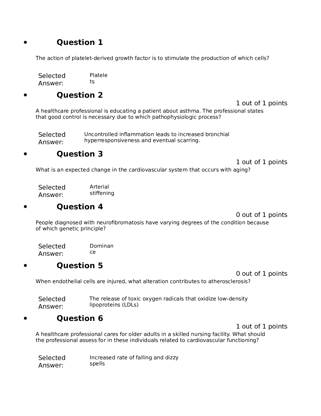
.png)
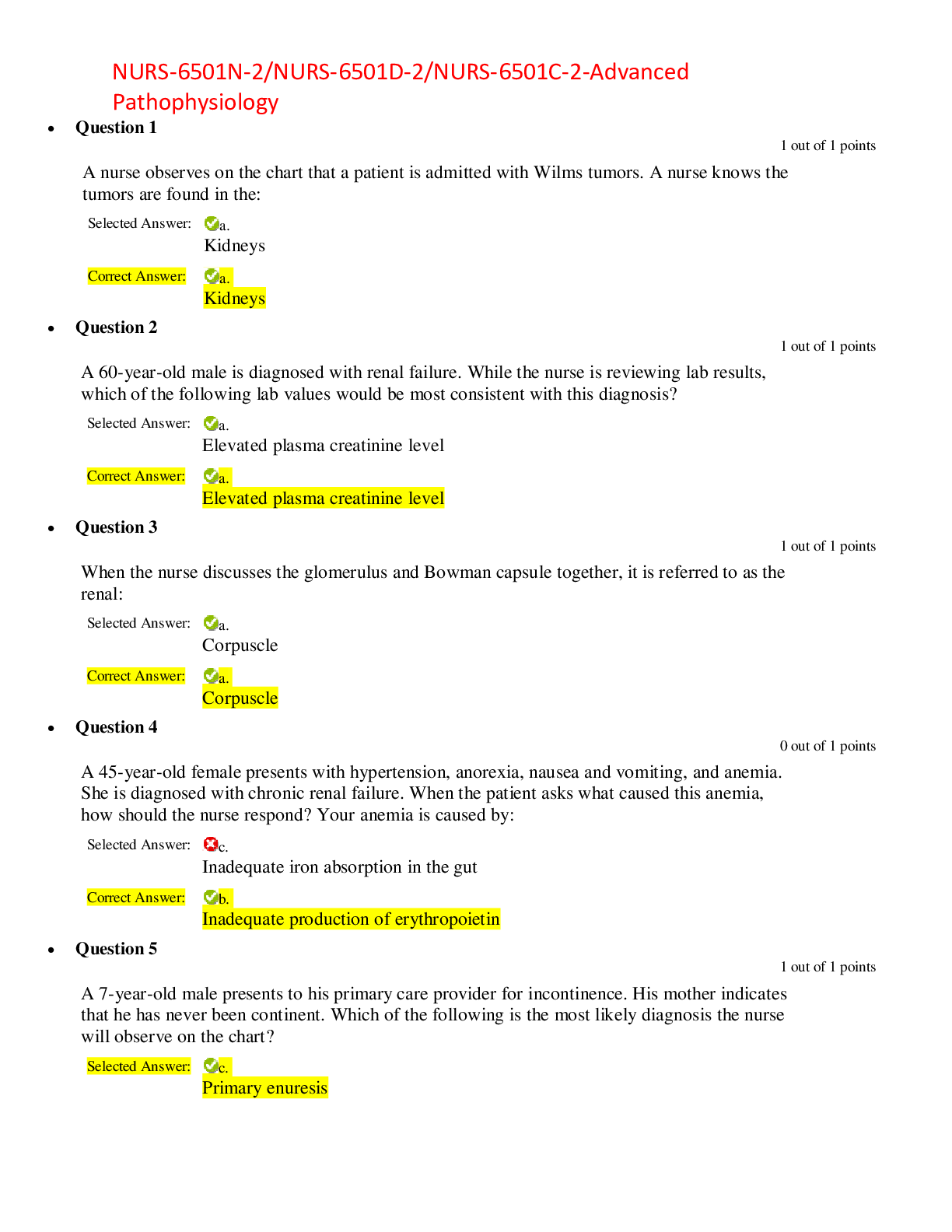
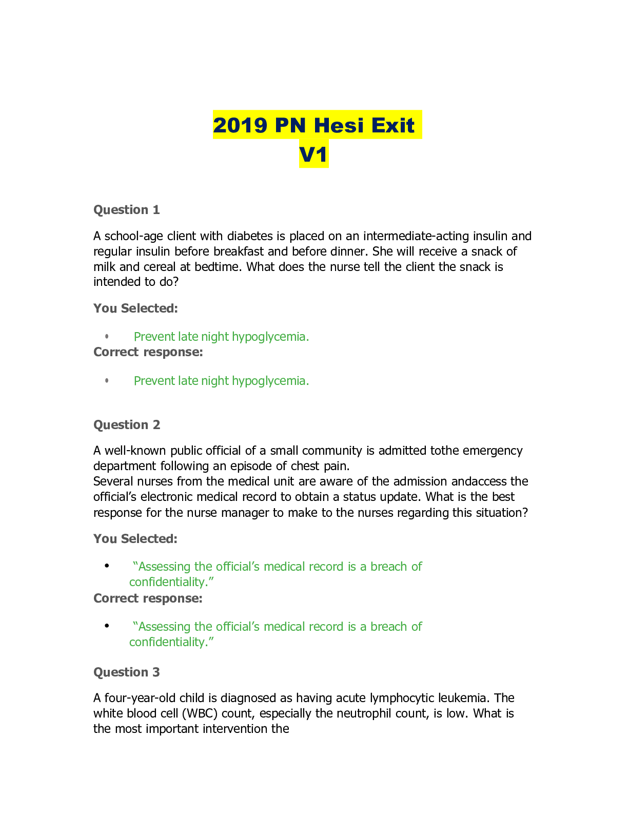

 – South University Savannah.png)
 - Prescription (102 Questions) – South University Savannah.png)
 Assessment question of Endocrinology (48 Questions) – South University Savannah.png)
 Dermatology (64 questions and answers) – South University Savannah.png)
 Prescription Gastroenterology (85 Questions) – South University Savannah.png)
 Prescription of Endocrinology (89 Questions) – South University Savannah.png)
 Assessment Eye, Ear, Nose and Throat (166 Questions) – South University Savannah.png)
 Cardiovascular Assessment (107 Questions and Answers) – South university Savannah.png)
 Respiratory Assessment (51 Questions) – South University Savannah.png)
 Assessment (23 Questions) – South University Savannah.png)
