Pathophysiology > CASE STUDY > NR 283 Week 6 RUA: Pathophysiological Processes Guidelines: Melanoma (GRADED) (All)
NR 283 Week 6 RUA: Pathophysiological Processes Guidelines: Melanoma (GRADED)
Document Content and Description Below
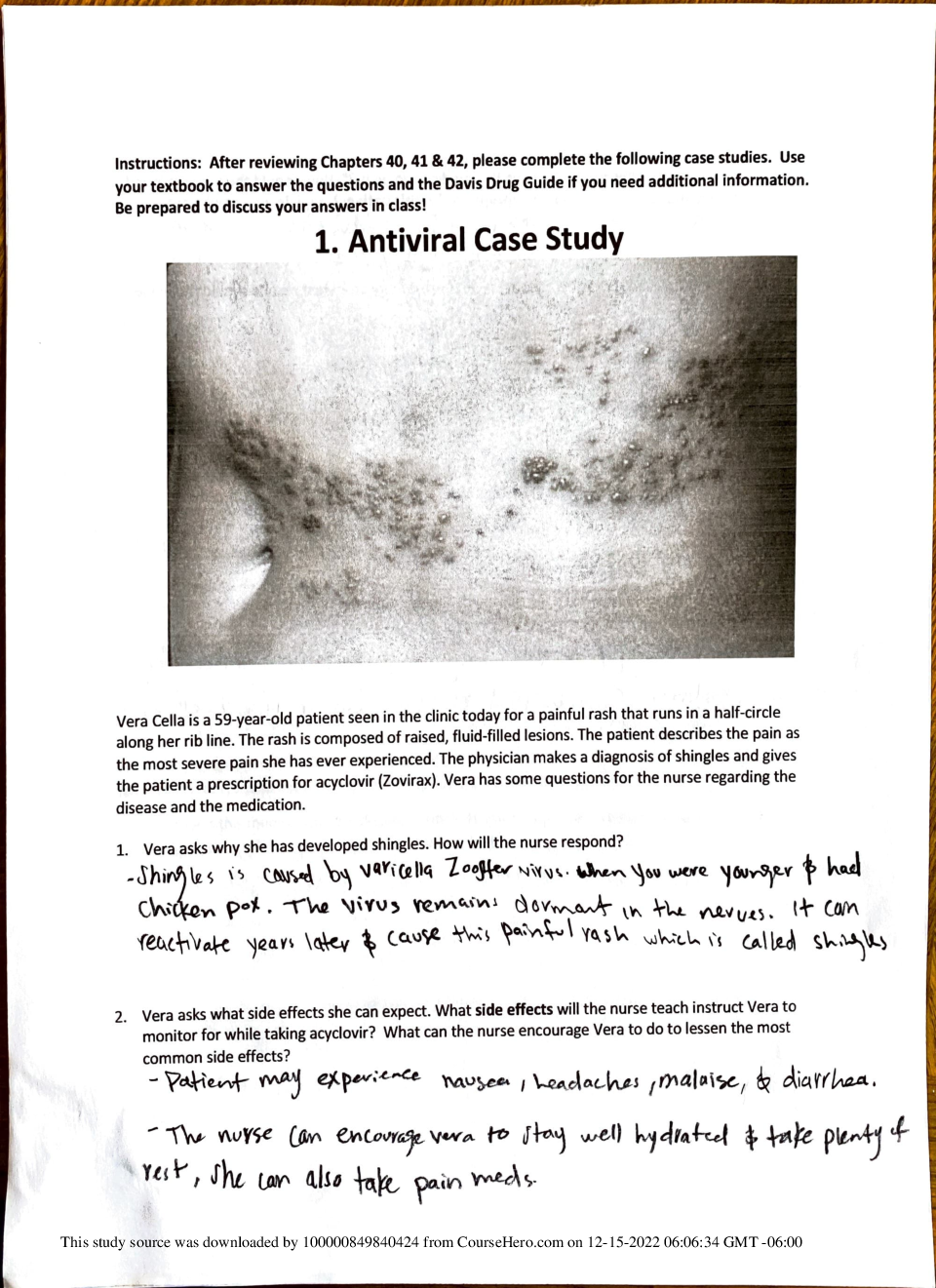
Melanoma Chamberlain College of Nursing NR283: Pathophysiology Spring Session A Melanoma Introduction of Disease Melanoma is a malignant form of skin cancer. While it is not the most com... mon form of skin cancer, it is the deadliest. Melanocytes are cells found on the border of the epidermis and dermis. When they are exposed to UV rays they produce more melanin which gives the skin a darker pigmentation. “The incidence of malignant melanoma has been rising steadily for at least the past 30 years, and it has now become the fifth most common tumor…” (Harting, 2014, p.53) While there are more cases being diagnosed, there are also many ways to prevent skin cancer. Etiology The biggest cause of skin cancer is the amount of UV radiation exposure. There are also two genes that are suspected in playing a role of susceptibility, but no evidence has proven that it is always true. (Porth & Gaspard, 2014) The more melanin that is present in the skin, the more protected a person is from the effects of UV exposure. UV exposure most commonly comes from the sun and from the lights in tanning beds. Even while using tanning bed, the same precautions need to be taken as would be when outside. During the winter UV rays can be amplified by reflecting off the snow. Long car drives also put people at risk because the windows amplify the UV rays. People who drive for a living have this increased risk, especially on the left side of the body because it faces the window. This means that those with fair skin, such as Caucasians, have a significantly higher risk of skin cancer. Physically, those with blonde or red hair and blue or green eyes are also at a higher risk. Individuals who have a lot of freckles and moles are also at an increased risk because UV radiation can cause mutation in those cells. It is also more common in men than women. A family history of malignant melanoma is significant as well as serious sunburns in childhood and previously having malignant melanoma. (Harting, 2014) Pathophysiology Processes Malignant melanoma is caused by the mutation of the pigment cells known as melanocytes. Melanocytes produce melanin which give rise to the pigment in the skin, including freckles and moles. The UV exposure causes damage to the genetic make-up of skin cells and over times these mutations start to cause uncontrolled growth, or cancer. This uncontrolled growth may start at dysplasia and eventually to anaplasia. (VanMeter & Hubert, 2014) The body is able to repair much of the damage but eventually the cells multiply too rapidly. There are several genetic factors that appear in some patients with cancer, but there is no conclusive evidence that it is linked to the melanoma directly. (Harting, 2014) Clinical Manifestations and Complications The most widely used self-diagnostic tool for malignant melanoma is ABCDE. A stands for asymmetry, where one half of the mole does not look like the other. B stands for border, which is usually irregularly shaped and jagged or notched. C is for coloring, which is usually brown of black and not uniform throughout. D is the diameter, about the size of a pencil eraser or larger is when it should be looked at. E is for evolving or changing in any of the previous letters. Other signs to look out for include a sore that doesn’t heal. bleeding, being raised, crusting, tenderness or itching. (Schub & Holle, 2017) If the melanoma is left untreated, it can spread to other parts of the body through the lymphatic system and can ultimately result in death. (Porth & Gaspard, 2014) Diagnostics A diagnosis comes from a biopsy of the suspected lesion. During the pathological examination, the tumor is staged using the Clark level. This level indicates the amount of local invasion from the epidermis, papillary dermis, papillary-reticular dermis, reticular dermis and finally the subcutaneous tissue. The standard tumor, nodes, metastasis (TMN) is used to stage the spread of the cancer. If there is a nodal involvement, further testing such as x-ray, PET scan and MRI can help to assess the extent. (Harting, 2014) A serum lactate dehydrogenase, liver function test and S-100 protein level may all be abnormally elevated. During treatment there may be a decrease in hemoglobin (anemia) and increase in WBC (infection). (Schub & Holle, 2017) References Harting, D. (2014). Malignant Melanoma. Radiation Therapist, 23(1), 51-76. Porth, C, M. & Gaspard, K. J. (2014). Essentials of pathophysiology: Concepts of altered states (4th ed.). Philadelphia, PA, United States: Lippincott Williams and Wilkins. Schub, T., & Holle, M. N. (2017). Melanoma. CINAHL Nursing Guide VanMeter, K. C. & Hubert, R. J. (2014). Gould's pathophysiology for the health professions (5th ed.). St. Louis, MO: Elsevier Saunders. [Show More]
Last updated: 1 year ago
Preview 1 out of 5 pages
Reviews( 0 )
Document information
Connected school, study & course
About the document
Uploaded On
Jun 23, 2022
Number of pages
5
Written in

Additional information
This document has been written for:
Uploaded
Jun 23, 2022
Downloads
0
Views
68


 (1).png)