*NURSING > EXAM > ARDMS Abdomen Ultrasound Registry Review Exam 272 Questions with Verified Answers,100% CORRECT (All)
ARDMS Abdomen Ultrasound Registry Review Exam 272 Questions with Verified Answers,100% CORRECT
Document Content and Description Below
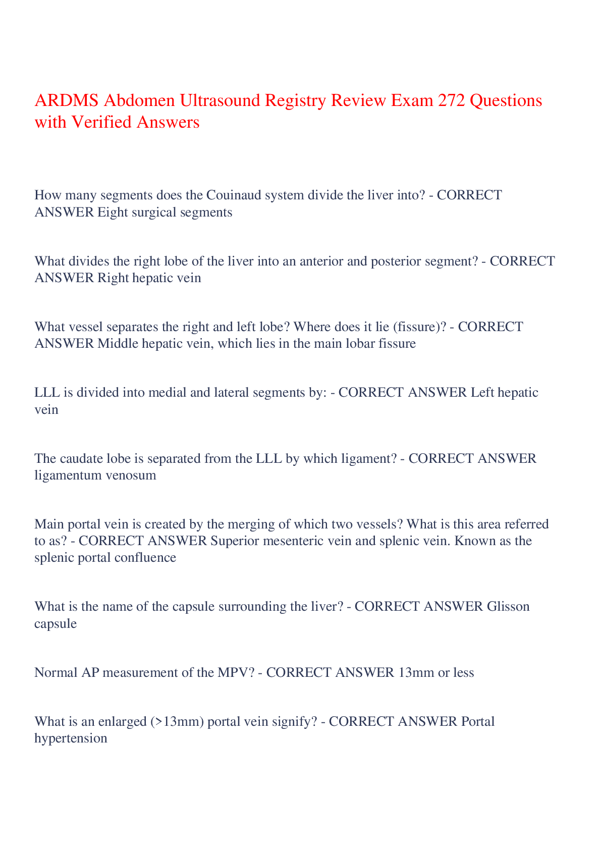
ARDMS Abdomen Ultrasound Registry Review Exam 272 Questions with Verified Answers How many segments does the Couinaud system divide the liver into? - CORRECT ANSWER Eight surgical segments Wha... t divides the right lobe of the liver into an anterior and posterior segment? - CORRECT ANSWER Right hepatic vein What vessel separates the right and left lobe? Where does it lie (fissure)? - CORRECT ANSWER Middle hepatic vein, which lies in the main lobar fissure LLL is divided into medial and lateral segments by: - CORRECT ANSWER Left hepatic vein The caudate lobe is separated from the LLL by which ligament? - CORRECT ANSWER ligamentum venosum Main portal vein is created by the merging of which two vessels? What is this area referred to as? - CORRECT ANSWER Superior mesenteric vein and splenic vein. Known as the splenic portal confluence What is the name of the capsule surrounding the liver? - CORRECT ANSWER Glisson capsule Normal AP measurement of the MPV? - CORRECT ANSWER 13mm or less What is an enlarged (>13mm) portal vein signify? - CORRECT ANSWER Portal hypertension Normal MPV flow? - CORRECT ANSWER Hepatopetal and monophasic w/ some respiratory variation Where do the hepatic veins drain? - CORRECT ANSWER IVC These veins are considered both interlobar and intersegmental - CORRECT ANSWER hepatic veins. They are located between the segments and the lobes normal hepatic vein flow - CORRECT ANSWER -Hepatofugal - away from liver -pulsatile, triphasic due to right atrial pressure changes -respiratory variation Narrowing or occlusion of the hepatic veins is indicative of: - CORRECT ANSWER Budd-Chiari syndrome The liver hilum is also know as - CORRECT ANSWER The porta hepatis flow pattern of the hepatic artery should be - CORRECT ANSWER low resistance since it is feeding the liver After birth the umbilical vein becomes - CORRECT ANSWER ligamentum teres aka round ligament -runs along with the falciform ligament -will usually be seen near left portal vein in left liver Where can the main lobar fissure be seen? - CORRECT ANSWER -in sag plane -will appear to connect the neck of the GB with the RPV -also separates right and left hepatic veins hepatic steatosis - CORRECT ANSWER fatty liver Causes of fatty liver disease - CORRECT ANSWER Fatty deposits within the hepatocytes. Once it becomes cirrhosis, it is non-reversible. 1. Alcoholic fatty liver disease 2. Non-alcoholic fatty liver disease: -obesity -starvation -chemotherapy -diabetes mellitus -hyperlipidemia -pregnancy -von Gierke disease (glycogen storage dx) -total parental nutrition -cystic fibrosis steatohepatitis - CORRECT ANSWER inflammation of the liver associated with fat precursor for chronic liver dx leading to fibrosis, cirrhosis, and HCC hepatomegaly size - CORRECT ANSWER >15cm need to correlate with clinical hx don't confused Riedel's lobe as hepatomegaly Fatty liver symptoms and labs - CORRECT ANSWER Symptoms: -usually asymptomatic Labs: -Increased LFTs (especially AST and ALT) Sono appearance of fatty liver dx and focal fatty infiltration - CORRECT ANSWER -Diffusely echogenic liver -Increased attenuation of sound beam -Walls of hepatic vasculature and diaphragm will not be easily imaged due to increased attenuation -Fatty changes will be diffuse or focal Focal fatty infiltration sono app: -hyperechoic area next to the GB, near the porta hep, or part of lobe may appear echogenic Focal fatty sparing sonographic appearance - CORRECT ANSWER -entire liver is involved with diffuse fatty infiltration with certain areas spared -Area of sparing can look like a solid hypoechoic mass -Hypoechoic area will be near GB, porta hep, or entire lobe may be spared -Can appear to look like pericholecystic fluid Two most common types of hepatitis: - CORRECT ANSWER A and B A: Fecal-oral route: contaminated water or food B: Contact with body fluids, mother-to-infant transmission, blood contact (IV drugs) Most common type of hepatitis in healthcare workers - CORRECT ANSWER Hep C -Spread by blood and body fluid contact Which type of hepatitis is the leading indication for liver transplantation in US? - CORRECT ANSWER Hepatitis C Wilson disease Hemochromatosis Autoimmune disorders Drugs All of the above can be causes for chronic ________ of the liver - CORRECT ANSWER Hepatitis What is Wilson disease? - CORRECT ANSWER -An inherited disorder that causes too much copper to accumulate in the liver, brain and other vital organs. -Also called hepatolenticular degeneration. -Causes fatty changes & fibrosis in the liver. Leads to chronic hepatitis Trademark feature: Copper ring around iris What is hemochromatosis? - CORRECT ANSWER Iron overload disease resulting in abnormal deposition of iron Can lead to fatty changes and chronic hepatitis Clinical signs and symptoms of hepatitis - CORRECT ANSWER -Nonobstructive Jaundice (related to hep on a cellular level NOT biliary obstruction) -Hepatosplenomegaly -Dark urine -F/N/V -Elevated LFTs -Fatigue -Chills Hepatic encephalopathy - CORRECT ANSWER -central nervous system dysfunction resulting from overexposure of brain to toxins -causes confusion and intermittent loss of consciousness What is refractory hypertension? - CORRECT ANSWER Hypertension that is unresponsive to medication - is related to renal artery stenosis Clinical signs and symptoms of renovascular hypertension and renal artery stenosis (RAS)? - CORRECT ANSWER -Refractory HTN (doesn't respond to meds -Very high systemic blood pressure (malignant HTN) -Abd bruit -Elevated creatinine and cholesterol -Unexplained CHF or pulmonary edema Most common cause of RAS and location? - CORRECT ANSWER Most common: Atherosclerosis -loc: Proximal segment 2nd most common: FMD -loc: distal 2/3 of artery What is the kidneys response when they receive less blood flow (due to RAS) - CORRECT ANSWER -Perceive the body as having reduced systemic BP -will activate renin-angiotensin aldosterone system >this will raise BP to try to increase flow to the kidneys, ultimately causing systemic HTN What can long standing RAS lead to? - CORRECT ANSWER -Renal parenchymal damage -Renal failure Most common indication for a renal transplant - CORRECT ANSWER End stage renal disease caused by diabetes Allograft - CORRECT ANSWER transplantation of healthy tissue from one person or cadaver to another person; also called homograft Where will a transplant kidney mostly be placed? - CORRECT ANSWER In pelvis The donor renal artery is usually connected to the recipients: (which artery)? What about the renal vein? - CORRECT ANSWER External iliac artery (EIA) and external iliac vein (EIV) Contraindications to renal transplant - CORRECT ANSWER -preexisting infection -serious cardiac dx -peripheral artery dx -metastatic dx Normal pancreas anatomy - CORRECT ANSWER -nonencapsulated -retroperitoneal structure between duodenal loop and splenic hilum Exocrine function of pancreas - CORRECT ANSWER Secretes: -trypsin -lipase -amylase Through the ductal system Endocrine function of pancreas - CORRECT ANSWER *non-ductal* Secretes: insulin via islets of Langerhans Normal AP measurement of pancreas - CORRECT ANSWER ≤3mm What supplies blood to the pancreas? - CORRECT ANSWER -Celiac axis -SMA -Splenic A Spatial relationship of pancreas - CORRECT ANSWER -Anterior to IVC -Head is medial to duodenum -CBD is posterior/lateral to panc head -GDA is anterior/lateral to panc head -SMA and SMV are posterior to neck of panc -SMA and SMV are anterior to uncinate process -Tail is anterior/medial to splenic hilum Spatial relationship of panc and its vessels - CORRECT ANSWER -Ao is posterior to body -Celiac axis arises from ao @ superior border of panc -SMA arises from ao @ inferior border of panc Branches of the celiac axis - CORRECT ANSWER Gives off left gastric artery and then divides into CHA and splenic artery splenic artery follows along superior border of body/tail of panc What does the common hepatic artery divide into? - CORRECT ANSWER Divides into proper hepatic A (PHA) and gastroduodenal arteries Where does PHA course? - CORRECT ANSWER superiorly from CHA toward the liver (anterior to portal vein) and left of the bile duct Two ducts of the pancreas - CORRECT ANSWER main duct: duct of Wirsung accessory: duct of santorini >50% of the population has complete regression of this duct What two ducts join to form the ampulla of vater, which empties into duodenum (at the major papilla)? - CORRECT ANSWER CBD and duct of wirsung Which is NOT a branch of the celiac axis? a. common hepatic artery b. right gastric artery c. splenic artery d. left gastric artery - CORRECT ANSWER b. right gastric artery in most people, the right gastric artery arises from the proper hepatic artery Superior mesenteric VEIN origin and termination - CORRECT ANSWER Origin: Ascends from small bowel Courses superior and then anterior to uncinate process Termination: splenic portal confluence -joins the splenic vein to form portal vein Inferior mesenteric VEIN origin and termination - CORRECT ANSWER origin: large bowel termination: ascends and terminates into splenic vein (which will eventually join SMV and become portal vein) The portal vein divides into the _________ and __________ branch inside the liver - CORRECT ANSWER left and right What is ALT and where is it found? - CORRECT ANSWER Alanine aminotransferase. Concentrated in the liver High levels of ALT indicate - CORRECT ANSWER Liver damage or disease Cholecystokinin (CCK) is produced by the __________ and released in response to __________ in the stomach - CORRECT ANSWER duodenum; food (especially fatty) Flow of bile - the pathway - CORRECT ANSWER liver → biliary radicles → right or left hepatic duct → common hepatic duct → cystic duct → GB → common bile duct (CBD) → ampulla of Vater (mixes w/ panc juice) → sphincter of Oddi → duodenum (mixes w/ chyme) Most stones in the CBD are located near the _______ ___ _____ - CORRECT ANSWER Ampulla of Vater Four main labs that are elevate with choledocholithiasis - CORRECT ANSWER 1. ALP 2. ALT 3. GGT 4. Bilirubin A patient with jaundice, pain, and fever secondary to stone lodge in cystic duct whhich compresses the common duct - CORRECT ANSWER Mirizzi syndrome biliary duct walls (not gb) should measure _____ or less - CORRECT ANSWER 4mm Most common cause of cholangitis - CORRECT ANSWER Bile duct obstruction resulting from choledocholithiasis Most common types of cholangitis - CORRECT ANSWER -acute bacterial -AIDS -oriental (recurring bacterial) -sclerosing Charcot triad - CORRECT ANSWER refers to biliary obstruction symptoms 1. Fever 2. Jaundice 3. RUQ pain Sono findings of cholangitis - CORRECT ANSWER 1. Biliary dilatation 2. Biliary sludge or pus 3. Choledocholithiasis 4. Bile duct wall thickening recent biliary or gastric surgery, emphysematous or prolonged acute cholecystitis, and fistula formation are associated with: - CORRECT ANSWER Pneumobilia (air in biliary tree) Sonographic appearance of pneumobilia - CORRECT ANSWER Echogenic linear structures within the ducts (both intra and extrahepatic) that produce ring-down artifacts and dirty shadowing Parasitic roundworm that is transmitted through fecal-oral route and makes its way from the small intestine to the biliary system - CORRECT ANSWER Ascariasis Sono findings of ascariasis - CORRECT ANSWER -worm noted in biliary duct as linear structure in sagittal plane -movement confirms diagnosis Cholangiocarcinoma what is it, prognosis, and what are its RFs. Most common manifestation (Type) - CORRECT ANSWER primary bile duct cancer prognosis: POOR RF: 1. primary sclerosing cholangitis 2. recurrent biliary infections 3. stone disease most common manifestation: Klatskin tumors what is a Klatskin tumor and where is it located - CORRECT ANSWER -the most common manifestation of cholangiocarcinoma at the junction of the left and right hepatic ducts causing dilation of intrahepatic ducts Jaundice, pruritis, unexplained weight loss, abd pain. Elevated bili and ALP. What malignancy is most likely? - CORRECT ANSWER Cholangiocarcinoma -- most commonly Klatskin tumor type sono findings cholangiocarcinoma - CORRECT ANSWER -dilated intrahepatic ducts that abruptly terminate -sometimes: solid mass noted within liver or ducts normal measurement of intrahepatic duct is less than: - CORRECT ANSWER 2mm Which anatomical landmark can help locate a gallbladder that is difficult to visualize? - CORRECT ANSWER Main lobar fissure A pancreatic mass causing hydrops of the GB without stones or pain is termed - CORRECT ANSWER Courvoisier's gallbladder exocrine function of pancreas - CORRECT ANSWER release digestive enzymes through duct of wirsung (main panc duct) into the small intestines duct of wirsung → ampulla of vater → sphincter of oddi → duodenum -amylase -lipase sodium bicarbonate -trypsin endocrine function of pancreas - CORRECT ANSWER islets of langerhans secrete insulin and glucagon into blood stream What triggers the sphincter of oddi to open? - CORRECT ANSWER CCK produced by the duodenum in response to the presence of chyme What does glucagon do? What does insulin do? - CORRECT ANSWER promotes the release of glucose by the liver to increase blood sugar levels insulin: stimulates body to use up glucagon to produce energy Which artery supplies blood to the head of the pancreas? how about the body and the tail? - CORRECT ANSWER Head: Gastroduodenal artery GDA body and tail: Splenic and SMA Which veins drain the pancreas? - CORRECT ANSWER splenic v, SMV, IMV, and portal Is the pancreas hyper, iso, or hypoechoic to the liver? - CORRECT ANSWER The pancreas is hyperechoic to the liver The main panc duct (duct of Wirsung) should not exceed ____ mm - CORRECT ANSWER 2mm What is another term for a peripancreatic fluid collection? - CORRECT ANSWER phlegmon With moderate and severe pancreatitis, the body will attempt to encapsulate the damaging digestive enzymes that leak from the pancreas, forming a _______ - CORRECT ANSWER pseudocyst Between which two organs is the lesser sac located? - CORRECT ANSWER pancreas and stomach 9 regions of the abdomen - CORRECT ANSWER right hypochondriac, epigastric, left hypochondriac, right lumbar, umbilical, left lumbar, right iliac, hypogastric, left iliac Plegmon - CORRECT ANSWER peripancreatic fluid resulting from inflammation of the pancreas -can occur with both acute and chronic pancreatitis pancreatic pseudocyst - CORRECT ANSWER collection of debris, fluid, pancreatic enzymes, and blood as a complication of acute pancreatitis. Body attempts to wall it off. Pseudocysts will occur in the lesser sac, which is the area between the pancreas and the _______ - CORRECT ANSWER stomach Medial to right lobe of liver, posterior to IVC, superomedial to the kidney, and lateral to the right crura. Which organ does this describe and on which side is it? - CORRECT ANSWER Right adrenal gland Where in the kidney are the loops of henle located? - CORRECT ANSWER In the medulla. Also called nephron loop. Nephrons are in the parenchyma and loops of henle dive into the medulla Two large antegrade diastolic and systolic waves followed by a small retrograde component that corresponds with the atrial contraction. This describes which liver vessels? - CORRECT ANSWER Hepatic veins What are the hepatic veins triphasic? - CORRECT ANSWER Due to the proximity to the heart Most common dysgenesis of the kidneys - CORRECT ANSWER Duplication: complete or incomplete What is the name for the CBD when it is in the liver? - CORRECT ANSWER Common hepatic duct Longus colli muscle is _______ to the thyroid. - CORRECT ANSWER posterior Which is more posterior in the body: aorta or IVC? - CORRECT ANSWER Aorta First branch off of the common hepatic artery? A. Cystic artery B. Pancreatic artery C. Duodenal artery D. Gastroduodenal artery - CORRECT ANSWER D. Gastroduodenal artery -Supplies the pancreas and duodenum with blood Pancreatic transplants usually require arterial resection using a Y-graft w/ two arms that are connected to: A. Common hepatic artery and splenic vein B. Common hepatic artery and splenic vein C. SMV and portal vein D. SMA and splenic artery - CORRECT ANSWER D. Superior mesenteric artery and splenic artery Why? Because branches of these arteries usually supply the native pancreas A single large bump bulging off of the lateral kidney. What is this normal variant? - CORRECT ANSWER Dromedary hump. Normal "bump" of normal renal parenchyma More commonly seen on the lateral left kidney. The _________ gland is anterior to the ear and is drained by the _______ A. parotid, Stensen duct B. submandibular, Stensen duct C. submandibular, Wharton duct D. sublingual, Wharton duct - CORRECT ANSWER A. parotid, Stensen duct Which of the following is a part of the renal parenchyma? A. Major calyces B. Pyramids C. Minor calyces D. Segmental arteries - CORRECT ANSWER B. Pyramids -they are in the medulla portion of the parenchyma all other choices are a part of the sinus *remember that the parenchyma includes the cortex AND medulla The pancreas is found in which retroperitoneal space? A. Posterior pararenal B. Retrofascial C. Anterior pararenal D. Perirenal - CORRECT ANSWER C. Anterior pararenal Which renal arterial branches are used to assess parenchymal resistance? A. interlobular or segmental B. Segmental or arcuate C. Segmental or interlobar D. Interlobar or arcuate - CORRECT ANSWER D. Interlobar or arcuate arteries -arcuates preferred but run parallel to capsule so interlobar are easier The normal gallbladder is usually less than _____ in transverse diameter A. 3mm B. 5cm C. 3cm D. 8cm - CORRECT ANSWER B. 5cm The renal pyramids are located in the: A. Medulla B. Cortex C. Sinus D. Calyces - CORRECT ANSWER A. Medulla Diffuse acute pancreatitis will typically result in A. A diffusely shrunken, hyperechoic gland B. A diffusely enlarged, hypoechoic gland C. A diffusely enlarged, hyperechoic gland D. A shrunken gland with diffuse areas of calcifications - CORRECT ANSWER B. A diffusely enlarged, hypoechoic gland Which has the potential for malignancy: serous (microcystic) or mucinous (macrocystic) cystadenoma? Where is it located in the pancreas? - CORRECT ANSWER Macrocystic, or mucinous cystadenoma has a malignant potential. Both cystadenomas usually arise from the body and tail of the panc Sono appearance of serous (microcystic) cystadenomas? - CORRECT ANSWER Mass that may appear solid and echogenic due to the small size of the cysts Sono appearance mucinous (macrocystic) cystadenomas? - CORRECT ANSWER -Multilocular cystic masses -may contain mural nodules and calcification -pancreatic duct may be dilated Insulinoma. Sono app? - CORRECT ANSWER a benign tumor of the pancreas from islet cells that causes hypoglycemia by secreting additional insulin Islet cells are the pancreatic cells that secrete insulin Sono: Usually solitary mass within panc Gastrinoma. What syndrome is it associated with? Sono app? - CORRECT ANSWER an islet cell tumor found within the pancreas -associated with zollinger-ellison syndrome -multiple masses within panc. Can be difficult to image Zollinger-Ellison syndrome - CORRECT ANSWER excessive secretion of acid by the stomach that leads to peptic ulcers In 80% of pancreatic transplants, a ______ transplant will also be performed at the same time - CORRECT ANSWER renal When the pancreas is transplanted at the same time as the kidney, the pancreas will be placed within the ______ side of the abdomen while the kidney is placed on the ______ side - CORRECT ANSWER panc: right kidney: left exocrine bladder drainage pancreatic transplant - CORRECT ANSWER -vasculature of donor pancreas is anastomosed to the recipient's common iliacs vessels -donor duodenum is anastomosed to the bladder and the recipient bladder is used to expel duodenum pancreatic juices Exocrine enteric drainage pancreas transplant - CORRECT ANSWER -Donor's duodenum is anastomosed to a loop of jejunum -Splenic artery and SMA are connected w/ donor iliac arteries. This forms a "Y" graft -Donor common iliac portion is anastomosed to the recipients CIA and EIA -transplants are usually located in RUQ and have vertical orientation **more common Which is the more common method for pancreatic transplants? Exocrine bladder drainage or exocrine enteric drainage? - CORRECT ANSWER Exocrine enteric drainage tx A transplant pancreas should be __________ and _________ just after transplant A. Enlarged and heterogeneous B. Homogeneous and hypoechoic C. Heterogeneous and hyperechoic D. Small and homogeneous - CORRECT ANSWER B. Homogeneous and hypoechoic A hypoechoic and heterogeneous pancreas post-transplant may be a sign of: - CORRECT ANSWER transplant rejection or pancreatitis a hypoechoic or heterogeneous gland with elevated resistive indices in a transplant pancreas may be due to: - CORRECT ANSWER Acute pancreatic transplant rejection A hyperechoic echotexture, atrophy, and calcifications of a transplant pancreas may indicate: - CORRECT ANSWER Chronic pancreatic transplant rejection The most common location of focal pancreatitis is within the: A. Head of panc B. Neck of panc C. Body of panc D. Tail of panc - CORRECT ANSWER A. Head of panc What is the most common islet cell tumor? A. Granuloma B. Gastrinoma C. Insulinoma D. Cystadenoma - CORRECT ANSWER C. Insulinoma Which cells perform the exocrine function of the pancreas? A. Whipple cells B. Islets of Langerhans C. Delta cells D. Acinar cells - CORRECT ANSWER D. Acinar cells von Hippel-Lindau disease - CORRECT ANSWER a hereditary disease that includes the development of cysts within the pancreas, kidneys, reproductive tract, brain, etc Gas or air produces a: A. clean shadow B. dirty shadow - CORRECT ANSWER B. dirty shadow. A clean shadow comes from sound absorbing materials such as stones Both acute and chronic splenic infarcts appears as wedge shaped mass in the spleen. An acute infarct will appear more _______ (hypoechoic/hyperechoic), while a chronic infarct will appear more _________ (hypoechoic/hyperechoic) - CORRECT ANSWER Acute: Hypoechoic Chronic: Hyperechoic Splenosis - CORRECT ANSWER the implantation of ectopic splenic tissue secondary to splenic rupture Most common benign tumor of the spleen? - CORRECT ANSWER Hemangioma Patients with a history of tuberculosis, histoplasmosis, or sarcoidosis have an increased risk of splenic ___________ - CORRECT ANSWER granulomas A disease that results from the inhalation of an airborne fungus. Can affect the lungs and then spread to other organs - CORRECT ANSWER Histoplasmosis A systemic disease that results in the formation of granulomas throughout the body - CORRECT ANSWER sarcoidosis A benign tumor-like malformation that consists of disorganized cells. Can be found in liver, spleen, breast, GI, etc - CORRECT ANSWER Hamartoma These benign tumors are associated with beckwith-wiedemann syndrome and tuberous sclerosis. Splenic ___________ - CORRECT ANSWER hamartoma Presence of Reed-Sternberg cells indicates which: Hodgkin's or non-Hodgkin's lymphoma? - CORRECT ANSWER Hodgkin's lymphoma Which has a poorer prognosis: Hodgkin's lymphoma or non-Hodgkin's? - CORRECT ANSWER Non-Hodgkin's has a poorer progonosis. -Hodgkin's is highly treatable -Non-Hodgkin's is not as treatable but is more common What is the exceeding rare PRIMARY malignant tumor of the spleen? - CORRECT ANSWER Angiosarcoma -lymphoma or leukemia is the most common tumor of the spleen but they are secondary spread Lymphangiomas are most commonly seen in what kind of patient? A. Women B. Men C. Children D. Pregnant women - CORRECT ANSWER C. Children What is a splenic lymphangioma? - CORRECT ANSWER A benign lesion from a congenital malformation of the lymphatic system -multicystic -can have hypo or anechoic locules and hyperechoic septations Blood disorder found more commonly in middle east, Mediterranean, and Caribbean Hispanic child in US - CORRECT ANSWER Sickle cell anemia -also very common in african americans Acute vs recurrent sickle cell crises affect on the spleen - CORRECT ANSWER acute: splenomegaly recurrent: spleen becomes fibrotic and atrophic Autosplenectomy (fibrosis and shrinkage). Association? - CORRECT ANSWER sickle cell anemia with multiple infarctions Patients undergoing a sickle cell crisis may have a decreased ___________ (labs) - CORRECT ANSWER hematocrit retroperitoneal organs - CORRECT ANSWER -Suprarenal (adrenal) glands -Aorta and IVC -Duodenum -Pancreas (except tail) -Ureters and Urinary bladder -Colon -Kidneys -Esophagus -Rectum "SAD PUCKER" Reticuloendothelial System (RES) - CORRECT ANSWER System of phagocytic cells including monocytes and macrophages spleen, liver, lungs are highly involved Tissue within the spleen responsible for its phagocytic function? A. Red pulp B. White pulp C. Culling pulp D. Pitting pulp - CORRECT ANSWER A. Red pulp Nephron begins to function by _____ weeks gestation - CORRECT ANSWER nine Maintain homeostasis by: -detoxify and filter blood -excrete metabolic waste -reabsorb amino acids, ions, glucose, and water -maintain normal pH, iron, and salt levels in blood -regulate blood pressure - CORRECT ANSWER Kidneys renunculi - CORRECT ANSWER -the two embryonic parenchymal tissue masses that combine to create the kidney -descend into fetal abdomen by 12 weeks -this is one of the reasons ectopic kidney locations are common Most common place for ectopic kidney to be located - CORRECT ANSWER pelvis (pelvic kidney) The _________ kidney is higher than the other side (right/left) - CORRECT ANSWER left -b/c liver is on right side making rk lower The renal pyramids are located in the - CORRECT ANSWER renal medulla The part of the kidney responsible for filtering and excretion - CORRECT ANSWER cortex The part of the kidney responsible for absorption - CORRECT ANSWER medulla minor calices, major calices, renal pelvis, and infundibula make up the: -where is this located? - CORRECT ANSWER renal collecting system -located in renal sinus Structure that collects urine before it empties into ureter - CORRECT ANSWER renal pelvis put the renal arteries in order from the abdominal aorta to the glomerulus -interlobular -arcuate -main renal artery -interlobar -afferent arterioles -segmentals -glomulerus -aorta - CORRECT ANSWER aorta→main renal artery→segmental→interlobar→arcuate→interlobular→afferent arterioles→glomulerus elevated bun and creatinine may indicate what? - CORRECT ANSWER renal disease most common congenital anomaly of urinary tract? - CORRECT ANSWER duplex or duplicated collecting system average range for kidney length in adults - CORRECT ANSWER 8-13 cm in length normal kidney cortex should be isoechoic or _____echoic to the liver or spleen - CORRECT ANSWER hypoechoic cortical thinning of the kidney is _______cm or less - CORRECT ANSWER 1cm or less most common cause of acute renal failure - CORRECT ANSWER Acute tubular necrosis (ATN) other causes: -RAS -infection -urinary tract obstruction -amyloidosis (amyloid build up) -henoch-schonlein purpura What is acute tubular necrosis (ATN)? - CORRECT ANSWER ischemic damage to the tubules, resulting in cell death and necrosis, ultimately resulting in renal failure causes: poison (antifreeze or others), drugs, and other harmful substances *reversible* sono: -enlarged -increased RI in parenchymal vessels -prominent pyramids The following are clinical signs of what? -elevated BUN and creatinine -oliguria -hypertension -leukocytosis -hematuria -edema -hypovolemia - CORRECT ANSWER acute renal failure The most common cause of chronic renal failure? - CORRECT ANSWER diabetes mellitus others: glomerulonephritis, chronic pyelonephritis, infection, UT obstruction In order to treat chronic renal failure, patients may be placed on dialysis or need a donor kidney. T/F - CORRECT ANSWER True The following are clinical signs of what? -malaise -elevated BUN and creatinine -fatigue -HTN -hyperkalemia (high potassium) - CORRECT ANSWER Chronic renal failure -small, echogenic kidneys -cortical thinning -loss of corticomedullary differentiation -possible renals cysts These are sono findings of: acute or chronic renal failure? - CORRECT ANSWER Chronic Which type of dialysis may cause some ascites in the body? A. hemodialysis B. hemofiltration C. Peritoneal dialysis - CORRECT ANSWER C. Peritoneal dialysis This is because it uses a solution that is instilled into abdomen through a catheter Exophytic - CORRECT ANSWER Grows outward from tissue This renal dx has a strong association with polycystic liver dx - CORRECT ANSWER autosomal dominant polycystic kidney disease (ADPKD) -with ADPKD, the cysts can be found in other organs as well, such as the liver, panc, and spleen A staghorn calculus, hydronephrosis, and a perinephric fluid collection are often seen with: - CORRECT ANSWER xanthogranulomatous pyelonephritis 1. This liver lobe is divided into 2 posterior and 2 anterior lobes (left/right) 2. This liver lobe is divided into 2 medial and 2 lateral segments (left/right) both are divided by portal branches - CORRECT ANSWER 1. right 2. left The pancreas _________ in echogenicity with age - CORRECT ANSWER increases -as a child it is hypo or isoechoic to the liver and increases w/ age -cystic fibrosis causes an increase in echogenicity In most people, the proper hepatic A bifurcates into left and right hepatic arteries. In 10% of people, the right hepatic A originates from the ______ and the LHA originates from the _____ - CORRECT ANSWER SMA ; left gastric artery Normal wall thickness of the bowel is: A. <3mm B. <3m C. <5cm D. <5mm - CORRECT ANSWER D. <5mm Distended bowel normal wall: 3mm Non-distended: 5mm Arteries that are dopplered within the testicular parenchyma? A. Deferential B. Gonadal C. Centripetal D. Cremasteric - CORRECT ANSWER C. Centripetal If there is vascular resistance, such as a vessel narrowing, the RI will: (increase/decrease) - CORRECT ANSWER Increase. RI is proportional to vascular resistance and compliance normal retroperitoneal lymph nodes usually measure less than _____ in length A. 0.5cm B. 1cm C. 1.5cm D. 2cm - CORRECT ANSWER B. 1cm Which are intERsegmental? portals or hepatic veins? - CORRECT ANSWER hepatic veins. They course between liver segments A renal hamartoma can also be called a - CORRECT ANSWER angiomyolipoma central stellate scar within a renal tumor is indicative of: - CORRECT ANSWER oncocytoma -2nd most common renal mass -male, 60s -asymp Vascular, hyperechoic benign mass in kidney with areas of calcifications. Appearance is similar to RCC - CORRECT ANSWER renal adenoma What vascular abnormality causes the following: -Hematuria -Proteinura -Possible L abd or flank pain -Pelvic pain -Left-sided testicular pain - CORRECT ANSWER Nutcracker syndrome. left renal vein compressed by SMA and Ao A renal artery ratio (RAR) greater than 3.5 and downstream tardus-parvus is indicative of: - CORRECT ANSWER Renal artery stenosis A waveform with a prolonged upstroke (systolic acceleration), and rounded peak is called? _______ _______. - CORRECT ANSWER Tardus parvus Allograft (homograft) A. Tissue from the patient taken from another area of the body B. Tissue from the same species but not genetically identical C. Tissue from a different species with similar genetic qualities D. Tissue from an identical twin of patient - CORRECT ANSWER B. Tissue from the same species but not genetically identical Term is commonly used in transplants, especially renal While vessels are the transplant renal vessels most commonly attached to? - CORRECT ANSWER Donor artery and vein usually connected to EIA and EIV Sonographically, the transplanted kidney should appear _______ to a normal kidney - CORRECT ANSWER similar Mild pelviectasis is considered ______ in the transplanted kidney - CORRECT ANSWER normal Waveform of the renal artery after transplant should yield ______ (low/high) resistance flow with ____________ (delayed/continuous) diastolic flow, while the renal vein should demonstrate ______ (continuous/pulsatile) flow away from the kidney - CORRECT ANSWER low, continuous, continuous lymphocele, urinoma, hematoma, abscess are associated with - CORRECT ANSWER post kidney transplant complications Most common vascular complication following renal artery transplant? - CORRECT ANSWER Renal artery stenosis anuria, oliguria, elevated creatinine and BUN, proteinuria, HPTN, and enlargement are associated with - CORRECT ANSWER kidney transplant rejection color aliasing and turbulent flow at level of anastomosis site may indicate: A. Normal post-transplant flow B. Renal artery stenosis C. Renal vein thrombosis D. AV fistula - CORRECT ANSWER B. Renal artery stenosis will also have elevated peak systole and a velocity greater than 200-250cm/s Most common cause of congenital hydronephrosis in infants/children: - CORRECT ANSWER UPJ obstruction -- ureteropelvic junction obstruction; also know as PUJ (pelvicureteric junction obstruction) Abnormal retrograde flow of bladder urine into the ureters and possibly to the kidneys; what is it caused by? - CORRECT ANSWER vesicoureteral reflux (VUR) -often caused by an abnormal angle of insertion of the distal ureter into bladder -can cause infection, scarring, and permanent damage Megacystitis is associated with which syndrome? - CORRECT ANSWER Prune belly syndrome (eagle barrett) absent abdominal muscles, undescended testis, urinary tract abnormalities: - CORRECT ANSWER Triad for prune belly syndrome Weigert-Meyer rule (in association with duplicated pelvicalyceal systems) - CORRECT ANSWER Upper pole moiety in duplex kidney is prone to obstruction b/c abnormal insertion into bladder. Lower pole moiety is prone to reflux. Nephroblastoma is also known as: - CORRECT ANSWER Wilms tumor Most common pediatric malignant abdominal mass that tends to mets to the liver and lungs. Also tends to invade the renal vein and IVC: - CORRECT ANSWER Nephroblastoma/Wilms tumor Remnant of embryonic development that extends from umbilicus to the apex of the bladder. Failure to close can result in UTI and palp abd mass between umbilicus and bladder. Seen in peds - CORRECT ANSWER Urachus Portion of ureter that connects to the renal pelvis is called the __________ _________. Portion of ureter that connects to the bladder is called the _________ __________ - CORRECT ANSWER ureteropelvic junction; ureterovesicular junction Most common area for urinary stone to become lodged: ___________ ________ - CORRECT ANSWER ureterovesicular junction Muscle that controls emptying of the urinary bladder - CORRECT ANSWER detrusor muscle trigone of the bladder is (superior/inferior)? - CORRECT ANSWER inferior urethra and ureters open here When distended, an abnormally thickened bladder wall will measure A. Over 4cm B. Over 4mm C. Less than 4mm D. 5-10cm - CORRECT ANSWER b. Over 4mm L x W x H x 0.56 is how you calculate ________ of the _________ - CORRECT ANSWER volume of the bladder Bladder that functions poorly due to neurologic disorder. pts may not feel the need to urinate (extremely distended bladder) or an unnecessary need to void - CORRECT ANSWER Neurogenic bladder With a neurogenic bladder, these structures may be seen on the wall - CORRECT ANSWER trabeculae ; these can also result from chronic bladder infection How can bladder diverticulum (OUTpouching) cause a UTI? - CORRECT ANSWER Urinary stasis within the diverticulum Nocturia and a wall thickness over 4mm are associated w/ - CORRECT ANSWER Cystitis Malignancy associated with gross hematuria and passing blood clots - CORRECT ANSWER Transitional Cell Carcinoma (TCC) of the bladder` Smooth/papillary mass projecting into bladder lumen. Non-mobile and will show vascularity. Malignant. What is this? - CORRECT ANSWER Transitional cell carcinoma How to differentiate TCC from a blood clot - CORRECT ANSWER Use color doppler and decub the patient Blood clots will not show vascularity and often they are mobile. TCC will have vascularity and is non-mobile TCC of the kidney is usually found in the: A. Cortex B. Medulla C. Minor calyx D. Renal pelvis - CORRECT ANSWER D. Renal pelvis -hypoechoic or isoechoic mass within the renal sinus -varying degrees of hydro -pt smoking history -gross hematuria -Mostly in renal pelvis Which malignancy is this associated with? - CORRECT ANSWER transitional cell carcinoma (TCC) of the kidney _______temia: elevation of blood urea nitrogen (BUN) and serum creatinine levels. - CORRECT ANSWER azotemia Which of the following renal conditions is associated with the development of cysts within the pancreas and liver? A. ARPKD B. ADPKD C. MCDK D. Acquired renal cystic disease - CORRECT ANSWER B. ADPKD Which of the following is not considered an intrinsic cause of hydronephrosis? A, Ureterocele B. Urethritis C. Urolithiasis D. Ureteropelvic junction obstruction - CORRECT ANSWER B. Urethritis Which of the following is not considered an extrinsic cause of hydronephrosis? A. Ureteral stricture B. Pregnancy C. Neurogenic bladder D. Uterine leiomyoma - CORRECT ANSWER A. Ureteral stricture Endocrine disorder that results from hypofunction of the adrenal cortex - CORRECT ANSWER Addison disease Controlled by hormones produced by hypothalamus, which are then released by the anterior pituitary gland: - CORRECT ANSWER Adrenal glands aka suprarenal glands A hormone secreted by the anterior pituitary gland, which controls the release of hormones by the adrenal glands A. Gonadotropin releasing hormone (GH) B. Vasopressin C. adrenocorticotropic hormone (ACTH) D. Somatostatin - CORRECT ANSWER C. Adrenocorticotropic hormone (ACTH) Zones of Adrenal Cortex (superficial to deep) - CORRECT ANSWER -Zona glomerulosa -Zona Fasciculata -Zona Reticularis (GFR) Aldosterone, androgens, and cortisol are produced by the adrenal: (cortex/medulla) - CORRECT ANSWER Cortex The adrenal medulla produces: - CORRECT ANSWER Catecholamines - epinephrine (adrenaline) and norepinephrine Aldosterone function - CORRECT ANSWER Regulate blood pressure by controlling sodium and water in body made by adrenal cortex Androgen function - CORRECT ANSWER Development of male characteristics made by adrenal cortex Cortisol function - CORRECT ANSWER -glucose metabolism -blood pressure regulation -immune function -inflammatory response The adrenal glands are supplied by which arteries and drain by which veins? - CORRECT ANSWER Blood supply: Suprarenal arteries (3) Drainage: Suprarenal veins (2) The right suprarenal vein drains _________ and the left suprarenal vein drains _________ - CORRECT ANSWER Right: directly into IVC Left: Into left renal vein The following are associated with: ___________ ____ -over production of ACTH -hyperpigmentation (darkening) -hypotension -weakness/fatigue -anorexia/weight loss -increased LFTS -hyperkalemia (potassium K) -hypernatremia (sodium Na) - CORRECT ANSWER Addison disease The kidneys will appear _______ (size) in the acute stages of Addison dx - CORRECT ANSWER enlarged -will atrophy in chronic stage Tumor of the adrenal gland (commonly adenoma) or pituitary causing hypercortisolism is associated with: - CORRECT ANSWER Cushing syndrome These findings are consistent with _________ __________: -buffalo hump -moon face -red or purple striae (stretch marks) on abd and thighs -hirsutism -hypertension - CORRECT ANSWER Cushing syndrome Syndrome that results from high levels of aldosterone secretion from adrenal cortex -can be caused by functioning tumor in adrenal cortex (likely adenoma) -HPTN -excessive thirst and urination -hypernatremia (sodium Na) -hyperkalemia (potassium K) -muscle cramps and weakness - CORRECT ANSWER Conn syndrome aka primary hyperaldosteronism Most common benign solid mass of the adrenal gland - CORRECT ANSWER adrenal adenoma -can be hyperfunctioning or nonhyperfunctioning solid, hypoechoic mass in the area of the adrenal gland is most likely to be a: A. carcinoma B. adenoma C. pheochromocytoma D. cyst - CORRECT ANSWER B. adenoma most common solid mass of adrenal gland hyperfunctioning benign adrenal mass that causes excessive release of epi and norepinephrine into blood, causing uncontrollable hypertension - CORRECT ANSWER pheochromocytoma Pheochromocytomas are large and hyperechoic masses arising from the adrenal ________ - CORRECT ANSWER medulla These symptoms are most consistent with an adrenal __________ - uncontrollable hypertension -tachycardia -headaches -anxiety -excessive sweating - CORRECT ANSWER pheochromocytoma Most common symptom of a simple cyst in the adrenal gland: A. Upper quadrant pain B. Asymptomatic C. Radiating pain D. Hematura - CORRECT ANSWER B. Asymptomatic However, large, infected, or hemorrhagic cysts can cause pain Accessory adrenal gland tissue found within the testes, epi, ovaries, and inguinal canal are called: - CORRECT ANSWER adrenal rests -associated w/ congestive adrenal hyperplasia and Cushing syndrome The adrenal glands are the ____ most common site of metastasis A. First B. Fourth C. Tenth D. The properties of the gland do not allow mets - CORRECT ANSWER B. Fourth Cancer of the adrenal _______ (cortex/medulla) tends to be large and invade adrenal vein and IVC - CORRECT ANSWER cortex This type of adrenal malignancy can mimic symptoms of Cushing syndrome and appearance of an adenoma. What is its sono appearance? - CORRECT ANSWER Adrenal carcinoma Sono: -solid, hypoechoic mass -large, heterogeneous mass Solid, large (average 8cm), heterogeneous mass arising from the adrenal medulla - CORRECT ANSWER Neuroblastoma A one year old infant presents to the department with a palpable abdominal mass, abdominal pain, and bone pain. You see a 10 cm mass in the abdomen between the right kidney and liver. What is this most likely to be? - CORRECT ANSWER Neuroblastoma of the adrenal gland This can happen to the adrenal glands after a traumatic birth or perinatal anoxia - CORRECT ANSWER Adrenal hemorrhage You see a rounded echogenic mass on a neonatal adrenal gland. You scan them again 3 days later and it is now heterogenous and hypoechoic. What are you thinking? - CORRECT ANSWER Hematoma secondary to adrenam hemorrhage Abdominal mass, jaundice, anemia, and an acute drop in hematocrit and blood pressure associated with the adrenal gland is indicative of: - CORRECT ANSWER adrenal hemorrhage Hormone responsible for regulating blood pressure by controlling the amounts of sodium and water in the body? A. Epinephrine B. Cortisol C. Aldosterone D. ACTH - CORRECT ANSWER C. Aldosterone The most common sonographic appearance of a neuroblastoma is: A. Hyperechoic mass B. Heterogeneous mass w/ calcs C. Anechoic mass D. Hypoechoic mass - CORRECT ANSWER B. Heterogeneous mass with calcifications The most common sonographic appearance of a pheochromocytomas is a(n): A. hyperechoic mass B. hypoechoic mass C. anechoic mass D. complex mass - CORRECT ANSWER A. Hyperechoic mass Which of the following abnormalities is associated with the production of milk of calcium bile A. hepatoma B. cirrhosis C. acute cholecystitis D. chronic cholecystitis - CORRECT ANSWER D. Chronic cholecystitis Varicoceles are more common on what side? Due to the relationship of the gonadal vascular anatomy to surrounding structures - CORRECT ANSWER left. It can be caused by nutcracker syndrome b/c the left renal vein is compressed by SMA and ao, which decreases drainage If a tumor ablation in the liver is performed and then contrast enhanced US is used. Will there be contrast uptake? - CORRECT ANSWER No -- ablation eliminates blood supply so a normal lesion will not take in contrast intraperitoneal accumulation of gelatinous ascites secondary to rupture of mucinous tumor. Commonly caused by ruptured mucinous tumor of appendix - CORRECT ANSWER Pseudomyxoma peritonei Potential collateral pathway where blood moves from coronary and short gastric veins into the esophageal veins. This is associated with: - CORRECT ANSWER cirrhosis the aorta is _______ to the left and right crura of the diaphragm while the ivc is ________ to the right crus - CORRECT ANSWER posterior;anterior [Show More]
Last updated: 10 months ago
Preview 1 out of 43 pages
Buy this document to get the full access instantly
Instant Download Access after purchase
Add to cartInstant download
We Accept:

Reviews( 0 )
$11.00
Document information
Connected school, study & course
About the document
Uploaded On
Sep 10, 2023
Number of pages
43
Written in
Additional information
This document has been written for:
Uploaded
Sep 10, 2023
Downloads
0
Views
52

