NURSING 602 Module 5 Questions for Self-Study,100% CORRECT
Document Content and Description Below
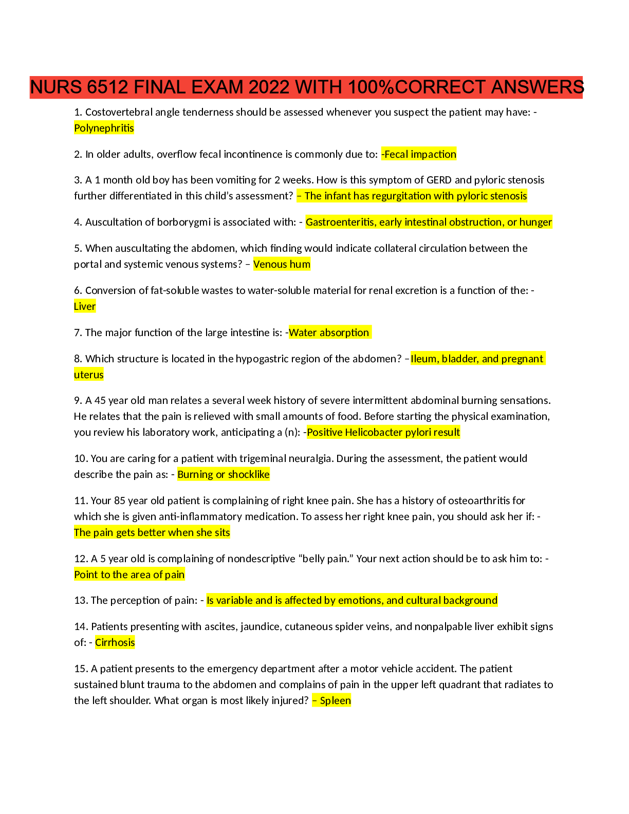
NURSING 602 Module 5 Questions for Self-Study Alterations in Oxygen/Ventilation Normal Mechanisms of Oxygen/Ventilation and Mechanics of Gas Exchange 1. Differentiate between anatomic, alveol... ar, and physiologic dead space a. Anatomic: consists of conducting airways from nose to respiratory bronchioles – minute ventilation b. Alveolar (functional dead space)- consist of ventilated, but under perfused areas of the lung, waster ventilation – gas exchange. c. Physiologic dead space: is the sum of anatomic and alveolar dead space 2. Outline the mechanics of inspiration and expiration as they affect inspiration and expiration a. Inspiration: the AP diameter of the vest increases. The external intercostal muscles and the diaphragm contract while the internal intercostals relax. Gas enters the lungs by high atmospheric pressure to lower b. Expiration: Lung pressure exceeds atmospheric pressure. Relaxation of diaphragmatic muscles causes the rib cage to decrease in size. VENTILATION DOES NOT EQUAL OXYGENATION 3. Discuss the concepts of chest and lung compliance and airway resistance a. Lung compliance is the elasticity of the lung and is impacted by the volume of air entering the lung. Compliance effects the lungs ability to expand and the easiness or difficulty of the lung to inflate. Emphysema can impair this expansion. Airway resistance – relationship between driving pressure and flow through the pulmonary tree is influenced by radius and the pattern of gas flow. The more narrow the lumen of the respiratory tree, the more resistance to airflow. Turbulent airflow (in upper airway) creates friction and increases resistance to air flow, but normal laminar flow in the smaller airways decreases resistance. 4. Describe the physiologic advantages of surfactant a. Surfactant is a combination of phospholipids and proteins. It also synthesizes in the type II cells of the lungs to prevent alveoli collapse at end expiration – the more expanded alveoli, the more increased lung compliance. b. Advantages: i. Increased lung compliance ii. Decreased work of breathing iii. Promotion of stability of the alveolus 5. Discuss the distribution of ventilation to perfusion the terms ventilation-perfusion inequality in the lung a. When a person is sitting up-right, the alveoli at the top of the lung has less room to expand and are not as well ventilated. The base of the lung is more ventilated and compliance is better. b. Regional differences are less in the supine individual, alveoli pressure exceeds venous pressure. Middle of the lung has higher venous pressure because its closer to the left atrium. Perfusion is continuous 6. What is hypoxic vasoconstriction and why is it an important consequence of mismatching of ventilation to perfusion? a. Pulmonary blood vessels supplying those vasoconstrict. It diverts pulmonary capillary blood flow away from hypoxia alveoli to better ventilated areas. Minimizes wasted perfusion 7. Outline the control of ventilation a. Center control in the brain, in the lower pons. The pneumotaxic center located in the upper pons is responsible for the regulation of inspiration of inspiratory volume and resp rate. b. Effectors control output and include the respiratory muscles and the diaphragm c. Increased CO2 in the blood, liberates hydrogen ion that diffuses into the CSF and lowers the pH leading to an increase in resp drive. d. Carotid receptors are stimulated primarily by a decrease in blood pH Classifications Lung Disease the Affect Oxygen/Ventilation 1. Differentiate among characteristics obstructive, restrictive, and vascular pulmonary diseases? a. Obstructive: Manifested by disorders that cause increased resistance to airflow. Reduction of airways produces obstruction and increases resistance. The resistance can be caused by processes within the airway lumen, in the airway wall or in the supporting structures that surround the airway. b. Vascular: disorders that affect the blood vessels in the lungs. Impairs gas transfer and oxygenation. c. Restrictive: Caused by decreased expansion of the lung. Alterations in the lung parenchyma, chest wall, or pleura can impede lung expansion. 2. What are the effects of TLC, FEV1/FVC and airway resistance in obstructive and restrictive pulmonary diseases? a. TLC: Obstructive – increased, Restrictive- decreased b. FEV1/ FVC: Obs. - <70%, Rest, - >85% Obstructive, Restrictive, and Vascular Lung Disorders 1. Outline the pathophysiology of asthma, chronic bronchitis, and emphysema a. Asthma: disease of airway inflammation, episodic airflow obstruction and increased bronchial responsiveness. Immune activation, IL4, 5, and 13 . Th2 effector response. Mast cells release histamines. i. Hyperplasia of mucous producing goblet cells ii. Mucosal edema iii. Hypertrophied bronchial muscles iv. Bronchospasm v. Narrowed airway lumens b. Chronic bronchitis: Inflammation of airways (mostly smaller). Increased mucous secretions in the airways, mucosal narrowing, decrease in functional cilia, hyperinflation c. Emphysema: a deficit of protease inhibitors with an imbalance of local antioxidant injury. Loss of lung elastic connective tissue decrease in alveolar capillary bed vessels – hypoxemia. 2. What are the causes and consequences of reduction of airway diameter in acute asthma attack? a. The causes of reduction of airway diameter is due to mucous plus getting stuck in the alveoli entrance on expiration, trapping air into the alveoli. Air trapping can cause pulses paradoxus (large decrease in stroke volume during inspiration). Negative pressure is transmitted to the heart and blood vessels and causes a decrease in left ventricular filling. Systolic pressure falls as cardiac output falls momentarily. 3. Describe the pathophysiology alterations that produce the clinical presentation/features associated of emphysema a. Decreased breath sounds- due to decreased airflow with prolonged expiratory time b. Hyperinflation of the lungs is prominent c. Wheezes on expiration d. Pursed lip breathing lessens CO2 retention e. Chronic hypoxemia often leads to polycythemia 4. How does a restrictive disease such as pulmonary fibrosis, pleural, chest wall and neurologic disease affect lung expansion and the rate and depth of ventilation? a. They restrict the normal expansion of the lung by inflammation and thickening of the interstitial of the alveolar wall. b. Breathing is rapid and shallow with an increase of resp rate, inspiratory crackles, digital clubbing of fingers and toes. 5. Describe the pathophysiology of pulmonary edema and its effects on oxygenation/ventilation a. Valvular dysfunction, CAD, or ventricular dysfunction causes an increase in left atrial pressure. Which then increases pulmonary capillary pressure. Protein rich fluids leak into pulmonary interstitial spaces causing the lung interstitial pressures to increase, and facilitates fluid leak into the alveoli. b. Reduced lung compliance, distensibility of the alveoli, reduce gas diffusion, loss of surfactant, reduced gas exchange. 6. What are the common causes of hypoxemia, why do they occur and what are their cardinal features? a. Common causes: pneumonia, PE, pulmonary edema b. Features: Blood gases- low PaO2 and low arterial oxygen saturation 7. Describe the causes and pathophysiology of pulmonary embolism a. Derive from venous thrombi, emboli in the lungs are small but can cause small infracts in the pulmonary bed. Massive emboli can cause cardiovascular collapse 8. Discuss the common clinical manifestation of lung cancer? a. Unproductive cough, hoarse voice, chest pain, dyspnea for pleural effusion. 9. Describe the pathophysiology of cystic fibrosis a. Genetic defect of chloride and sodium transport. Increased water reabsorption leads to dehydrated mucus, reduction in ciliary movement prevents the upward movement of mucus and the removal. Mucus plugs form, accumulate and can lead to airway obstruction. Impairs airway clearance that leads to resp failure. 10. Describe the effects of aging on the pulmonary system a. Conducting airways – calcification, change in size b. Lung parenchyma – enlarged terminal airspaces, ventilation/ perfusion mismatching c. Bellows apparatus – increased rigidity of the chest wall decreased resp muscle strength d. Ventilatory control - markedly decreased response to hypercapnia and hypoxia e. Cardiovascular system – decreased HR and CO decreased responsiveness to hypoxemia [Show More]
Last updated: 1 year ago
Preview 1 out of 9 pages
Buy this document to get the full access instantly
Instant Download Access after purchase
Add to cartInstant download
We Accept:

Reviews( 0 )
$12.00
Document information
Connected school, study & course
About the document
Uploaded On
Oct 03, 2021
Number of pages
9
Written in

Additional information
This document has been written for:
Uploaded
Oct 03, 2021
Downloads
0
Views
34
.png)