NURSING 602 Module 11 Questions for Self-Study,100% CORRECT
Document Content and Description Below
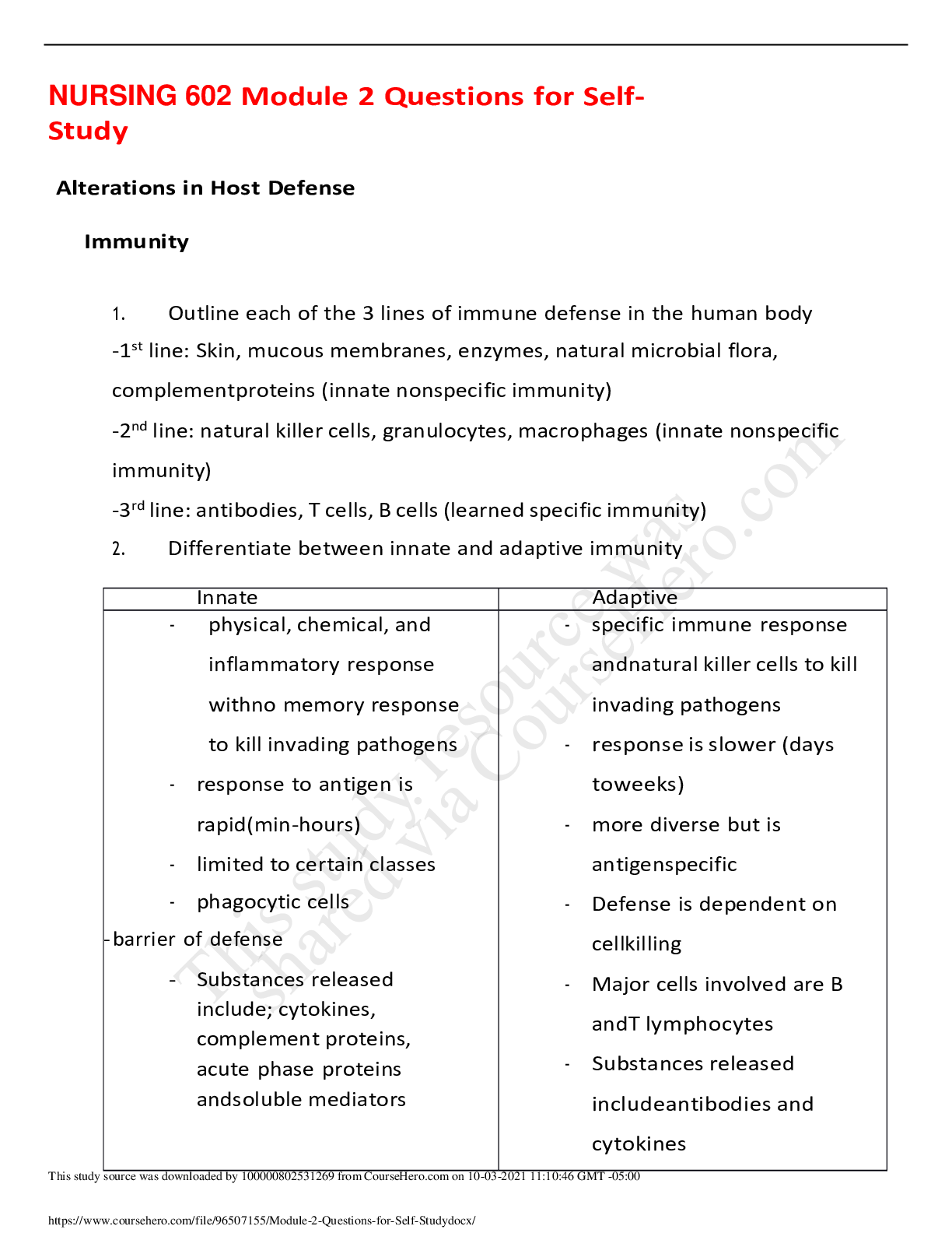
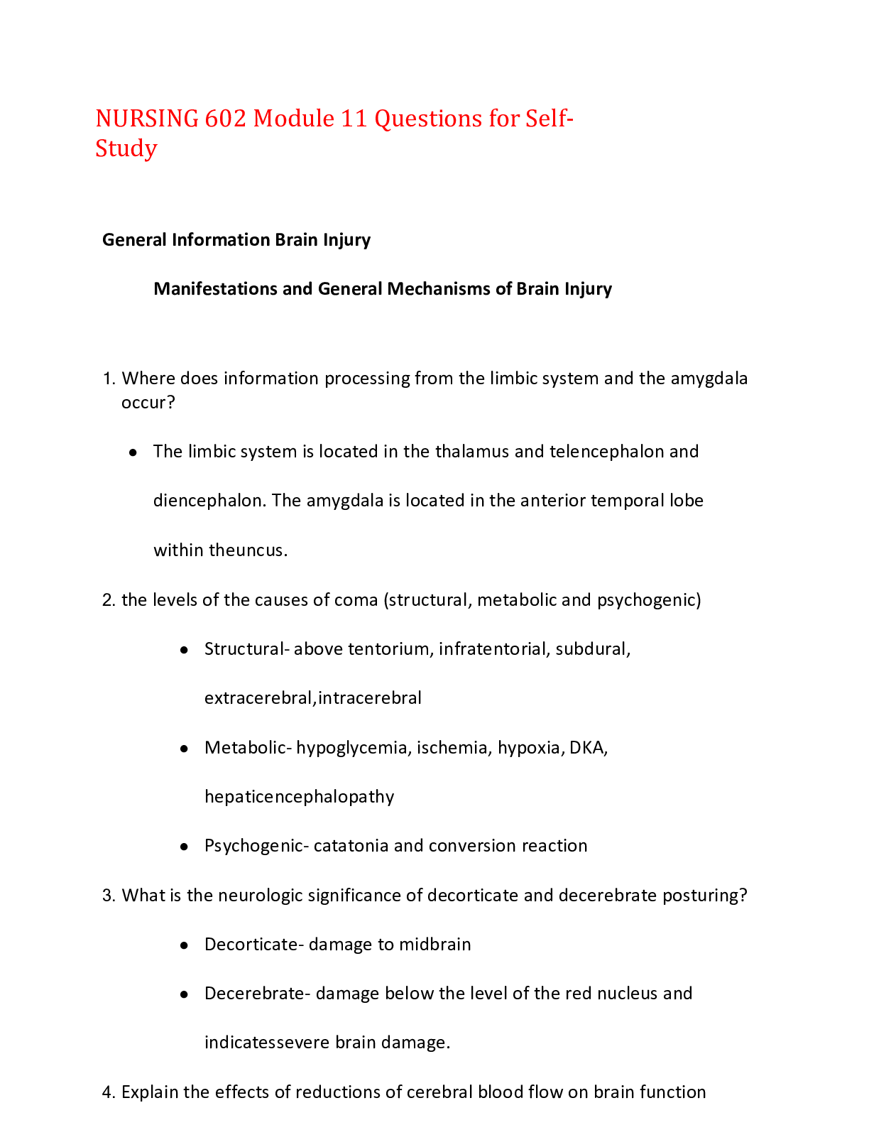
NURSING 602 Module 11 Questions for Self-Study General Information Brain Injury Manifestations and General Mechanisms of Brain Injury 1. Where does information processing from the limbic ... system and the amygdala occur? ● The limbic system is located in the thalamus and telencephalon and diencephalon. The amygdala is located in the anterior temporal lobe within the uncus. 2. the levels of the causes of coma (structural, metabolic and psychogenic) ● Structural- above tentorium, infratentorial, subdural, extracerebral, intracerebral ● Metabolic- hypoglycemia, ischemia, hypoxia, DKA, hepatic encephalopathy ● Psychogenic- catatonia and conversion reaction 3. What is the neurologic significance of decorticate and decerebrate posturing? ● Decorticate- damage to midbrain ● Decerebrate- damage below the level of the red nucleus and indicates severe brain damage. 4. Explain the effects of reductions of cerebral blood flow on brain function ● If reduction of less than 40%, it harms the brain and ionic function as well as electrical conduction. Below 10% leads to imminent cell death. 5. What is the significance of decorticate and decerebrate posturing? ● Decorticate- damage to midbrain ● Decerebrate- damage below the level of the red nucleus and indicates severe brain damage. 6. What factors determine the extent of neurological damage after a brain injury? ● Amount of blood flow reduction, regional location of reduction. Metabolic state of the brain prior to the precipitating event. Hypoxic and Ischemic Brain Injury 1. What is the difference between the effects of hypoxic and focal ischemic brain injury on neurons? ● Neurons are tolerant to hypoxia and are shown as euphoria, drowsiness, and impaired problem solving Intracranial Pressure 1. Identify the effects of increased intracranial pressure on the contents of the skull. Which content is the most deformable if ICP is increased? ● An increase within the skull results in a decrease in one or more other components. ● Interstitial fluid 2. Differentiate between vasogenic and cytoxic cerebral edema on brain cells ● Vasogenic- swelling due to extravasation of electrolytes, proteins, and fluid into extracellular space ● Cytoxic- swelling of tissue due to cellular energy failure. Lack of ATP causes sodium accumulation in cell with osmotic draw into cell Brain Herniation 1. What are the causes of brain herniation? ● Unrelieved growing brain masses, large areas of continuous bleeding, or expanding edema in the brain that increases pressure. 2. Identify the 4 the areas of brain herniation ● Subfalcine- leg weakness ● Central- altered LOC, decorticate posturing, rostral-caudal deterioration ● Uncal- hemiparesis, visual loss, ipsilateral pupil dilation, respiratory arrest ● Tonsillar- quadriparesis, constricted pupils, ataxic breathing, changes in HR and BP 3. Outline the clinical presentation/features associated with each area of brain Herniation ● Refer to #2 Transient Ischemic Attack and Stroke 1. Differentiate between a transient ischemic attack (TIA), and stroke associated Ischemic neurological deficit ● TIA- temporary loss of cerebral perfusion with no permanent loss of function. Its rapid and sensory loss lasts 2-60 min ● Ischemic- Sudden occlusion of cerebral artery. Persists for more than 24 hours. 2. Discuss the pathophysiology of excitotoxic brain injury? What are the consequences on neuronal cell function? ● Excessive glutamate accumulation at synapses due to failure of energy requiring re-uptake mechanisms. ● Cell function is impaired by ischemia, so accumulation occurs 3. Differentiate among the 4 types of stroke and the common causes of each ● Hemorrhagic- caused by severe long-standing hypertension ● Thrombotic- caused by a large vessel occlusion ● Embolic- thrombi release due to cardiac source (a-fib) ● Lacunar- occlusion of small penetrating arteries. Commonly in the basal ganglia, pons, or cerebellum 4. What are the common clinical manifestations of a blockage of the anterior, middle, or posterior cerebral arteries? ● Anterior- paralysis, sensory loss, aphasia, cognitive disorders ● Middle- hemiplegia, aphasia, altered mental status ● Posterior- visual defects, memory defects, and preservation 5. What is meant by the ischemic core, ischemic penumbra, and zone of hyperperfusion and luxury perfusion? ● Ischemic penumbra- core of tissue surrounding the occluded blood vessel that gets no perfusion ● Zone of hyperperfusion- triggers the release of vasodilation ● Luxury perfusion- zone where blood flow exceeds the needs of the brain tissue Traumatic Brain Injury 1. Outline the pathophysiology of concussion/mild traumatic brain injury (TBI) ● Rupture of cellular and vascular membranes, damage to axons, metabolic derangements, neuroinflammation 2. Describe the clinical presentation/features of mild TBI ● concussion / mild TBI- loss of consciousness of less than 30 min, alternation of mental status, fatigue and headache, sleep disturbances, dizziness 3. Describe the 3 grades of concussion ● Grade 1: transient confusion, no LOC, inability to maintain a coherent train of thought, inability to goal-directed movement ● Grade 2: transient confusion, no LOC, amnesia of no more than 15 min, mental status abn. ● Grade 3: Loss of consciousness Coup Contrecoup Injuries 1. Discuss the mechanisms of coup vs. contrecoup traumatic brain injuries. How might the clinical presentation be different for coup vs. contrecoup injuries? ● Coup- localized impact to the skull. ● Contrecoup- brain shifts inside the skull and meninges and may be from bruising or bleeding. 2. Describe the common causes of secondary brain injury resulting from ischemia ● Edema, altered vasoregulation. Inflammation, seizures, and cerebral hypoxia Intracranial Hemorrhage 1. Differentiate between causes of epidural, subdural, and intracerebral hematoma ● Epidural- skull fracture ● Subdural- tears of the bridging veins. Falls, assaults, MVA ● Intracerebral- contusions deep in the brain secondary to head trauma 2. Outline the differences in the clinical presentation of epidural, subdural, and intracerebral hematoma ● Epidural- unconsciousness followed by a lucid interval (talk and die syndrome), confusion, dizziness, drowsiness, enlarged pupils, LOC ● Subdural- slurred speech, severe headache, weakness, numbness, seizure, drowsiness, LOC, N/V ● Intracerebral- concussion but motor skills of stroke Seizure Disorders 1. Identify the major pathophysiologic causes of seizures ● Imbalance between excitability and inhibition in the brain 2. Differentiate between types of partial seizures and generalized seizures ● Partial- simple partial seizures occur when there is abnormal activity in one region of the brain. Complex has changed in level of consciousness ● generalized - affect the entire surface of the brain. 3. Describe the consequences of status epilepticus on metabolic demands of the brain ● Metabolic demands of the brain leads to subsequent brain hypoxia and will produce brain damage Brain Tumors 1. Discuss the clinical manifestations of space occupying brain lesions (such as GBM) ● Headache, confusion, decline in brain function and memory loss, personality changes, speech difficulty, incontinence, balance difficulties 2. Detail the pathophysiological causes for the clinical presentation/features signs associated with space occupying lesions in the brain ● Composed of poorly differentiated neoplastic astrocytes and found in the white matter or cerebral hemispheres. Dementias 1. Outline the initial clinical presentation of Alzheimer’s disease, Creutzfeldt-Jakob disease, frontotemporal dementia, vascular dementia, and Lewy body dementia ● Alzheimer's disease- memory loss, impaired learning ● Creutzfeldt-Jakob disease- dementia/ mood/anxiety/movement disorders ● Frontotemporal dementia- apathy: poor judgement; speech issues ● Vascular dementia- may be sudden onset; apathy, falls focal weakness ● Lewy body dementia- visual hallucinations, REM sleep disorder; parkinsons 2. Discuss pathophysiology Alzheimer’s disease ● Presence of neuronal intracellular neurofibrillary tangles are a hallmark (tau protein). Formation of extracellular amyloid plaques. The inflammation of amyloid proteins result in activation of the complete cascade and generation of acute phase proteins 3. Discuss the cognitive impairment associated with the 7 stage of Alzheimer’s disease ● Individuals lose ability to respond to environment; loss of ability to speak and control movement Review of Spinal Cord Tracts 1. Differentiate between upper motor neuron disorders and lower motor neuron disorders ● Upper motor neurons ● Lower motor neuron Spinal Cord Injury 1. Differentiate between the typical damage that occurs with a non-penetrating vs. a blunt spinal cord injury ● Non-penetrating- compression on the spinal cord or roots. ● Blunt spinal cord injury- fractured vertebral body moved backward compression the spinal cord 2. What are the mechanisms of injury and pathophysiology associated with flexion and a hyperextension spinal cord injury? ● Flexion- head is bent acutely forward and two adjacent cervical vertebrae are forced together. ● Hyperextension- the head is hyperextended and stress is placed on the laminae and pedicles of the cervical vertebrae 3. Differentiate between the cause and clinical features of central cord, Brown-Sequard, anterior cord, and posterior cord syndromes ● Central cord- damage to the center of the spinal cord. Shows deceased in lower extremity sensation and alteration in bladder control ● Brown-sequard- caused by blunt or penetrating trauma, disc or bone herniation or tumors. The is ipsilateral loss of motor function, vibratory sense and proprioception ● Anterior cord- causes by crushing or compression injuries. Loss of motor function, pain, and temperature sensation ● Posterior cord- injury to the posterior cord. Loss of deep touch, position sense, vibration sense and proprioception (may make walking difficult) Selected Movement Disorders Parkinson’s Disease 1. Discuss the pathophysiology of Parkinson’s disease ● Loss of pigmented dopaminergic cells of the SN. The Sn influences motor planning so a loss contributes to the involuntary movements of Parkinson's disease 2. What are the 3 cardinal signs and symptoms of Parkinson’s disease? ● Tremor, bradykinesia, and rigidity 3. Other than the cardinal signs and symptoms what other clinical features are associated with Parkinson’s disease? ● dysphagia , drooling, mask-like facial expressions, fatigue, psychosis, and speech impairment Myasthenia Gravis 1. Describe the pathophysiology and clinical presentation of Myasthenia gravis ● Shallow and abnormally wide or absent synaptic cleft. Ptosis, diplopia, weakness chewing, difficulty swallowing 2. Discuss the pathophysiology underlying the common clinical presentation/features of Myasthenia gravis ● Decrease release with acetylcholine combined with fewer functional synaptic end-plates results in impulse transmission and muscle fatigue Joint Disorders 1. Differentiate between the pathophysiology of rheumatic arthritis (RA), osteo (RA) arthritis and gout ● RA- abnormal activations of B cells, T cells, and innate immune effectors. Release of enzymes causes damage to the synovial membranes of borth large and small joints. ● Osteoarthritis- affects chondrocytes where there are injuries die to mechanical load and frequency of use ● Gout- overproduction of uric acid or reduced ability to get rid of it. The uric acid crystals deposit in joints 2. Discuss the pathophysiology underlying the common clinical presentation/features of RA, OA, and gout ● RA- weakness, fatigue, malaise, and low-grade fever is due to systemic inflammation. ● OA- stiffness, pain, and swelling are caused by cartilage becoming thin or articular bone becomes dense and scleotic ● Gout- fever, elevated sed rate, elevated uric acid levels. Phagocytosis of the uric acid crystals initiates are inflammatory response and causes the release of cytokines 3. What joints are commonly affected by RA, OA, and gout ● Peripheral joints ● OA- DIP joints of fingers, thumb, joint of feet, cervical and lumber spine, and Heberden, Bouchards and hips and knees ● Gout- metatarsophalangeal joint [Show More]
Last updated: 1 year ago
Preview 1 out of 23 pages
Buy this document to get the full access instantly
Instant Download Access after purchase
Add to cartInstant download
We Accept:

Reviews( 0 )
$14.00
Document information
Connected school, study & course
About the document
Uploaded On
Oct 03, 2021
Number of pages
23
Written in

Additional information
This document has been written for:
Uploaded
Oct 03, 2021
Downloads
0
Views
22