Pharmacology > STUDY GUIDE > PHARMACOLO N3365 Complex Exam 3 (GRADED A) Questions and Answers Solutions Study Guide (All)
PHARMACOLO N3365 Complex Exam 3 (GRADED A) Questions and Answers Solutions Study Guide
Document Content and Description Below
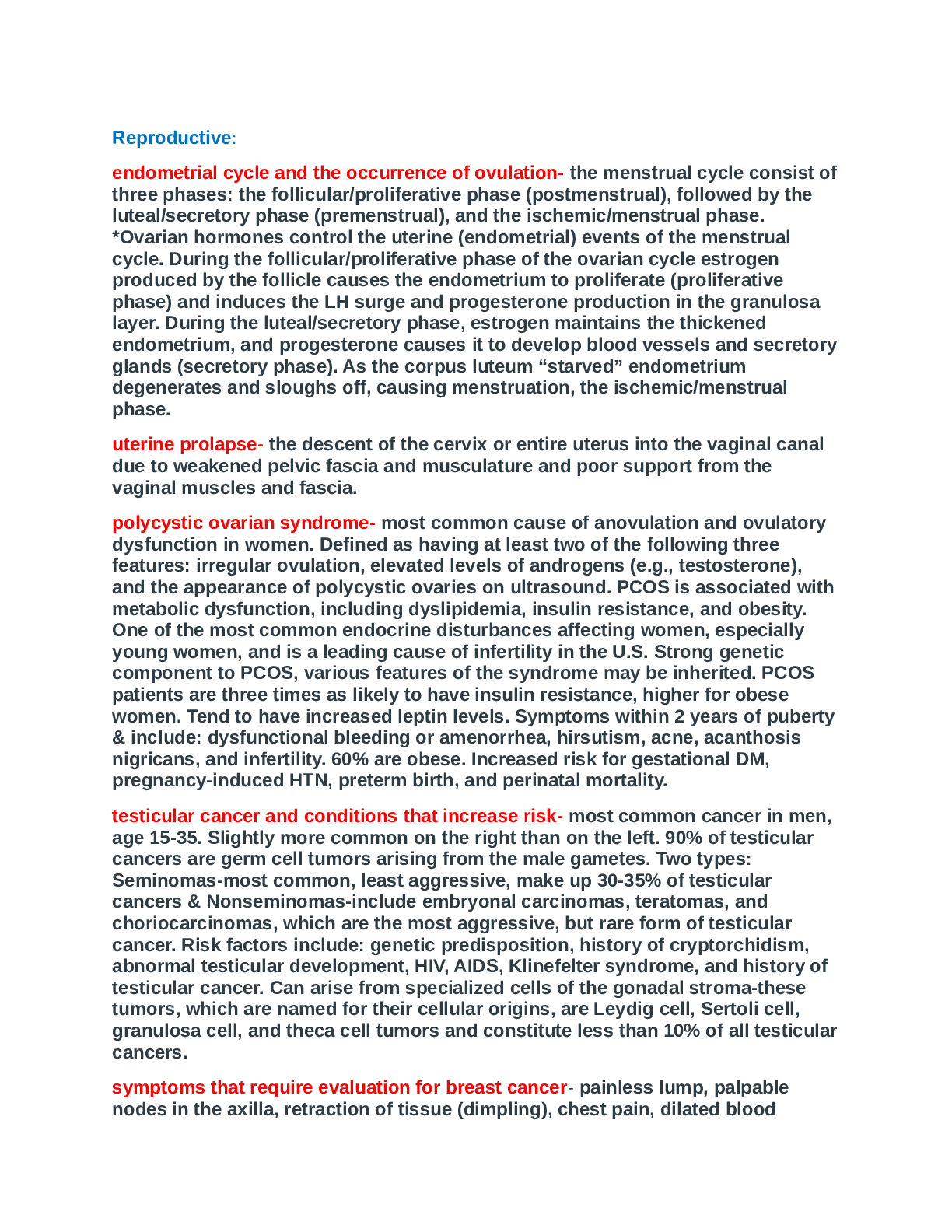
PHARMACOLO N3365 Complex Exam 3 (GRADED A) Questions and Answers Solutions Study Guide Increased Intracranial Pressure: • Pearson: Ch. 11, p. 709-715 • ATI: Ch. 3, pp. 17-24 Patho: • Comp... onents of the Skull – brain, CSF, blood • Monroe Kellie Doctrine • Normal ICP – 5-15 mmHg ABG Assessment: • PCO2 • PaO2 • Hyperventilation Clinical manifestations of Increased ICP (ATI p 75): • Severe headache, Nausea /Vomiting • Seizures • Deteriorating LOC, restlessness, irritability • Pupillary response – dilated, fixed or sluggish, pinpoint nonreactive • Altered respirations: ▪ Cheyne-Stokes respirations ▪ Central neurogenic hyperventilation ▪ Apnea ▪ Arterial blood gases ▪ When to intubate? • Abnormal Posturing/deterioration in motor function: ➢ Decerebrate ➢ Decorticate ➢ Flaccity • Cushing’s Triad—late finding—(severe hypertension, widening pulse pressure, bradycardia) • Cranial nerve dysfunction Interventions: ICP Monitoring Devices: • Intraventricular catheter: “Ventriculostomy”. A fluid-filled catheter: inserted into the lateral ventricles (usually on the right side) through a burr hole. With this device you are able to release CSF fluid through a sterile drainage system (VERY important to maintain sterility here…it’s the brain we are talking about!!) with a 3-way stopcock with transducer connected to a bedside monitor. • Subarachnoid screw or bolt: A hollow, threaded screw or bolt placed into the subarachnoid space through a twist-drill burr hole in the front of the skull, behind the hairline. The bolt is connected by fluid- filled tubing to a transducer leveled at the approximate location of the lateral ventricles • Epidural or subdural sensor: non-invasive because it does not penetrate the dura Level at Foramen of Monroe for calibration of ICP Considerations with ICP monitoring device: • The head is shaved around the insertion site—the site is cleansed with antibacterial solution • Local anesthetic can be used—if client’s GCS is 8-11 • Sterility – This is a very sterile procedure and it’s important to maintain sterility during duration of monitoring. This is to reduce CNS infection • Maintain integrity of system at ALL times—risk for serious, life-threatening infection • Inspect insertion site at least every 24 hours for redness, swelling and drainage. Change dressing per facility and/or as needed. • ICP monitoring equipment must be balanced and recalibrated per facility protocols • Observe ICP waveforms. ICP greater than 15 for a sustained amount of time is considered increased ICP. • Neuro checks as ordered. Typically q15min initially and then gradually q1h (ATI) Cerebral perfusion pressure (CPP): • Normal – 70-95 • MAP-ICP=CPP • DBP+1/3(SBP-DBP)=MAP Non-Pharmacological Nursing Interventions: What Increases ICP • Hypercarbia • ETT suctioning coughing • Extreme neck or hip flexion/extension • HOB less than 30 • Increased intra-abdominal pressure (tight clothing, Valsalva) What decreases ICP • HOB at least 30 • Maintain body midline • Maintain patent airway – mechanical ventilation as necessary • Maintain C-Spine until cleared • Provide a calm environment – Limit visitors if necessary! • Monitor fluid and electrolytes Pharmacological Interventions (ICP): Osmotic Diuretics • Mannitol • Increases cerebral oxygen delivery • Fluid moves from tissues into the blood vessels which reduces ICP by decreasing the total brain fluid content. • Contraindicated in renal disease • Osmotic properties begin to take effect within 15-30 minutes • Insert an indwelling catheter Serum Osmo • (Sodium/2) + (BUN/3) + (Glucose/18) • Normal 265-295 Hypertonic Saline • Produces movement of water out of edematous brain cells into the blood vessels • Requires frequent BO and serum NA levels due to the risk of fluid overload Corticosteroids • Used to treat vasogenic edema surrounding tumors and abcesses • Stabilizes the cell membrane and inhibits the formation of pro-inflammatory mediators • Improves neuronal function by improving cerebral blood flow and restores auto regulation • Causes hyperglycemia • Risk for increased infections • Risk for GI bleed Sedation Barbiturates • High dose • Decreases ICP and cerebral edema • Needs to be closely monitored with brain waves – this is done with a PSA/BIZ (brand names may be called different things) monitor. Electrodes are placed across the forehead. Monitors brain waves. Usually 40-60 is where we want our patients. (this is from my practice, this might have changed in the last couple of years) • Pentobarbital/Thiopental – client may be placed in barbiturate coma to decrease cellular metabolic demand until ICP can be decreased. • Dosage is adjusted to keep the patient completely unresponsive • Mechanical ventilation, cardiac and hemodynamic monitoring and ICP monitoring required Anti-Seizure Medications • Phenytoin is used prophylactically to prevent or treat seizures. • Fosphenytoin – IV form • Suppresses seizures without depressing entire CNS • Client specific dosing and based on therapeutic lab results • Must monitor for drug interactions Opioids • Avoid opioids in those clients who are not mechanically ventilated • Can cause inaccurate neurological assessment • Respiratory depression Diagnostics: • CT • MRI • **CT and MRI are used to diagnose the cause of IICP if not known. Such as hydrocephalus or space occupying lesions.** • A lumber puncture is generally not performed with increased ICP because of the risk of herniation. If an LP is performed, a low pressure shunt is formed at the site and CSF can escape. As CSF pressure drops, herniation can occur. Outcomes/Complications: • Patient can progress to brain death quickly if not treated early!!! • Neuro Assessment is key • Herniation Head Injuries ATI p. 75-76 Some points to remember in regards to head injuries/trauma • Initial GCS • Stabilize C-Spine Causes of Head Injury: • MVC’s • Drug and/alcohol use • Sports injuries • Assault • Gunshot wounds • Falls Types of brain injuries: • Concussion • Contusion • Diffuse Axonal Injury Intracranial hemorrhage: Bleeding in the epidural, subdural or intracerebral spaces following a traumatic event. • Epidural • Subdural ▪ Acute Subdural hematoma ▪ Subacute subdural hematoma ▪ Chronic subdural hematoma • Intracerebral – also known as a hemorrhagic stroke Surgical Intervention: (Module Obj. 5) ATI p. 77 Craniotomy • During a craniotomy a bone flap (piece of the skull) is removed allowing access to hematoma, tumor..etc • Removal of nonviable brain tissue • Removal of brain tumors • Evacuation of epidural or subdural hematomas • Decreases ICP Potential Complications: • Permanent neurological deficits • Seizure disorders • Infection • Death Post –Op Nursing Interventions: • Administer medications to reduce cerebral edema • Monitor ICP • Infratentorial – keep patient flat and on either side for 24-48 hours (prevents pressure on neck incision site) • Supratentorial – maintain HOB at 30 • Assess dressing for drainage Interdisciplinary Care: • PT/OT • Speech therapy • Social Services • Rehabilitation facilities • Rehab can take months to years. Patient and support system must be prepared for varying outcomes Skull Fractures: • Fracture of the base of the skull, typically involving the temporal bone, occipital bone, sphenoid bone, and/or ethmoid bone. • Occur following a forceful head injury • Monitor from drainage from the ears or eyes for CSF fluid which indicates a basilar skull fracture. A “halo” sign (yellow stain surrounded by blood on a paper towel; fluid will test positive for glucose) • No NGT placement with basilar skull fractures or facial fractures Battle’s Sign – bruising over the mastoid process Raccoon eyes – Periorbital edema and ecchymosis Findings: • Amnesia • Loss of consciousness – length of time of unconsciousness is significant • CSF leak Stroke: Cerebral Vascular Accident (CVA): ATI p. 81-84 Disruption in cerebral blood flow secondary to the following • Hemorrhagic • Ischemic • Thrombotic • Embolic Transient Ischemic Attack (TIA): Stroke symptoms without lasting deficits Can be warning of impending stroke Risk Factors: AV malformations A-Fib Cerebral aneurysm Hyperlipidemia DM Oral Contraceptives Obesity Smoking HTN Cocaine use Atherosclerosis Clinical Manifestations: • Slurred speech • Visual disturbances • Dizziness • Weakness to extremities • Manifestations based on area that was affected (deprived of oxygen) Left Sided Hemisphere Right Sided Hemisphere Expressive and receptive aphasia (inability to speak and understand language) Responsible for visual and special awareness and proprioception (sense of self and movement) Agnosia – inability to recognize objects Overestimation of abilities Alexia – difficulty reading Unilateral neglect – more common in right sided strokes Agraphia – difficulty writing Loss of depth perception Right extremity paralysis or weakness Poor impulse control and judgement Slow cautious behavior Weakness or paralysis to left side Depression anger, frustrates easily Visual changes Hemianopsia – loss of visual field in one or both eyes Diagnostics: MRI/MRA Lumbar Puncture GCS Nursing Interventions: • Monitor for fever • Monitor for s/s of decreased LOC • Assist with communication skills • Assist with ADL’s as needed • Assist with immobility • Maintain skin integrity • Maintain safe environment Pharmacological Interventions: • Anticoagulants – controversial and not recommended due to risk of intracerebral bleeding • Thrombolytic (tPA) – give with 4.5 hours • Antiplatelets – low dose ASA given within 24-48 hours to prevent further clot formation • Other antiplatelets not recommended • Antiepileptics – not commonly given unless seizure activity develops • Gapapentin can be given for paresthetic pain in affected extremity Interdisciplinary Care: • Speech therapy • PT/OT • Social Services for rehab Complications: • Dysphagia – risk for aspiration • Not all patients will have dysphagia but should be evaluated prior to reestablishing oral intake • Unilateral neglect – observe affected extremities for injury Spinal Cord Injures ATI p. 87-92 Involves the loss of motor function, sensory function, reflexes and control of elimination Quad=all four extremities and trunk Injuries below T1 result in paraplegia (lower extremities) Risk Factors: The majority of Spinal cord injuries are traumatic events • Male gender • Extreme sports • Impact sports ( football, diving) • Acts of violence • Alcohol drug abuse • Cancer, arthritis • Falls Clinical manifestations: • Lack of sensation – inability to feel light touch • Absent deep tendon reflexes • Flaccidity of muscles • Hypotension • Shallow respirations • Spinal Shock • Neck or back pain Nursing Interventions: • What is the priority? AIRWAY!!! Always and forever! • Provide oxygen and suction as needed • Intubation and mechanical ventilation if necessary • Incentive spirometry • Monitor neurological status – report any decline in neurological function • Fluid support and nutritional support are key as patient will more than likely be NPO for several days. We want bowel and elimination to be as normal as possible. A simple UTI can lead to urosepsis in these patients. Often times, patients with spinal cord injuries survive the trauma only to succumb to massive infections. • Muscle strength and tone – monitor patient for increasing loss of muscle strength. Encourage passive ROM • Mobility – those with complete injuries will not regain mobility. Those with incomplete injuries can regain some function with the assistance of various types of braces. • Sensation Surgical Interventions: • Halo fixation • Spinal Fusion • Laminectomy Outcomes/Complications: • Skin breakdown. • GI • Spastic neurogenic bladder • Flaccid neurogenic bladder • Bladder and bowel training needed • Orthostatic Hypotension • Spinal Shock Autonomic Dysreflexia: • Sudden onset of excessively high blood pressure, severe headache, pallor below the level of the spinal cord lesion • Other S/S – blurred vision, diaphoresis, restlessness, nausea • Stimulation of the PNS – bradycardia, flushing above spinal cord lesion, nasal stuffiness • Secondary to stimulation of the SNS and inadequate compensation of the PNS. • Does not occur with patients with lesions below T6 because PNS is able to neutralize SNS response Nursing Interventions for Autonomic Dysreflexia: • Sit client up to decrease blood pressure • Determine cause • Treat potential causes Diagnostics for SCI: CT, MRI, X-Rays – Used to determine extent of damage and location of blood and/or bone fragments UA, CBC, ABG…etc – used to evaluate for internal bleeding and gas exchange if phrenic nerve is involved Pharmacological Interventions: Glucocorticoids Vasodilators Vasopressors Stool softeners Atropine Anticoagulants Muscle relaxants Analgesics Cholinergics Interdisciplinary Care: ➢ PT/OT ➢ Reestablish mobility with either wheelchair, braces or crutches ➢ Patients with SCI will need a varying degree of assistance regarding home, ADL’s, in home therapy. Patient will more than likely be able to live alone. ➢ Social services ➢ Referral to support groups ➢ Wheelchair accessible home Neurogenic Shock: ATI p. 88 & 92 • The first sign of ANY type of shock is what? -hypotension • Neurogenic shock occurs where is a total loss of communication within the SNS which maintains muscle tone in our blood vessels • Occurs within 24 hours of a SCI • Peripheral vasodilation What to look for? • Hypotension • Dependent edema • Loss of temperature regulation Clinical manifestations: • Bradycardia • Hypotension • Dependent edema • Loss of temperature regulation Interventions: • Monitor VS for hypotension and bradycardia • IVF’s • Atropine • Vasopressors Meningitis: ATI p. 31-32 KNOW the differences in Viral and Bacterial • Viral: o Mumps, measles, herpes, arbovirus (west Nile) o No vaccine!! • Bacterial: o Otitis media o Pneumonia o Sinusitis o Microorganisms: Neisseria meningitides, Streptococcus pneumoniae, Haemophilus influenzae Meningitis is the inflammation of the meninges (protect the brain and spinal cord) Other risk factors: Immunocompromised Direct contamination with CSF Invasive procedure (LP), skull fx, penetrating wound Overcrowded living conditions – dorms, prisons, nursing homes Prevention: VACCINATE!!!! • Hib (haemophilus influenza type b) • PPSV (PCV 13 or 23) “pneumonia vaccine” • Meningococcal (MCV4) Clinical Manifestations: Excruciating, constant headache Assessment Findings: Nuchal rigidity (stiff neck) Fever and chills Photophobia N/V Hyperactive DTR Tachycardia Seizures Red macular rash (meningococcal) Restlessness, irritability + Kernig’s Sign +Brudzinski’s Diagnostics: Culture everything CBC CSF analysis CSF will have elevated WBC, and protein. In bacterial there will be decreased glucose CT/MRI may be done to identify potential mass or cause of ICP Interventions: • Droplet precautions • Minimize exposure to bright light • HOB at 30 • Seizure Precautions • Decrease environmental stimuli Medications: • Antibiotics • Phenytoin • Analgesics • Prophylactic antibiotics are given to those who are in close proximity to the patient….roommates Complications: • Increased ICP: o Decreased LOC o Pupillary changes o Impaired extraocular movements o HOB elevated 30 o Avoid coughing or straining • SIADH: o Abnormal stimulation to the hypothalamic area of the brain—excess secretion of antidiuretic hormone (vasopressin) o Dilute blood o Concentrated urine o Administer demeclocycline o Restriction of fluid o Daily weights • Septic emboli: o Can develop in the hands and feet o Leads to gangrene, Disseminated intravascular coagulation (DIC) or Stroke o Monitor circulatory status of extremities and coagulation labs Brain Tumors: ATI p. 49-51 • Occur in any part of the brain • Cerebral tumors are the most common • Benign • Malignant Tumors apply pressure to surrounding brain tissue Although the exact cause of brain tumors is unknown, some risk factors are noted to have a predisposition • Genetics • Environmental agents • Exposure to ionizing radiation • Exposure to electromagnetic fields • Previous head injury Clinical Manifestations: • Dysarthria • Dysphagia • Vertigo • Hemiparesis • Cranial nerve dysfunction • Papilledema Positive Babinski sign Positive Romberg Diagnostics: • CT/MRI • LP – should not be performed if increased ICP is suspected. Can cause herniation • EEG • Labs to determine electrolytes, renal and endocrine function • Biopsy Interventions/Pre-op Education: • Continue anti-epileptics if given pre-operatively • Stop ASA 72 hours before surgery • Power of attorney should be implemented • Report use of any supplements • No ETOH, tobacco, anticoagulants or NSAIDs 5 days before surgery • Maintain airway! • Monitor neurological status • Maintain safety – assist with ambulation and provide assistive devices as needed • Seizure precautions Medications: • Non-opioid analgesics • Corticosteroids to reduce cerebral edema • Anticonvulsants • H2 antagonists • Antiemetic’s • Post-op – covered under craniotomy section Other interdisciplinary care: • PT/OT • Social Services • Home care • Chemo or Radiation may be still be needed after surgery • Palliative Care Complications: • SIADH: o Fluid retained—overproduction of vasopressin or antidiuretic hormone (ADH) from the posterior pituitary gland o Hypothalamus has been damaged—no longer regulate the release of ADH o Fluid restriction o Administer oral conivaptan o Treatment of hyponatremia o Disorientation, headache, vomiting, muscle weakness, decreased LOC, irritability, loss of thirst, and weight gain o If severe or untreated—seizures/coma • Diabetes Insipidus: o After supratentorial surgery o Large amounts of urine are excreted—deficiency of ADH o Hypothalamus has been damaged and no longer regulate ADH o Massive fluid replacement o Careful attention to lab values o Replacement of essential nutrients [Show More]
Last updated: 1 year ago
Preview 1 out of 15 pages
Reviews( 0 )
Document information
Connected school, study & course
About the document
Uploaded On
Jun 03, 2021
Number of pages
15
Written in

Additional information
This document has been written for:
Uploaded
Jun 03, 2021
Downloads
0
Views
40


.png)
 Questions and Answers Straighterline.png)


.png)