NURSING. > EXAM > NURS 2115 Exam 1 Study Guide Ch. 14, 62, 72 Questions with Answers,100%CORRECT (All)
NURS 2115 Exam 1 Study Guide Ch. 14, 62, 72 Questions with Answers,100%CORRECT
Document Content and Description Below
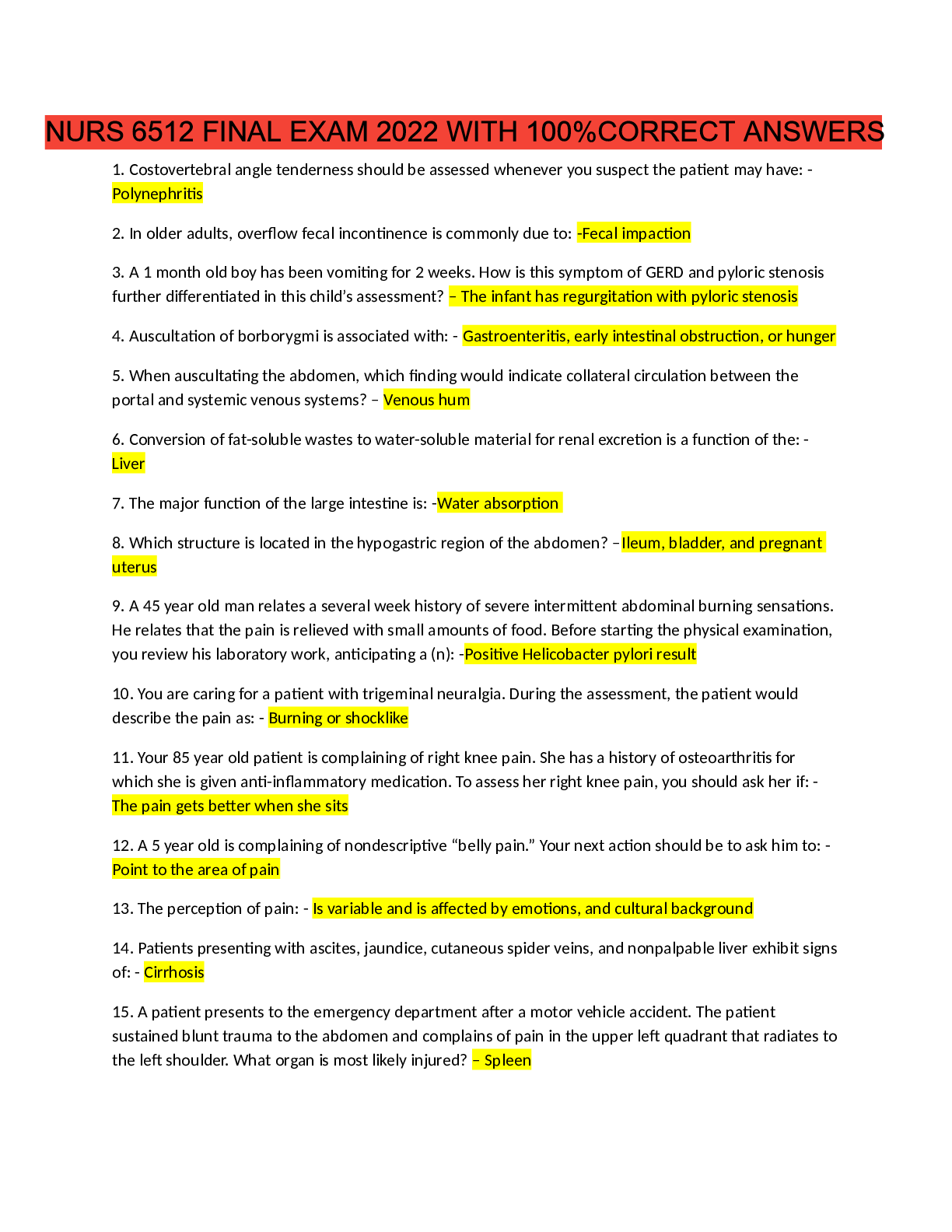
NURS 2115 Exam 1 Study Guide Ch. 14, 62, 72 Questions with Answers 1. Primary survey: you must know A-G assessment and interventions • A: airway and alertness/c-spine stabilization o Alertness as... sessment: ▪ AVPU method: ❖ A: alert ❖ V: responds to voice ❖ P: responds to pain ❖ U: unresponsive o Airway assessment: ▪ Assess for partial or complete obstruction, we need to know whether their airway is effective or ineffective ▪ Assess for hypoxia, brain damage, hypercarbia, respiratory or cardiac arrest ▪ Assess for patency: ❖ Vocalization ❖ Tongue ❖ Loose teeth or foreign objects ❖ Bleeding ❖ Vomitus or other secretions ❖ Edema ❖ Burns ▪ Look, listen, and feel ▪ Avoid yes/no questions to avoid head shaking ▪ Try to get the patient to where they can see your face to decrease desire to move their neck o Interventions: ▪ Stabilize c-spine; assume there is a c-spine injury until proven otherwise ▪ Reposition the head ▪ Heimlich/abdominal thrusts ▪ No trauma: head-tilt-chin lift ▪ Suspected trauma: jaw thrust-we want to avoid manipulating the c-spine ▪ Removal of loose objects or debris ❖ Logroll patient ❖ Stabilize head ▪ Suctioning: g-tube ▪ Insertion of oral or nasal pharyngeal airway ❖ Nasal: doesn’t initiate gag reflex ❖ Oral: initiates gag reflex ▪ ETT insertion/RSI protocols • B: breathing and ventilation o Assessment: ▪ LOC ▪ Spontaneous respirations ▪ Rate, depth, pattern: too fast, slow, or not present ▪ Symmetric chest rise and fall ▪ Presence and quality of bilateral breath sounds ▪ Color ▪ Presence of indicators of work of breathing: ❖ Nasal flaring ❖ Retractions ❖ Head bobbing ❖ Expiratory grunting ❖ Accessory muscle use ▪ JVD ▪ Tracheal position ❖ A tension pneumothorax is indicated and very serious ▪ Chest wall integrity ❖ Sucking chest wound: open wound to chest that has caused a lung to collapse and the patient is breathing through the nose but is also getting oxygen in the chest through the wound, causing increased intrathoracic pressure o Interventions: ▪ Tension pneumothorax: ❖ Oxygen administration: 15L/min with non-rebreather ❖ Prepare to assist with needle thoracentesis if tension pneumothorax is present: i. 14 gauge needle is inserted into chest to release the pressure built up in the collapsed lung that is putting pressure on the other lung ❖ Apply 4x4 dressing and wound and tape it on 3 sides because if all 4 sides are taped there is no way for the oxygen to escape from the chest and decrease the intrathoracic pressure ❖ You can also use an impregnated/Vaseline dressing on the wound instead of a 4x4 gauze • C: circulation and control of hemorrhage o Assessment: ▪ Reduction in circulating blood volume: primary cause of shock ▪ Quick radial and peripheral pulse assessment: rate and quality ▪ Skin color ▪ Capillary refill ▪ Uncontrolled bleeding ▪ Shock s/sp** ▪ Muffled heart tones ❖ Main sign of pericardial tamponade o Interventions: ▪ Pericardial needle aspiration: used for pericardial tamponade ▪ Control external bleeding ▪ Obtain vascular access: as we start IVs, draw blood samples as well ▪ Consider I/O insertion ▪ Administer IVFs ❖ 20mL/kg of crystalloids (NS/LR) ❖ Repeat bolus if needed ▪ Consider blood transfusion ❖ 10 mL/kg ▪ Obtain labs ▪ Initiate cardiac compressions if HR is <60/min in kids • D: disability-neuro status o Assessment: ▪ Brief neuro assessment ▪ Interpret findings based on age and developmental level ▪ Glasgow coma score (GCS) ▪ AVPU ▪ Pupils o Interventions: ▪ Conduct further investigation during 2nd survey ▪ Initiate drug therapy: ❖ Mannitol: cerebral swelling ▪ Consider intubation: if not able to maintain airway because of neuro reasons ▪ Glucose? • E: expose/environmental control o Remove clothing: ▪ Save for forensics ▪ Safety: ❖ Don’t stick your hands in their pockets ❖ Don’t cut through forensics ❖ Watch for glass and other things that may be in the bed o Keep patient warm: ▪ Warm blankets ▪ Warm IVFs ▪ Overhead lights ▪ Temp of room • F: full set of vitals and family presence o Vitals: ▪ Temp, pulse, respirations, BP o Family: family is allowed to be present because if something goes wrong, it helps with acceptance/closure later on for them to see you doing everything you can to help save their loved one • G: get resuscitation adjuncts o L: labs o M: monitor cardiac rhythm o N: naso/orogastric tube: insert especially if they were intubated o O: oxygenation and ventilation analysis ▪ Pulse ox, ETCO2 monitoring o P: pain assessment and management 2. Secondary survey: you must know H-I assessment and interventions • H: history and head to toe o MIST: ▪ M: mechanism of injury ▪ I: injuries sustained ▪ S: signs and symptoms in the field ▪ T: treatment in the field o SAMPLE: ▪ S: signs and symptoms ▪ A: allergies ▪ M: medications ▪ P: pertinent/past medical history ▪ L: last intake/output; meal ▪ E: events leading up to incident o Get history from anyone who is able to give you info ▪ Patient, family, EMS, etc o Spend all the time you need to find every injury and all potential injuries that the patient has or may have ▪ Ex: car injury: blunt injury to abdomen, no anatomical/neurological issues but bruising on outside, need to check for underlying issues that may be causing the abdominal bruising ▪ Bones and muscles may be compressing internal injuries ▪ Car crash assessment: ❖ Impact 1: hit tree/object ❖ Impact 2: hit dash/windshield/road ❖ Impact 3: when your insides hit going the speed you hit • I: inspect posterior surfaces: o Maintain c-spine control o Support extremities with expected injuries o Logroll the patient o Observe/palpate 3. Heat cramps, heat exhaustion, heat stroke. S/SP? What type of treatment interventions will be done? What is the priority of care? • Heat cramps: severe cramps in large muscle groups fatigued by heavy work o S/SP: ▪ Brief, intense cramps that occur during rest after exercise or heavy labor ▪ Nausea ▪ Tachycardia ▪ Pallor ▪ Weakness ▪ Diaphoresis o Treatment: ▪ Rest and fluids • Heat exhaustion: due to prolonged exposure to heat, body is unable to supply fluids to vessels to induce perspiration needed for cooling o S/SP: ▪ Profuse diaphoresis** ▪ Elevated body temp: >100 but <104** ▪ Fatigue, lightheaded ▪ N/V/D ▪ Impending doom ▪ Tachypnea, tachycardia ▪ Hypotension ▪ Dilated pupils ▪ Mild confusion ▪ Ashen color o Treatment: ▪ Place in cool area ▪ Remove constrictive clothing ▪ ABCs ▪ Monitor for arrhythmias ▪ Oral fluids if not nauseated ▪ IV fluids if nauseated • Heat stroke: perspiration-regulating mechanism in hypothalamus has failed causing excessive retention of body heat o S/SP: ▪ Temp >104** ▪ unable to perspire** ▪ Dry, hot, and flushed skin ▪ AMS (confusion to coma) ▪ Tachycardia ▪ Hypotension o Treatment: ▪ ABCDs ▪ Rapid cooling ❖ Cool sheets, ice packs, sponge baths, iced saline lavage, cooling blankets, IVs ▪ Treat until core temp has been lowered to 102 ▪ Thorazine IV: suppress shivering ▪ Monitor for rhabdomyolysis and DIC 4. You will be given several patients and will have to determine who needs to be seen first… you must go by your ABCD’s and most critical/emergent!!!!! 5. In trauma patients, what S/SP relate to the most life threatening conditions? • Head trauma: o S/SP of increased ICP: ▪ Decreased LOC ▪ Pupil dilation ▪ Vomiting ▪ Slurred speech ▪ Inability to track objects o Ataxia o Bulging fontanelle o Posturing o Seizures o Bradycardia o Hypotension, tachycardia, irregular respirations • Thoracic trauma: o Pneumothorax, hemothorax, or tension pneumo o Pneumothorax: air accumulated in the pleural space ▪ Tachypnea ▪ Tachycardia ▪ Pale ▪ Unequal chest rise and fall ▪ Diminished or absent breath sounds o Hemothorax: blood accumulates in the pleural space ▪ Signs or shock ▪ Dyspnea ▪ Tachypnea ▪ Diminished or absent breath sounds ▪ Dullness to percussion* o Tension pneumo: air enters the pleural space on inspiration but cannot escape ▪ Tracheal deviation ▪ JVD ▪ Severe respiratory distress ▪ Faint peripheral pulses ▪ Hypotension ▪ Bradycardia ▪ Altered LOC o Flail chest: 2 or more consecutive ribs on the same side that have been fractured in at least 2 places o Pulmonary contusion: a “bruise” of the lung tissue with hemorrhage & extravasation of plasma and protein into the alveolar and interstitial spaces ▪ Results in atelectasis and consolidation, pulmonary shunting, and hypoxemia ▪ Respiratory distress ▪ Tachypnea ▪ Localized rales or wheezes ▪ Hemoptysis ▪ Hypoxemia • Pericardial tamponade: collection of blood in the pericardial sac o Restricts diastolic ventricular filling & blood flow in and out of the ventricles o Venous pressure rises o CO and arterial BP fall o Dyspnea o Cyanosis o Signs of shock o Decreased BP o JVD o Muffled heart tones** o Bradycardia o Dysrhythmias: PEA, asystole • Myocardial penetrating injury: 60-90% die before getting to the hospital • Injury to aorta: rapid deceleration • Abdominal trauma: o S/SP of shock o Kids can compensate for 25% of blood volume loss without any evidence of S/SP o Stabilize penetrating item in body • Pelvic trauma: o Lacerated or ruptured bladder is usually consequence of blunt trauma o Bladder injuries frequently associated with pelvic fractures o Hematuria: blood at meatus can be assumed that there is pelvic or bladder injury o Abdominal pain o Voiding problems 6. Fluid resuscitation • Shock/burns/trauma: 20 mL/kg bolus • Sepsis: 30 mL/kg bolus • Burn patients: 30-50mL/kg of urine output • Extreme blood loss: blood transfusion 10mL/kg • Crystalloids: NS/LR • 3:1 ratio: it takes 3 drops of NS/LR to replace 1 drop of blood for volume • It takes 1 drop of blood to replace 1 drop of blood for volume 7. Why is mechanism of injury significant? • It is often a predictor of traumatic injury patterns • Can indicate maltreatment of the patient 8. Blunt vs. penetrating trauma • Blunt: does not disrupt the skin o Presentation may be subtle* o Can be sneaky injuries because they do not always show the damage due to no cuts or skin disruption o Ex: ▪ MVC, motorcycle, bicycle crash ▪ Auto vs. pedestrian ▪ Recreational injuries • Penetrating: disrupts the skin o If the penetrating object is still there, leave it in place ▪ It can be shunting blood ▪ Removing could cause major blood loss or hemorrhaging 9. Be able to triage a patient as emergent, urgent, or non-urgent or level I,II, or III • Emergent: highest priority o Need immediate intervention or life, limb, or vision may be a problem o Hypovolemic shock, coding, asthma attack, ABC problems • Urgent: need prompt management o Can wait up to 2 hours without compromise, reassess in 2 hours for changes o Kidney stones, lacerations with controlled bleeding, possible fractures • Non-urgent: minor o Can wait more than 2 hours without compromise o Coughs, colds, sprains • Vital may give a better idea on how to triage 10. What is the Five-Level Emergency Severity Index (ESI)? • Gets the right patient to the right resources at the right time • Incorporates: o Concepts of illness severity o Resource utilization: ▪ EKG, labs, x-ray, IVFs, etc • A: Requires immediate life-saving intervention? o Yes: level 1 • B: High risk situation? Or confused/lethargic/disoriented? Or severe pain/distress? o Yes: level 2 • C: How many different resources are needed? o None: level 5 o One: level 4 o Many: assess danger zone vitals • D: danger zone vitals? o <3 months: HR: >180; RR: >60; SaO2: <92% o 3 months-3 years: HR: >160; RR: > 40; SaO2: <92% o 3-8 years: HR: >140; RR: >30: SaO2: <92% o >8 years: HR: >100; RR: >20; SaO2: <92% o Yes: consider level 2 o No: level 3 • ESI level 1: o Cardiac arrest o Respiratory arrest o Severe respiratory distress o Critically injured trauma patient who presents unresponsive o Overdose with a RR of 6 • ESI level 2: o 3 broad questions to determine ESI 2 or not: ▪ Is this a high risk situation? ▪ Is this patient confused, lethargic, or disoriented? ▪ Is the patient in severe pain or distress? 11. How should family presence during a resus or trauma be handled? What is the ENA recommendation? • Families are allowed to be in the room because it is beneficial to the patient • Nurses should identify with patient which family members should be present should a life-threatening event occur • Support family member during event • Assist families in understanding the event as it unfolds, providing support during the crisis • Gives family a sense of acceptance later on if something goes wrong. They know that you did everything you could to save their family member 12. Hypothermia: how are these types of injuries handled or treated? Nursing interventions? • Core temp is 95 degrees or less • Shivering suppressed below 90 degrees • Death occurs when core temp falls below 78 degrees • Nursing interventions: o Monitor: ▪ Vitals, CVP, urine output, ABGs, blood chem, chest x-rays freq ▪ Core body temp: esophageal bladder or rectal thermistor ▪ Continuous ECG o Re-warming/risk for afterdrop ▪ Passive external: mild- 90-95 degrees ❖ Over bed heaters to extremities and increases blood flow to acidotic, anaerobic extremities ▪ Active external: mild- 90-95 degrees ❖ Forced air warming blankets ▪ Active internal (core): used for mod-severe- <82.5-90 degrees ❖ Cardiopulmonary bypass, warm fluid admin, warm humidified O2 by ventilator, and warmed peritoneal lavage ▪ Discontinue rewarming efforts when core temp is 93 degrees o Correct dehydration and acidosis ▪ Correct hypotension and dehydration with IVF ▪ Admin sodium bicarb to correct acidosis o Avoid defib: ▪ Temp <90 degrees may experience v-fib if moved or touched ❖ Avoid moving patient 13. Snake bites: how are these types of injuries treated? Nursing interventions? • Treatment: o Initial first aid at site: ▪ Lie down, remove constrictive clothing, provide warmth ▪ Clean wound, cover with light sterile dressing ▪ Immobilize body part below heart level ▪ ABCs o Anti-venom is the only way to truly get rid of the pain and save them o Crotalidae (Cro-fab): given for pit vipers • Nursing interventions: o Remove constrictive clothing/jewelry o Assess extremity swelling o Immobilize affected extremity at/below heart level o Give tetanus and analgesia as necessary o You can give a sedative (Ativan) to take off the edge o Do NOT use ice or tourniquets 14. Can shock occur in a burn patient? Why or why not? • Yes, burn damage causes the inflammatory response to cause a change in capillary permeability o The capillaries have larger holes and allows fluids to leak out of the intravascular space causing the blood volume to decrease o The larger molecules (albumin) also leaks out of the intravascular space 15. Why do electrical burn patients often experience acute renal failure? Treatment? • Characterized by entry and exit wound • Their renal tubules cannot receive blood because the tissue is burned and reduces vasculature • Treatment: o Obtain serum creatinine: degree of muscle injury in early phase of care o IVF titrated to a higher target of urine output: 75-100 mL/hr ▪ Add 50 mEq of sodium bicarb/L of IVF o Surgical treatment o Sequential surgical debridement may be necessary 16. Know the different burn depths based on S/SP. Nursing interventions for burn patients. • First degree: superficial: only the epidermis is effected o Sunburns, low-intensity flash, superficial scald o S/SP: ▪ Tingling ▪ Hyperesthesia (hypersensitivity) ▪ Pain soothed by cooling ▪ Peeling ▪ Itching o Appearance: ▪ Red/pink ▪ Blanches with pressure ▪ Dry ▪ Minimal or no edema ▪ Possible blisters o Nursing interventions: ▪ Oral pain meds ▪ Cool compresses ▪ Skin lubricants • Second degree: partial thickness: o Scalds, flash flame, contact o Superficial: extends through epidermis to the upper layer of dermis o Deep: extends through epidermis to the deeper layers of the dermis ▪ S/SP: ❖ Pain ❖ Hyperesthesia ❖ Sensitive to air currents ▪ Appearance: ❖ Blistered, mottled red base ❖ Disrupted epidermis ❖ Weeping surface ❖ Edema ▪ Nursing interventions: ❖ Recovery 2-3 weeks ❖ May require some grafting • Third degree: full thickness: epidermis, dermis, and sometimes subq tissue; may involve some connective tissue and muscle o Flame, prolonged exposure to hot liquids, electric current, chemical, contact o S/SP: ▪ Insensate ▪ Shock ▪ Myoglobinuria (red pigment in urine) and possible hemolysis ▪ Possible contact points (entry/exit points in electrical burns) o Appearance: ▪ Dry ▪ Coagulated blood vessels may be visible ▪ Eschar: black + brown, yellow, white, red, pale ▪ Edema ▪ Damage all the way through the nerves o Nursing interventions: ▪ Eschar may slough ▪ Grafting necessary • Fourth degree: full thickness plus fat, fascia, muscle, and/or bone: o S/SP: ▪ Shock ▪ Myoglobinuria and possibly hemolysis o Appearance: ▪ Charred o Nursing interventions: ▪ Amputation likely ▪ Grafting is not beneficial • Nursing interventions/care: o On the scene: ▪ Remove threat ▪ ABCDEs ❖ 100% humidified oxygen if carbon monoxide poison suspected ▪ Cool the burn ▪ Remove restrictive objects ▪ Initiate at least 1 large-bore IV ▪ Stabilizing patient ▪ Cover wound with dry, clean cloth or gauze o Emergent: ▪ Provide 100% humidified oxygen ▪ Assess breath sounds, RR, rhythm, depth, and chest symmetry ▪ Observe for erythema or blistering of lips or buccal mucosa, singed nasal hairs, burns of face, neck, or chest; increasing hoarseness, soot in sputum or tracheal tissue in respiratory secretions ▪ Monitor ABG values, pulse ox, and carboxyhemoglobin levels ▪ Monitor vitals, hemodynamics, and urine output ❖ 30-50 mL/hr ❖ 75-100 mL/hr in electrical burn patients ▪ Elevate burned extremities ▪ When patient goes into burn shock aka capillary leak syndrome ▪ Abnormal labs: ❖ K+: intracellular K+ is released into the body causing an elevation ❖ HCT: falsely elevated because fluid level is lowering but RBCs aren’t moving ❖ BUN/creat: elevated because fluids can’t remove toxins ❖ Output: decreased i. Is it sufficient? ii. Make sure it’s at least 50mL/hr ❖ ABG: respiratory alkalosis because of hyperventilation ❖ HR: tachycardia ❖ BP: hypotension ❖ CVP: decreased until intravascular volume is replenished ❖ GI mucosa: high risk for curling’s ulcer ▪ LR are usually used; can replenish what is being lost with the ▪ Airway: ❖ Confined space: predisposed to carbon monoxide poisoning i. May appear as O2 sat within range but pt is actually suffocating ii. May be flushed ❖ Burns to face or neck? i. Was there smoke that occurred in the face? ii. Risk for laryngeal edema or airway closure? 1. Inflammatory response acts the same way in your laryngeal tissue causing change in capillary permeability ❖ Singed nasal hairs? ❖ Dyspneic? ❖ Stridor? ▪ Circumferential burns: ❖ Escharotomy o Acute: occurs when the patient responds to fluid resuscitation ▪ What occurs? ❖ Diuresis ❖ Tissues are starting to heal ❖ Capillaries are not leaking as much ▪ Manifestations? ❖ Copious urine output ❖ Improved BP ❖ Elevated CVP ▪ Fluid replacement? ❖ Not as high of a priority ❖ Still needs to be managed ▪ Colloids appropriate? ❖ In the emergent phase: contraindicated ❖ Acute phase: may be appropriate ❖ Gelatinous solutions used to maintain high osmotic pressure ▪ Pain control: ▪ Debridement: removed cellular debris ❖ Enzymatic debridement: special enzymes applied to wound to allow for easy removal of debris i. Removes some of healthy and alive skin ❖ Manual: i. Tweezers ❖ Shower: hydrotherapy ▪ Grafting: ❖ Promotes healing ❖ Homographs: human skin ❖ Heterographs: animal skin ❖ Autografts: graft from your own body ▪ Pressure dressings/garment: ❖ Prevent contractures with hypertrophic scarring ❖ Minimize hypertrophic scarring ❖ Improves venous return to the heart: decrease venous stasis o Rehab: ▪ Main focus: ❖ Prevent infection ❖ Nutrition: calories, calories, calories i. Hypermetabolic ❖ Hypothermia: pt is going to be cold i. Needs extra calories to heal ii. Keep room and space warm bc they can’t regulate body temp: room should be 80-85 degrees ❖ Fluid and electrolytes: i. Continuous monitoring ❖ Interprofessional care: i. Psychosocial ii. Social work iii. PT/OT iv. Dietician 17. Understand basic principles of burn dressing changes • Clean the wound: mild soap, water, and a wash cloth • Apply the prescribed topical agents • Cover wound with several layers of dry dressings with lighter dressing over joints to allow for mobility • Circumferential dressing should always be applied distally to proximally in order to promote return of excess fluid to central circulation • Occlusive dressings, gauze, and topical antimicrobial agent may be used over areas with new skin grafts • Dressings that adhere to the wound bed may be removed more comfortably and with less damage to healing tissue by moistening the dressing with water • The patient may participate in removing the dressings • Wounds are then cleaned and debrided to remove any remaining topical agent, exudate, and nonviable tissue • Sterile scissors and forceps may be used to trim loose eschar and encourage separation of devitalized tissue • Document: color, odor, size, exudate, signs of re-epithelialization, any changes from previous dressing change, and other key characteristics 18. How would you treat a patients with a chemical spill? • Irrigate immediately: skin should be flushed with a constant stream of water as patient’s clothing is removed • Prolonged lavage with tepid water is important • Decontamination shower: deluge in ED o Wear proper PPE to prevent cross contamination • Attempt to determine the identity and characteristics of agent to specify future treatment • Standard treatment appropriate for size and location of wound is initiated o Antimicrobial, debridement, tetanus prophylaxis, antidote admin • May require plastic surgery for further wound management • Patient must have affected area reexamined at 24 and 72 hrs and in 7 days 19. What is the problem with circumferential burns? What is the priority of care? • Burn occurs all the way around a body area: limb, torso, etc • Eschar forms that becomes inelastic tissue and is not compliant • Eschar does not allow for swelling and tissue to leak out, which could obstruct arterial blood flow • Priority: o Providing a way for the edema to be released o Escharotomy: incision cut down the length of the extremity or affected area to allow for tissues to expand underneath the eschar 20. Parkland formula • Tells us the amount of fluids to administer in the first 24 hrs post burn • Pt weight in kg x %TBSA x 2-4 mL= amount of fluids to give • First 8 hrs: give ½ of the total amount of fluids o Give amount from the time of the burn, not the time brought in • Next 16 hrs: give the other ½ of the fluids 21. Pain management in the burn patient • Want patient comfortable to decrease stress response • Provide a long-acting analgesic agent that will provide even coverage o Helpful to use escalating doses o PCA pump o Opioids o NSAIDS o Anxiolytics o Anesthetic agents • Educate patient and family about burn pain and its relationship to the depth of injury as well as pain management plan • Nonpharmacologic therapies 22. Rules of nines to determine extent of burn • How to calculate the total body surface area burned to know how much fluids to give • Anterior of head: 4.5% • Whole head: 9% • Anterior of arm: 4.5% • Whole arm: 9% • Anterior chest: 18% • Posterior chest: 18% • Whole abdominal area: 36% • Anterior leg: 9% • Whole leg: 18% • Perineal area: small amount 23. Organ perfusion depends on MAP. Be sure you understand the concepts related to MAP regarding the CO, total blood volume, and vascular bed tone. Calculate MAP. • Total volume and cardiac output are DIRECTLY related to MAP o Increase in total volume or CO= increase in MAP o Decrease in total volume or CO= decrease in MAP ▪ Ex: hypovolemia • Vascular bed size is INVERSELY related to MAP o Increase in size= decrease in MAP o Decrease in size= increase in MAP o Systemic vascular resistance (SVR): the tension that increases or decreases on the vessels to help move the blood along our blood system ▪ Increase in SVR (vasoconstriction)=increase in MAP ▪ Decrease in SVR (vasodilation)=decrease in MAP o Afterload: what your heart has to pump against to get blood out ▪ Afterload is affected by SVR • Calculate MAP: systolic BP + 2(diastolic BP) ÷ 3 o MAP should be >70 mm/Hg for good perfusion 24. Know the different stages of shock and what S/SP you would expect a patient to exhibit in the different stages. Be sure to include the different types of shock. What is happening with the compensatory mechanisms in the different stages of shock? • Shock: widespread abnormal cellular metabolism that occurs when the human need for oxygenation and tissue perfusion is not met to the level needed to maintain cell function o Syndrome characterized by decreased tissue perfusion and impaired cellular metabolism o It is a “syndrome” • Classifications of shock: o Low blood flow: inadequate volume ▪ Cardiogenic shock ▪ Hypovolemic shock: you don’t have blood in the right places o Maldistribution of blood flow: you have blood, but not in the right places ▪ Neurogenic shock ▪ Septic shock ▪ Anaphylactic shock • Stages of shock: o Initial: very quick and not usually recognized because the body is not having to compensate yet ▪ Slight variation in MAP: decrease of 5-10 mm/Hg ▪ Metabolism changes from a decrease in aerobic to an increase in anaerobic ▪ Lactic acid begins to accumulate and must be removed by blood and broke down by the liver ▪ Increase in HR ▪ Heart and respiratory rate increased or slight increase in diastolic BP ▪ Adaptive responses of vascular constriction and increased HR ▪ S/SP: ❖ HR and RR slightly increased o Compensatory (nonprogressive): where we want to recognize shock** ▪ Neural, hormonal, and chemical compensatory mechanisms activated ▪ Can last for hours ▪ Homeostasis is maintained ▪ S/SP: ▪ FIRST SIGNS: subtle changes in LOC, HR, and RR** ❖ Oriented to person, place, and time ❖ HR: >100/min ❖ RR: >20/min ❖ Decrease in MAP 10-15 ❖ Restless, agitated, apprehension, change in LOC ❖ Cool, clammy skin ❖ Decrease in urine output ❖ Decreased bowel sounds ❖ Increased blood glucose ❖ Decreased reflexes ❖ Dilated pupils ❖ Respiratory alkalosis in beginning stages of ABG ❖ Thirst ❖ Hyperkalemia ❖ Metabolic acidosis ❖ Tissue hypoxia in nonvital organs o Progressive: ▪ Begins as compensatory mechanisms begin to fail ▪ S/SP: ❖ HALLMARK: decreased cellular perfusion and altered capillary permeability** ❖ Sustained decrease in MAP of >20 mm/Hg from baseline ❖ BP is too low to sustain vital organs Heart, brain, and lungs are starting to become hypoxic ❖ Vital organs can only tolerate this for a short time before permanent damage occurs due to hypoxia ❖ Patient is third spacing: going to look very swollen, they’re sick as crap i. Third spacing messes up their capillary permeability and things can’t get out of the vessels ii. It sounds like wet lungs when auscultating, alveoli can’t receive oxygen like it needs and everything is leaking out of the lungs ❖ The gut can get so dry and crack and bacteria can invade i. Give prophylactic meds to keep from cracking ▪ Interventions have to be very aggressive to help ▪ Conditions causing shock need to be corrected within ONE hour of the onset progressive stage o Refractory: ▪ Body is unable to respond effectively to interventions and shock cont. ▪ Remaining cells perform metabolic function anaerobically ▪ Too much cell death and tissue damage result from too little O2 to tissues ▪ Waste products are not being eliminated ▪ S/SP: ❖ Liver and kidney labs are going to be extremely elevated ❖ Lactic acid will be elevated i. Byproduct of anaerobic activity o Leads to SIRS and MODS o There is no clear cut divisions between the stages** • What is happening with compensatory mechanisms during different stages: o Initial: no compensatory mechanisms necessary yet o Compensatory: all systems start to attempt preventing deterioration of circulation by attempting to return CO and arterial pressure to normal limits ▪ Neural compensation: ❖ Decrease reflexes, change in LOC, pupils dilated, resp alkalosis in beginning stages on ABG ▪ Catecholamine effect: epi and norepi cause increase in HR ❖ Body thinks the BP drops a little, speeds up HR and RR to keep organs perfused ▪ Endocrine compensation: ❖ Increase HR: causes O2 need in heart to decrease ❖ Contractility strength decreases ▪ Aldosterone: shunts blood from organs ❖ Periphery and skin get shunted to keep blood in heart, lungs, and brain: skin is cool and clammy ❖ Kidneys decrease producing urine to prevent loss of blood volume and cause a decrease in urinary output ❖ GI decreases motility and causes a decrease in bowel sounds i. May complain of thirst, or dry coating of paste in mouth ▪ Chemical compensation: ❖ Increased glucose levels: body tries to give you more energy ❖ EARLY and EXPECTED finding ▪ Plasma shifts: patient starts 3rd spacing, fluids shifts from intravascular to interstitial o Progressive: compensation mechanisms start to fail ▪ Kidneys: no urinary output at all ▪ GI: no bowel sounds ▪ Chart on slide 21 ▪ Hypotension: SNS stimulation ❖ B-adrenergic: myocardium i. Decreased contractility ii. Increased HR iii. Results in decreased CO ❖ A-adrenergic: widespread profound vasoconstriction i. Capillaries, renal, lungs, viscera ▪ Capillaries: increased hydrostatic pressure ❖ Shift of fluid from intravascular to interstitial ❖ Results in decreased circulating volume ▪ Renal: renin-angiotensin activation ❖ Results in increased arteriolar constriction ▪ Lungs: arteriovenous shunting ❖ Leads to hypoxemia ❖ Results in decreased oxygen availability ▪ All compensatory mechanisms lead to progressive tissue hypoxia ❖ Leading to anaerobic metabolic ❖ Resulting in metabolic acidosis o Refractory: patient is unresponsive to all comp mechanisms 25. How do you treat the different types of shock? Be able to prioritize treatment • Hypovolemic shock: occurs when there is a loss of intravascular fluid, volume is inadequate to fill the space o Too little circulating blood volume causes a decrease in MAP so the body’s total need for oxygenation is not met (cellular hypoxia) o PRELOAD issue: amount (volume) of blood in the heart before it ejects into the body; indirect measure of volume o Causes: ▪ Absolute and direct loss: result of external fluid loss ▪ Relative and indirect loss: result of internal shifting of fluid-third spacing o Treatment: ▪ Establish and maintain patent airway/c-spine control until x-ray is good ▪ Admin high flow O2- 15L/min with nonrebreather or bag mask ▪ Establish 2 large bore IVs ▪ Control any external bleeding ▪ Assess for life threatening injuries ▪ Consider vasopressor therapy only after nothing else has worked and hypovolemia is corrected ❖ Epinephrine ▪ Foley and NG insertion ▪ Treat arrhythmias ▪ Fluid resuscitation: ❖ Crystalloids: NS/LR are first line treatment ❖ Colloids: volume expanders NOT touched until everything is tried with crystalloids i. Hespan, albumin, dextran ❖ Replace 20mg/kg via fluid bolus as fast as possible i. Use 14/16 gauge IV and 2 IVs if possible ii. Go straight for AC iii. Use larger tubing if possible iv. Avoid pig tails and hep locks v. Can infuse with a pressure infuser bag, inflate to 200 vi. Rapid infuser: warms fluids to prevent hypothermia, volume overload, and coagulation problems ❖ 3:1 rule: 3 mL of saline to replace 1 mL of blood lost ❖ 1:1 rule: blood infusion, if no improvement after fluid replacement ▪ Drug therapy: only given after fluid resus doesn’t work ▪ Trendelenburg position o Chronotropic means numbers ▪ Positive chronotropic effect: increase HR ▪ Negative effect: decreases HR ❖ Beta blockers, calcium ch blockers o Inotropic: strength of contractility ▪ Positive: stronger contraction of the heart ❖ Digoxin ▪ Negative: weakened strength of contraction • Cardiogenic shock: special kind of shock during which the heart does not adequately pump enough blood into the body o Actual heart muscle is unhealthy o Contraction is directly impaired o Decreased blood flow to tissues o Affects patients whose area of infarction involves at least 40% of the left ventricle o S/SP of left failure: ▪ Pulmonary congestion ▪ Acute pulmonary edema ▪ Decreased cardiac output ▪ Atrial gallop (S4) ▪ Ventricular gallop (S3) ▪ Wheezing breath sounds ▪ Weight gain o S/SP of right failure: ▪ Low cardiac output ▪ JVD ▪ Dysrhythmias ▪ S3, S4 ▪ Decreased breath sounds ▪ Cause of RV failure along us often underlying lung disease o Treatment: ▪ Fix underlying problem ▪ May give fluids depends on what part of the heart is involved ▪ Drug therapy: lots of drugs ❖ Beta blockers ❖ TPA ❖ Blood thinners ▪ Oxygen: 2-6 L/min ▪ Pain: IV morphine • Distributive shock: caused by a loss of sympathetic tone, blood vessel dilation, pooling of blood in venous and capillary beds, and increased capillary permeability o All leads to decrease in MAP o Not a volume problem, it’s a vessel problem o Problem with sympathetic tone: vessels are too big, and blood cannot move like it needs to o SVR isn’t present to squeeze the vessel and it doesn’t pump effectively o Blood pools and can cause problems with capillary permeability and coagulation issues • Anaphylactic shock: acute and life-threatening allergic reaction o S/SP: ▪ Wheezing: decrease in wheezing is not good ▪ Flushed: huge histamine release ▪ Cyanotic around mouth ▪ Hives/rashes ▪ Anxious ▪ Tachypneic o Treatment: ▪ Airway is #1 priority ▪ First line of treatment: epinephrine IV or subq ▪ Second treatment: ❖ Nebulizers ❖ Benadryl for itching ❖ Solumedrol for swelling ▪ Intubation may be necessary if airway is completely closed • Neurogenic shock: hemodynamic phenomenon that occurs after a spinal cord injury at T5 or above o Results in massive vasodilation without compensation as a consequence of the loss of SNS vasoconstrictor tone ▪ Leads to pooling of blood in vessels o Early S/SP: ▪ Hypotension** ▪ Bradycardia** ▪ SNS is messed up ▪ Poikilothermia: patient’s body temp takes on room temp because of issue with SNS and hypothalamus o Treatment: ▪ Keep trauma bay at 75 degrees ▪ Maintain c-spine ▪ Don’t give fluids ▪ First line of treatment: give vasoconstriction drug to improve BP ❖ Levophed, epinephrine 26. SIRS, sepsis, septic shock: what is the difference? • SIRS: (systemic inflammatory response syndrome) systemic inflammatory response to a variety of initial insults including infection, ischemia, infarct, and injury o Causes: trauma, burns, pancreatitis, other o Causes increased capillary permeability that results in fluids body’s ability to adequately provide perfusion, oxygen, and nutrients o Characterized by a generalized inflammation in organs remote from initial insult o Normally, inflammatory process is contained within a confined environment • Sepsis: systemic response to infection causing life threatening organ dysfunction due to a dysregulated immune response to infection o Life-threatening condition that arises when the body’s response to infection causes injury to itself and its organs o Occurs generally with respiratory, GI, renal, or skin infections o • Septic shock: sepsis with the presence of circulatory, cellular, and metabolic dysfunction associated with a higher rate of mortality than sepsis alone o Hypotension despite adequate fluid resuscitation and requires vasopressors to support perfusion o Organ failure • SIRS is an inflammatory response that results from an initial insult and is usually contained to a specific area o Collection of signs that the body is reacting to a range of injuries or illnesses • Sepsis is a systemic response to infection and begins to injure its own tissues and organs o Must have 2 or more signs of SIRS plus an infection is suspected or confirmed 27. Why will Ms. Heyer make such a big deal about SIRS and sepsis? 28. What is a sepsis bundle? • Sepsis bundle: what needs to be done in a certain amount of time • Time 0: time patient presents with sepsis • 1: measure lactic acid level o Remeasure if >2 • 2: obtain blood, sputum, urine, wound drainage, and tips of IV caths cultures before you give antibiotics and get them immediately o Use aseptic technique • 3: admin broad spectrum antibiotics • 4: begin rapid admin of crystalloids for hypotension or lactic acid > 4 o 30 ml/kg to flush out infection • 5: apply vasopressors (levophed) if hypotension during or after fluid resus to maintain MAP >65 29. Lactic acid level. What is normal? What do abnormal values indicate? • Lactic acid: byproduct of anaerobic activity • Normal value: 2-4 • Abnormally high: indicates poor tissue perfusion and higher risk for mortality 30. What is procalcitonin and what does it indicate? • PCT: substance produced by many cells in the body, often in response to bacterial infection • Indicated localized infection o Assess patient for systemic infection • Normal value: 0.05-1.9 • >2: indicates systemic infection and lines up with SIRS • >10: indicates septic shock 31. Be sure to understand the different S/SP of hyperdynamic sepsis compared to hypodynamic sepsis, as well as treatment. • EARLY: Hyperdynamic sepsis/septic shock (warm) S/SP: o Normal/slight hypotension o Tachycardia, tachypnea o Respiratory alkalosis o High CO and low SVR o Warm, flushed skin o Hyperthermia/hypo o Hyperglycemia • LATE: Hypodynamic sepsis/septic shock (cold) S/SP: o Hypotension o Tachycardia, tachypnea o Low CO and high SVR o Cool, pale skin o Hypothermia o Hypoglycemia • Treatment: o Initial care o Sepsis bundle o Fluid management: ▪ 30mL/kg bolus over 30 min ▪ NS/LR ▪ Trendelenburg o Pharmacologic: ▪ If BP doesn’t come back up to viable levels after fluid resus ▪ Norepinephrine or dopamine ❖ Don’t give epinephrine, phenylephrine, or vasopressin initially ▪ Inotropic meds ▪ Sedation and neuromuscular blockades to reduce metabolic demand ▪ RBC transfusion: RBCC <7 ▪ DVT prophylaxis with low-dose unfractionated heparin or low-molecular weight heparin o Ventilator or supplemental oxygen support o Nutrition: ▪ Have high metabolic demands so patients need g-tube or TPN o Glycemic control: ▪ Initial glucose level is elevated, try to maintain tighter control so you don’t waste energy ▪ IV insulin to keep <180 o CRRT: give slow dialysis if they’re more severe to clean out the infection 32. What is DIC? Treatment for DIC? • Bleeding disorder resulting from an increased tendency to clot • Bleeding from everywhere because you’re burning up your clotting factor so quickly that you cannot replaced it fast enough to stop the bleeding • Trigger: physiological disequilibrium activating coagulation and fibrinolysis • Because of rapid thrombin formation, clotting factors are used up at a rate greater than replenishment • S/SP: o Bleeding from nose, gums, and injection sites o Purpura, petechiae (bleeding), and ecchymosis (bruise) o Labs: high PT, PTT o Hypovolemic shock may occur • Treatment: o Treat underlying condition triggering DIC o Heparin: stops the clotting factor cascade to allow time for the clotting factor amount to build back up ▪ Not uncommon to give ▪ Maybe given in early stages of hyperdynamic septic shock to prevent DIC o Blood, platelets, FFP, Amicar transfusion 33. How would you know a patient is adequately perfused? • Adequate urinary output • Skin perfusion • MAP > 65 • Strong pulses • Warm, dry skin • Normal BP, HR, RR 34. What is SIRS? MODS? How would you know a patient is experiencing SIRS or MODS? • SIRS: systemic inflammatory response syndrome: systemic inflammatory response to a variety of insults including infection, ischemia, infarct, and injury o Manifests 2 or more of the following: ▪ Temp > 100.4 or < 97 ▪ HR > 90/min ▪ RR > 20 or PaCO2 < 32 ▪ WBC > 12,000 or > 10% bands o Causes increased capillary permeability that results in fluids seeping from the capillaries o Capillary instability and vasodilation interrupt the body’s ability to adequately provide perfusion, oxygen, and nutrients o Characterized by a generalized inflammation in organs remote from initial insult o Normally, inflammatory process is contained within a confined environment • MODS: o Failure of more than 1 organ system as a result of uncontrolled inflammatory response such that homeostasis could not be maintained without intervention o the sequence of cell damage caused by massive release of toxic metabolites and enzymes ▪ Metabolites released from dead cells o Manifestations: ▪ Initially: dyspnea and respiratory failure, need IVF and vasoactive agents to support BP and CO ▪ Hypermetabolic state: ❖ Hyperglycemia ❖ Excess lactic acid ❖ Increased BUN ❖ Metabolic rate: 1.5-2x the basal rate ❖ 7-10 days: hepatic and renal dysfunction i. Elevated bilirubin and liver function tests o MODS occurs first in the liver, heart, brain, and kidney o Labs: PT, PTT, INR decreasing indicate patient is going into DIC o Results from SIRS o Microthrombi form: the patient is going into DIC o Myocardial depressant factor from the ischemic pancreas o Most common cause: sepsis and septic shock 35. Don’t forget about studying your drug list! This is also a crucial step! ACTIVELY STUDY!!!! • 1. Isotonic Crystalloids (NS, LR) o Fluid resuscitation/replacement ▪ Burn victims ▪ Hypovolemic shock ▪ Sepsis/septic shock • 2. Volume expanders (Hespan, Dextran, Albumin) • 3. Sympathomimetic Drugs: o Alpha agonists: ▪ Phenylephrine: vasopressor o Beta agonists: ▪ Epinephrine, norepinephrine (Levophed), dopamine, dobutamine, • 4. Dobutamine (Dobutrex): blood pressure support for cardiogenic shock and heart failure o Treatment: ▪ Cardiogenic shock o Improves contractility, increases stroke volume and CO • 5. Dopamine (Intropin): treats symptoms of shock by providing blood pressure support and improving blood flow o Treatment: ▪ Sepsis: achieve MAP of 65 or higher ▪ Cardiogenic shock ▪ SOFA score: drop greater than 2 is indicative of organ dysfunction ❖ MAP < 70 i. 2: dopamine less or equal to 5 ii. 3: dopamine equal or greater than 5 iii. 4: dopamine greater than 15 o Improves contractility, stroke volume and increases CO and BP by vasoconstriction o This drug is used for different reasons based on 3 dosing levels • 6. Epinephrine (Adrenalin) o Anaphylactic shock: first line of treatment o Hypovolemic shock: ONLY AFTER fluids and nothing else has worked o Neurogenic shock o Septic shock: don’t give initially • 7. Norepinephrine (Levophed) o Give to septic shock only if BP doesn’t rise o First line of treatment for neurogenic shock • 8. Vasopressin (Pitressin) o Increases blood pressure by vasoconstriction in patients in shock o Not initials treatment for sepsis/septic shock • 9. Phenylephrine (Neo-Synephrine) o Increases blood pressure by vasoconstriction in patients in shock • 10. Vasodilator Drugs: o Sodium nitroprusside (Nipride) ▪ Reduce preload and afterload, and oxygen demand of heart in shock and sepsis o Nitroglycerin (Tridil): same • 11. H-2 Receptor Blockers o Famotidine: prevention of stress ulcer formation in patients with severe burns • 12. Corticosteroids o Anaphylactic shock o Obstruction of airway o Not allowed 6-8 hours after snake bite • 13. Diphenhydramine (Benadryl) o Anaphylactic shock • 14. Lovenox o DVT prophylaxis in sepsis/septic shock • 15. Afterload o What does this mean? What drugs affect afterload? • 16. Preload o What does this mean? What drugs affect preload? • 17. Inotrope o What does this mean? Do these drugs have an inotropic effect? • 18. Chronotrope o What does this mean? Do these drugs have a chronotropic effect? • 19. N-acetylcysteine (Mucomyst): o Antidote for acetaminophen overdose • 20. Opioids • 21. Fluid bolus o 20 ml / kg o 3:1 Rule • 22. How do you administer a fluid bolus? • 23. What fluids are used for a fluid bolus? • 24. What are some of the complications / problems associated with large volume • administration? • 25. Parkland formula • 26. Silver sulfadiazine (Silvadene): o Burn wounds • 27. Sulfamylon: o Burn wounds o Diffuses through eschar and avascular tissue (cartilage) [Show More]
Last updated: 11 months ago
Preview 1 out of 32 pages
Reviews( 0 )
Document information
Connected school, study & course
About the document
Uploaded On
Jun 14, 2023
Number of pages
32
Written in

Additional information
This document has been written for:
Uploaded
Jun 14, 2023
Downloads
0
Views
33


.png)
.png)
