Cardiac Telemetry Exam 56 Questions with Verified Answers,100% CORRECT
Document Content and Description Below
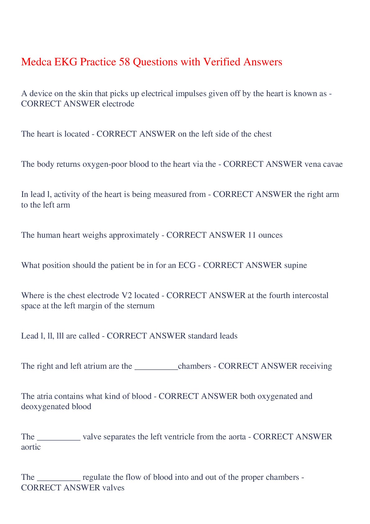
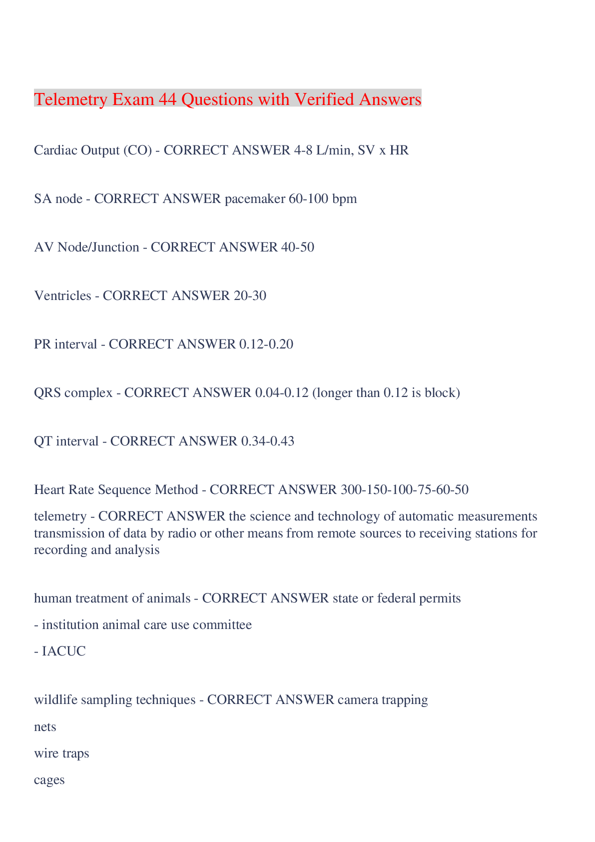
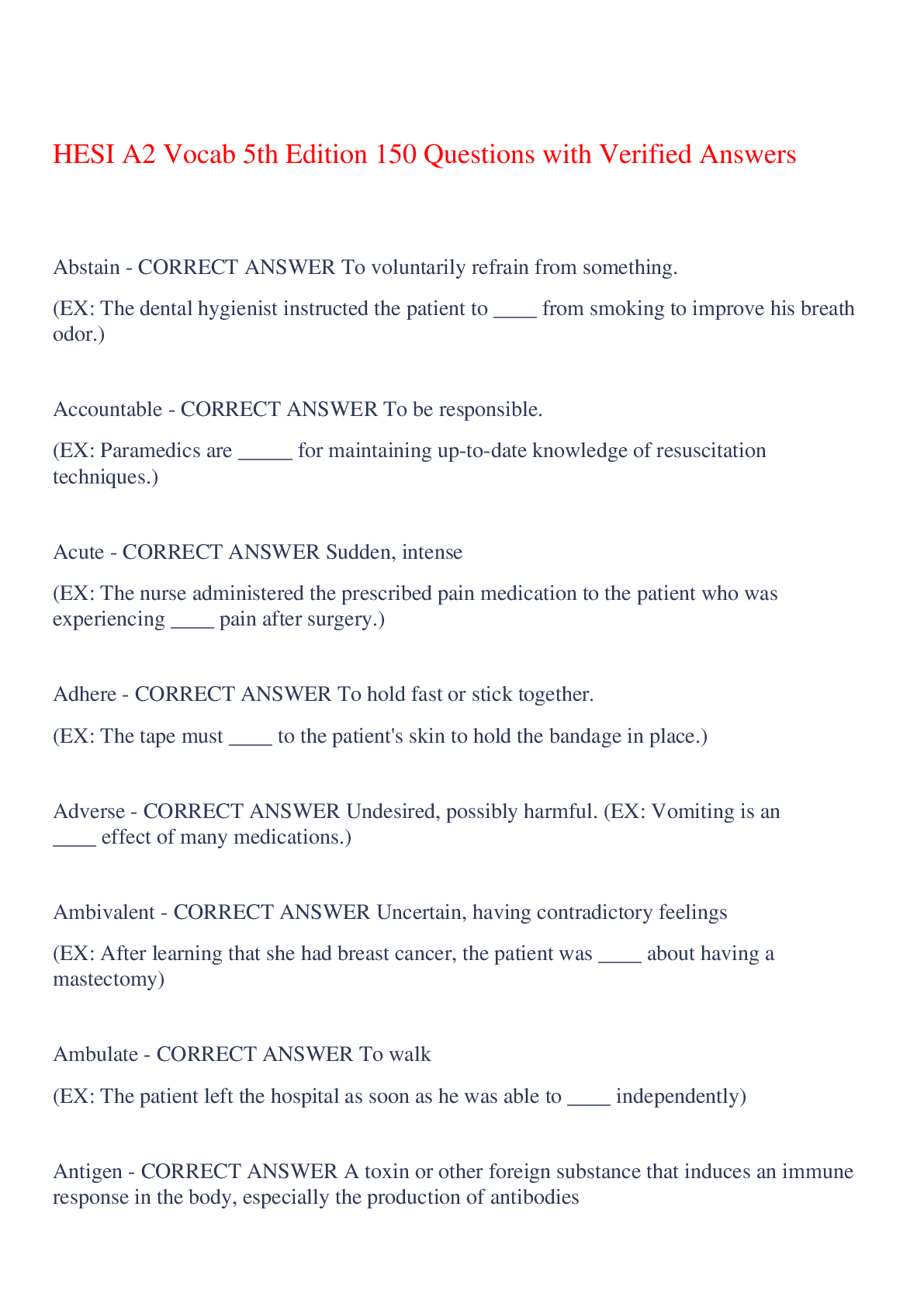
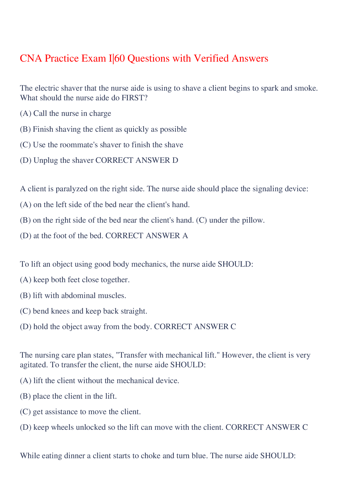
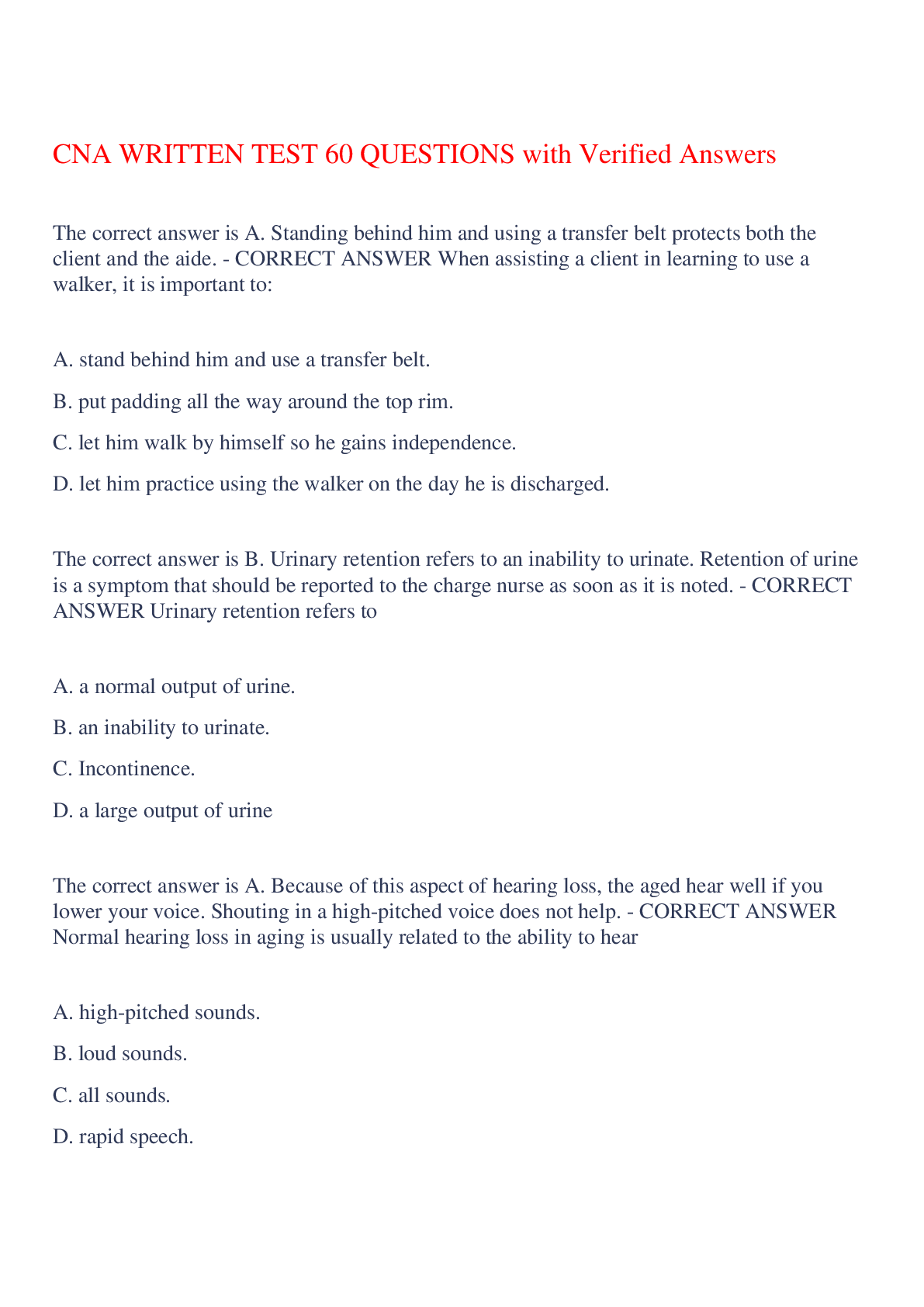
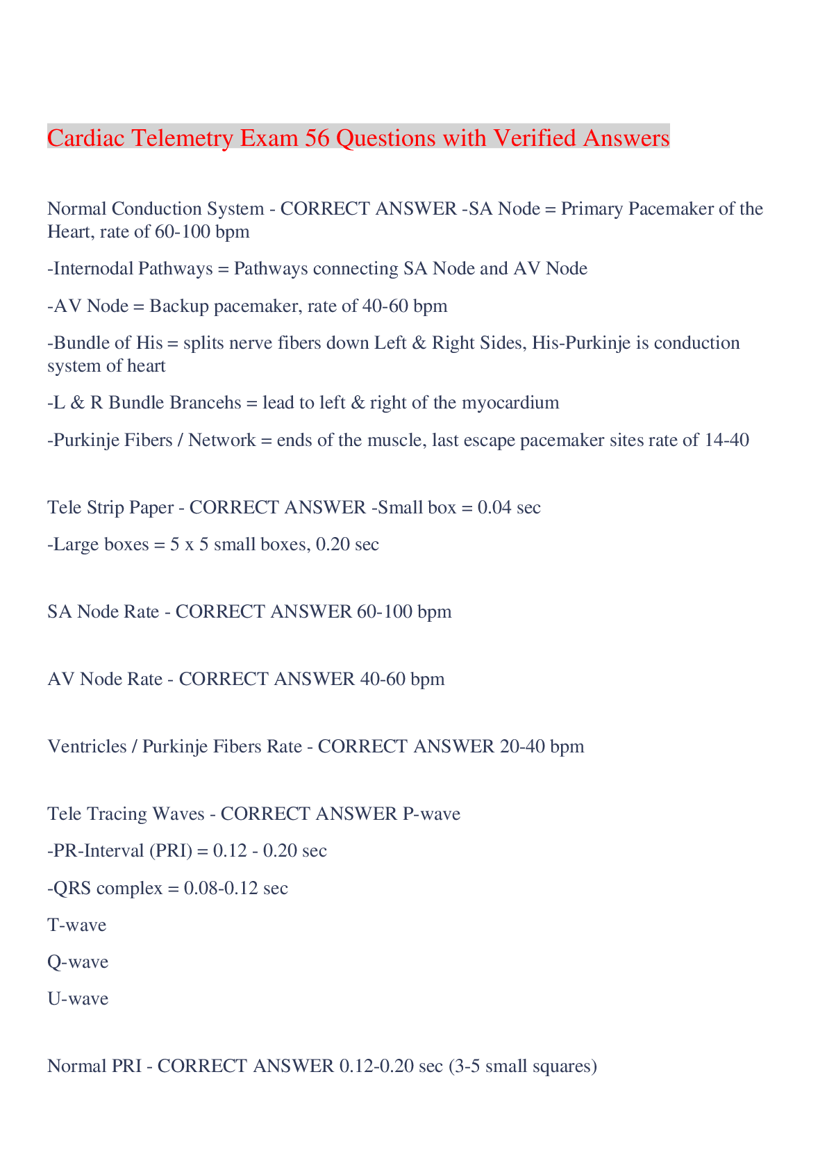
Cardiac Telemetry Exam 56 Questions with Verified Answers Normal Conduction System - CORRECT ANSWER -SA Node = Primary Pacemaker of the Heart, rate of 60-100 bpm -Internodal Pathways = Pathways co... nnecting SA Node and AV Node -AV Node = Backup pacemaker, rate of 40-60 bpm -Bundle of His = splits nerve fibers down Left & Right Sides, His-Purkinje is conduction system of heart -L & R Bundle Brancehs = lead to left & right of the myocardium -Purkinje Fibers / Network = ends of the muscle, last escape pacemaker sites rate of 14-40 Tele Strip Paper - CORRECT ANSWER -Small box = 0.04 sec -Large boxes = 5 x 5 small boxes, 0.20 sec SA Node Rate - CORRECT ANSWER 60-100 bpm AV Node Rate - CORRECT ANSWER 40-60 bpm Ventricles / Purkinje Fibers Rate - CORRECT ANSWER 20-40 bpm Tele Tracing Waves - CORRECT ANSWER P-wave -PR-Interval (PRI) = 0.12 - 0.20 sec -QRS complex = 0.08-0.12 sec T-wave Q-wave U-wave Normal PRI - CORRECT ANSWER 0.12-0.20 sec (3-5 small squares) -Indicates AV conduction time -Measure from P wave start to QRS wave -Consider heart blocks if abnormal Normal Etiology SA Node - CORRECT ANSWER Firing of SA Node causing depolarization of Atria. The impulse travels from SA node, through internodal pathways to AV node, where it is delayed for short amount of time. (PRI) Tele Tracing QRS Complex - CORRECT ANSWER Q-Wave = 1st Negative Deflection R-Wave = 1st Postive Deflection S-Wave= Negative Deflection following R-Wave Normal QRS Complex - CORRECT ANSWER 0.08-0.12 sec (2-3 small squares) Duration of this will lengthen when electrical activity takes a long time to travel through the heart. Normal conduction, AV node -> His-Purkinje system -> fast duration of this complex Consider BBB or Ventricular origin if higher Q-T interval - CORRECT ANSWER 0.35 - 0.43 sec Heart rate - CORRECT ANSWER bradycardia <60 Normal 60-100 tachycardia <100 Tele tracing measurements - CORRECT ANSWER HR PR Interval QRS Complex Regularity Telemetry Interpretation - CORRECT ANSWER 1. What is the Rate? (atrial, ventricular) 2. Is it Regular? (R to R) 3. Are there P waves? (PRl <0.20? Does every QRS complex have a P wave? 4. Width of QRS complex? (< 0.12) 6-Second Strip Interpretation method - CORRECT ANSWER 1. count 30 small squares 2. Can do for atrial or ventricular rate (R to R usually suffices) 3. Multiply # of R waves (in the 30 sq.) by 10 to get the HR HR = # of R waves x 10 Sinus Rhythms (SA Node) - CORRECT ANSWER -Normal Sinus Rhythm (NSR) -Sinus Bradycardia -Sinus Tachycardia (ST) -Sinus Arrythmia Normal Sinus Rhythm - CORRECT ANSWER Rate: 60-100 P-waves: 0.12-0.20 Regularity: Regular R-R Sinus Bradycardia - CORRECT ANSWER Rate: <60 P-waves: 0.12-0.20 Regulartiy: Regular R-R Sinus Tachycardia (ST) - CORRECT ANSWER Rate: >100 P-waves: 0.12-0.20 Regularity: Regular R-R Sinus Arrhythmia - CORRECT ANSWER Rate: Varies P-Waves: 0.12-0.20 Regularity: Irregular R-R Junctional Rhythms (AV Node/Junction) - CORRECT ANSWER -Junctional Escape -Premature Junctional Contraction (PJCs) -Accelerated Junctional Rhythm Junctional Rhythm Pathophysiology - CORRECT ANSWER SA node does not control the heart's rhythm, may be a block in pathway. AV node takes over as pacemaker. Atria still contract before ventricles d/t backwards conduction of AV to atria. Junctional Rhythm ECG Presentation - CORRECT ANSWER -Usually with lost P wave or inverted P wave, closer to QRS -May have Retrograde P wave (depolarization from the AV node back to the SA node) -Narrow PRI Junctional Escape Rhythm - CORRECT ANSWER -Rate: 40-60 bpm b/c AV as backup pacemaker, beats late in timining -P-waves: Inverted, following QRS, or lost -QRS 0.10 sec or less -Rhythm: Regular -Narrow PRI <0.12 Accelerated Junctional Escape Rhythm - CORRECT ANSWER -Rate: >60 bpm bc AV fires above this -Same as Junctional Escape Premature Junctional Contractions (PJCs) - CORRECT ANSWER -Random, early Junctional beats -QRS 0.10 sec or less -Typically followed by a long, compensatory pause PJC vs Junctional Escape beat - CORRECT ANSWER Ventricular Rhythms - CORRECT ANSWER -Idioventricular Rhythm / agonal (~20-40) -Accelerated Idioventricular rhythm (60-100) -Premature Ventricular Complex -Ventricular Tachycardia >100 -Ventricular Fibrillation -Torsades de Pointes Idioventricular Rhythm - CORRECT ANSWER -Rate: <50 bpm, slow -Impulses originating from the ventricles, escape p, no SA/AV impulse -Indicates ventricles producing escape beats -No P-waves -Wide QRS >0.12 Accelerated Idioventricular Rhythm - CORRECT ANSWER -Rate: 50-100 bpm When 3 or more ventricular beats appear in sequence. Same rules as idioventricular except faster rate. -QRS wide >0.12 Premature Ventricular Complex (PVC) - CORRECT ANSWER Occur when a ventricular site generates an impulse before next regular sinus beat. Frequent PVC's may cause heart to skip a beat. -Wide QRS -QRS shape can be WIDE & bizarre -No P wave -Bigeminy, Trigenminy, Quadrigeminy types -Couplet (2 PVCs consecutive) -Triplet (3 PVCs consecutive) PVC Bigeminy - CORRECT ANSWER every other beat is PVC PVC Trigeminy - CORRECT ANSWER every 3rd beat is PVC PVC Couplet and Triplet - CORRECT ANSWER Quadrigeminy - CORRECT ANSWER every 4th beat is PVC Ventricular Tachycardia (VT) - CORRECT ANSWER -Rate: >100 Impulses originate from ventricles. -Wide QRS d/t origination in the ventricles -Can be monomorphic (QRS complexes of same height) or polymorphic (QRS varies in shape and size- if QT long may be Torsades) Ventricular Tachycardia Monomorphic - CORRECT ANSWER Ventricular Tachycardia Polymorphic - CORRECT ANSWER Ventricular Fibrillation (VFib) - CORRECT ANSWER D/t chaos in ventricles bc no organized electrical activity to generate muscle activity, ventricles quivering, pulselessness, no blood circulating -No normal EKG waves present -No HR -Emergency condition Torsades de Pointes - CORRECT ANSWER -Type of VT -"corkscrew" appearance -QTc prolong pt (like those on antipsychotics, are at risk) -Mg is the antidote -QRS complexes vary in shape and amplitude & appear to wind around baseline Atrial Rhythms - CORRECT ANSWER -Atrial Fibrillation (Afib) -Atrial Flutter / AF RVR -Wandering Atrial Pacemaker -Multifocal Atrial Tachycardia Atrial Fibrillation (Afib) - CORRECT ANSWER -No P waves Atria quiver, fibrillatory waves. Fast firing. -Irregular Ventricular Beats (R to R) -Rate >100 is Afib with Rapid Ventricular Response Atrial Flutter (AFlutter) - CORRECT ANSWER -Classic "Saw tooth pattern" -Often regular (d/t typical 4:1 atrial pulses ratio), but can be irregular Multifocal Atrial Tachycardia (MAT) - CORRECT ANSWER When many non-SA sites firing impulses. -Rate: >100 bpm -P-waves vary in shape, see at least 3 dif. types -PRI varies -Ventricular rhythm is regular (R to R) Wandering Atrial Pacemaker (WAP) - CORRECT ANSWER -Similar to MAT but Rate <100 bpm -P waves present & vary in shape (at least 3 dif.) -Irregular atrial rhythm, regular ventricular rhythm Premature Atrial Contractions (PACs) - CORRECT ANSWER early beats with similar P-waves, see compensatory pause SupraVentricular Tachycardia (SVT) - CORRECT ANSWER -Narrow QRS complex (means impulse originates above the ventricles) -Rate >150 bpm see lost P waves *ST is 100-150 usually ST vs VT - CORRECT ANSWER -VT rate >100, impulses originate from ventricles -VT has wide QRS d/t origination in the ventricles Heart Blocks - CORRECT ANSWER -First Degree AV Block -Second degree, Type 1 (Mobitz 1 / Wenkebach) -Second degree, Type 2 (Mobitz 2) -Third Degree / CHB -Bundle Branch Block First Degree AV Block - CORRECT ANSWER -PRI prolongation >0.20 *All PRI measurements are the same, all are conducted -P-waves before every QRS Second Degree AV Block, Type 1 - CORRECT ANSWER -PRI longer & longer, until QRS dropped Second Degree AV Block, Type 2 - CORRECT ANSWER -Dropped QRS, no prolongation Third Degree AV Block aka CHB - CORRECT ANSWER -CHB = Complete Heart Block -complete dissociation of P-waves & QRS complese "husband and wife that don't talk anymore" ventricles don't get atrial impulses, so see p waves doing their own thing without correlation Bundle Branch Block - CORRECT ANSWER -Delay in either Left or Right bundle branches. -Need 12 lead EKG to Specify -Defined by WIDE QRS complex, sometimes with bunny ears, but otherwise normal rhythm Tachy Rhythms - CORRECT ANSWER 1. ST - Sinus Tachycardia 2. AFib RVR 3. SVT - SupraVentricular Tachycardia 4. VT - Ventricular Tachycardia 5. Torsades de Pointe Brady Rhythms - CORRECT ANSWER 1. Sinus Bradycardia 2. Junctional (Junctional Escape) 3. Ventricular (Ventricular Escape) [Show More]
Last updated: 5 months ago
Preview 1 out of 9 pages
Instant download
Instant download
Reviews( 0 )
Document information
Connected school, study & course
About the document
Uploaded On
Dec 08, 2023
Number of pages
9
Written in
Additional information
This document has been written for:
Uploaded
Dec 08, 2023
Downloads
0
Views
29


