Pathophysiology > EXAM > NURS548 Advanced Pathophysiology Midterm Review Latest Solution 2022 (All)
NURS548 Advanced Pathophysiology Midterm Review Latest Solution 2022
Document Content and Description Below
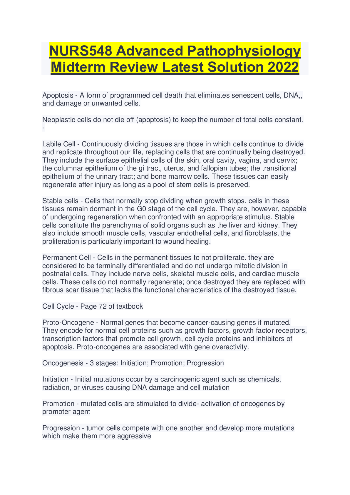
NURS548 Advanced Pathophysiology Midterm Review Latest Solution 2022 Apoptosis - A form of programmed cell death that eliminates senescent cells, DNA,, and damage or unwanted cells. Neoplastic c... ells do not die off (apoptosis) to keep the number of total cells constant. - Labile Cell - Continuously dividing tissues are those in which cells continue to divide and replicate throughout our life, replacing cells that are continually being destroyed. They include the surface epithelial cells of the skin, oral cavity, vagina, and cervix; the columnar epithelium of the gi tract, uterus, and fallopian tubes; the transitional epithelium of the urinary tract; and bone marrow cells. These tissues can easily regenerate after injury as long as a pool of stem cells is preserved. Stable cells - Cells that normally stop dividing when growth stops. cells in these tissues remain dormant in the G0 stage of the cell cycle. They are, however, capable of undergoing regeneration when confronted with an appropriate stimulus. Stable cells constitute the parenchyma of solid organs such as the liver and kidney. They also include smooth muscle cells, vascular endothelial cells, and fibroblasts, the proliferation is particularly important to wound healing. Permanent Cell - Cells in the permanent tissues to not proliferate. they are considered to be terminally differentiated and do not undergo mitotic division in postnatal cells. They include nerve cells, skeletal muscle cells, and cardiac muscle cells. These cells do not normally regenerate; once destroyed they are replaced with fibrous scar tissue that lacks the functional characteristics of the destroyed tissue. Cell Cycle - Page 72 of textbook Proto-Oncogene - Normal genes that become cancer-causing genes if mutated. They encode for normal cell proteins such as growth factors, growth factor receptors, transcription factors that promote cell growth, cell cycle proteins and inhibitors of apoptosis. Proto-oncogenes are associated with gene overactivity. Oncogenesis - 3 stages: Initiation; Promotion; Progression Initiation - Initial mutations occur by a carcinogenic agent such as chemicals, radiation, or viruses causing DNA damage and cell mutation Promotion - mutated cells are stimulated to divide- activation of oncogenes by promoter agent Progression - tumor cells compete with one another and develop more mutations which make them more aggressive Sequence of events in cellular response to inflammation includes leukocyte - 1. margination and adhesion 2. transmigration 3. Chemotaxis 4. activation and phagocytosis non-osseous, malignant neoplasm of tissues derived from mesoderm - Sarcoma Benign tumor of adipose tissue - lipoma Benign neoplasm arising in glandular tissue - adenoma malignant tumor of bone - osteosarcoma benign tumors - tissue name + "oma" Malignant tumor-epithielial tissue - tissue name + "Carcinoma" malignant tumor- mesenchymal tissue - tissue name + "sarcoma" What happens during the vascular stage of acute inflammation - Prostaglandins and leukotrienes affect blood vessels. arterioles & venules dilate increased blood flow to the injured area redness and warmth result capillaries become more permeable allows exudate to escape into tissues swelling and pain result Serous exudate - watery fluid low in protein content that result from plasma entering the inflammatory site Hemorrhagic exudate - Occurs with severe tissue injury that causes damage to blood vessels or when there is significant leakage of red cells from capillaries fibrinous exudate - contains large amounts of fibrinogen and form a thick and sticky meshwork, much like fibers of a blood clot. Membranous exudate - develops on mucous membrane surfaces and are composed of necrotic cells enmeshed in fibro-purulent exudate. Purulent exudate - contains pus, which is composed of degraded white cells, proteins and tissue debris. Process that occurs when there is injury to tissue - leukocytes mobilize to the area via the bloodstream. Once they reach the compromised area, the 1st process is "pavementing", whereby the leukocytes collect along the endothelial lining of the blood vessel. This allows them to "get out of the flow of traffic", in preparation of making a "left hand turn" out of the blood vessel. Once they make this left hand turn and leave the blood vessel (emigration), they are directed to the site of injury (it is very dark inside) by way of chemical signals (chemotaxis). Now they can "clean up" the area and remove unwanted debris via the process of phagocytosis....... [Show More]
Last updated: 1 year ago
Preview 1 out of 9 pages
Instant download
Instant download
Reviews( 0 )
Document information
Connected school, study & course
About the document
Uploaded On
Jul 14, 2022
Number of pages
9
Written in

Additional information
This document has been written for:
Uploaded
Jul 14, 2022
Downloads
0
Views
38


 LATEST UPDATE 2022.png)