*NURSING > QUESTIONS & ANSWERS > uWORLD Cardio Pathophysiology QuestionBank, 100% proven pass rate. (All)
uWORLD Cardio Pathophysiology QuestionBank, 100% proven pass rate.
Document Content and Description Below
uWORLD Cardio Pathophysiology QuestionBank, 100% proven pass rate. uWORLD: A 67 yo man comes to the ED due to progressive SOB and chest tightness. he has no lightheadedness or syncope. The patie... nt takes lisinopril for HTN and metformin for Type 2 DM. He has smoked a pack of cigarettes daily of the last 40 years. The blood pressure cuff is inflated to 140 mmHg and the pressure is released very slowly. At 120 mmHg, intermittent Korotkoff sounds are head only during expiration. At 100mmHg, Korotkoff sounds are heard throughout the respiratory cycle The physical examination finding can be seen in the following? - ☑☑Pericardial disease > Pulsus paradoxus is defined by a decrease in systolic blood pressure of > 10 mmHg with *inspiration*. It is most commonly seen in patients with cardiac tamponade but can also occur in severe asthma, chronic obstructive pulmonary disease, and constrictive pericarditis > Inspiration causes an increase in systemic venous return, resulting in increased right heart volumes. Under normal conditions, this results in expansion of the right ventricle into the pericardial space with little impact on the left side of the heart. However, in conditions that impair expansion into the pericardial space )eg, acute cardiac tamponade), the increased right ventricular volume occurring with inspiration leads to *bowing of the inter ventricular septum* toward the left ventricle. This leads to a decrease in left ventricular (LV) end-diastolic volume and stroke volume, with a resultant decrease in systolic pressure during inspiration. A patient has pulses paradoxus. The patient is tachypeneic and unable to speak in full sentences. Examination revels prolonged expiration and prominent bilateral wheezing. Heart sounds are nonroman. Chest imaging shows a normal-sized heart and hyper inflated lungs with a flattened diaphragm. Beside ECG revels no intrapericardial fluid accumulation or pericardial thickening. Which of the following physiologic changes is most likely to provide immediate relief in this patient? - ☑☑cAMP accumulation in smooth muscle cells > Asthma and chronic obstructive pulmonary disease (COPD) exacerbation are the most frequent causes of pulses paradoxus in the absence of significant pericardial disease. Beta-adrengeric agonists control acute asthma and COPD exacerbations by causing bronchial smooth muscle relaxation via increase intracellular cAMP. uWORLD: A 35 yo man is evaluated for progressive fatigue and SOB. Recently, he has noticed bilateral leg swelling and abdominal distention despite overall weight loss. He does not use tobacco, alcohol, or illicit drugs. Despite treatment, the patient dies several weeks later. Autospy revels significant endocardial thickening with dense fibrous deposits around the tricuspid and pulmonary valves as well as moderate pulmonary valve stenos. The left-sided cardiac chambers and valves are normal. Measuring the levels of which substances would have helped in diagnosing this patient? - ☑☑Urinary 5-hydroxyindolearcretic acid > Carcinoid syndrome typically presents with episodic flushing, secretary diarrhea and wheezing. It can lead to pathognomonic plaque-like deposits of fibrous tissue on the right-sided endocardium, causing tricuspid regurgitation and right-sided heart filature. Elevated 24 hour urnary 5-hydroxyindoleacetic acid can confirm the diagnosis. > The autospy findings - endocardial thickening and fibrosis of tricuspid and pulmonary valves - are chracerstic of *carcinoid heart disease.*. Carcinoids are well-differentiated neuroendocrine turbos found most commonly in the distill small intestine and proximal colon, with a strong propensity for metastasis to the liver. These tumors secrete seers products (including histamine, serotonin, and vasoactive intestinal peptide) that are metabolized in the liver. In patients with liver metastasis, these hormones are relased directly into the systemic circulation, leading to carcinoid syndrome. > Carcinoid heart diseases is caused by excessive secretion of *serotonin*, which stimulates fibroblast growth and fibrognesis. Pathognomic plaque-like *deposits of fibrous tissue* occur most commonly on the *endocardium*, leading to *tricuspid regurgitation*, pulmonic valvulpathy, and right-sided heart failure )eg ascites, peripheral edema). Endocardial fibrosis and thickening are generally limited to the right heart as vasoactive products and inactivated distally by pulmonary vascular endothelial monoamine oxidase. FYI - Measuring of plasma cortisol: Used for diagnosing adrenal insufficiency and Crushing syndrome - Measuring of homocysteine: elevation may contribute to arterial and venous thrombosis and to the development of atherosclerosis. - Measuring of plasma phenylalanine: May be elevated in patients with phenylalanine hydroxylase deficiency, resulting in central nervous system damage/intellectual disability : - Measuring of porphobilinogen: May be elevated in the porphyria, which are caused by deficiencies of the heme synthesis enzymes. The porphyries may produce cutaneous lesions, skin photosensitivity, or attacks of abdominal pain and neurological disturbances (acute intermittent porphyria) - Measuring of vanillymandelic acid: by product of norepinephrine and epinephrine, and can be used to detect *neuroblastoma* and other tumors of neural crest origin uWorld: A 62 yo woman comes to the hospital with intermittent but progressive substernal chest pain over the last 36 hours. Medical history includes HTN and hyperlipidemia, but the patient has been poorly compliant with her medication regimen and outpatient follow-up. She previously smoked a pack daily for 30 years but quit last year. Her blood pressure is 130/75 mmHg and pulse is 73/min. ECG on admission shows normal sinus rhythm with a 2-mm ST segment elevation in leads V2-V5. The patient is treated with medical management. However, on the fifth day of hospitalization she dies suddenly, despite adequate resuscitation. Which is the most likely cause of death in the patient? - ☑☑Profound hypotension > Rupture of the left ventricular free wall is a catastrophic mechanical complication of anterior wall myocardial infarction (MI) that usually occurs within the first 5-14 days after MI. Rupture leads to hemoperricardium and cardiac tamponade, causing profound hypertension and shock with rapid progression to pulseless electrical activity and death. > This patient most likely died from profound hypotension due to *ruptured left ventricular free wall*. Her initial presentation of substernal chest discomfort and ST segment elevation in anterior precordial leads in consistent with acute *anterior wall myocardial infarction (MI)*. Free wall rupture is a catastrophic mechanical complication that usually occurs within the first 5-14 days after a large anterior transmural MI (from left anterior descending occlusion). During this timeframe, the infarcted myocardium is substantially weakened by coagulative necrosis, neutrophilic and macrophage infiltration, and enzymatic lysis of myocardial connective tissue. Abrupt rupture of the left ventricle leads to *hemopericardium* and *cardiac tamponade*. Patients have sudden onset of chest pain and *profound hypotension and shock*, with rapid progression to pulseless electrical activity and death. FYI: > Carotid artery occlusion = can lead to ischemic symptoms (hemiparesis, vision changes) but does not typically cause sudden death. > Hypertensive emergency can lead to aortic dissection = that typically presents with tearing chest or back pain. Hypertension and shock can occur with retrograde extension of the dissection into he pericardial cavity or contrary arteries, with resultant hemoperricardium or MI, respectively. However, this patient's presentation of intimal normal blood pressure and ST segment elevation is more suggestive of acute MI with subsequent cardiac rupture. uWORLD: A 18 yo women is referred to cardiologist after a heart murmur is discovered during routine checkup. The patient is healthy and has no symptoms. Past medical history is unremarkable. She runs daily and wants to start actively training for a half marathon. She is concerned that the murmur is a sign of heart disease and would prevent her from pursuing her athletic activities. She has no family history of sudden cardiac death. Auscultation reveals a midsystolic click that is followed by a short late-systolic mummer at the cardiac apex. The mummer disappears with equating. This patient's condition is most likely related to an abnormality involving which of the following tissues? - ☑☑connective tissue > Cardiac auscultation in patients with mitral valve prolapse (MVP) typically reveals a non ejection (eg. mid systolic) click and mid- to late systolic mummer of mitral regurgitation. MVP is most often caused by defects in mitral valve connective tissue proteins that predispose to myxomatous degeneration of the mitral leaflets and chord tendineae. > The presence of a midstylic click following to a mid- to late systolic mummer at cardiac apex that disappears with squatting is most consistent with *mitral valve prolapse* (MVP) with mitral regurgitation (MR). The click results from sudden tensing of the charade tendineae as they are pulled taut by the ballooning valve leaflets. The mummer is due to malalignment of the valve margins during systole. *Manevuvers* that change left ventricular (LV) volume and cavity size can change the timing and intensity of the murmur. *Squatting* from a standing position *increases venous return* and *LV volume*, helping to bring the valve leaflets into a more normal anatomic arrangement. This, in turn, decrease the degree of MVP, causing a delay in the onset of click during systole, and systolic mummer typically becomes shorter or disappears. FYI > Hypertrophic cardiomyopathy (HCM) also presents with a systolic mummer at the cardiac apex that decreases in intestnity with squatting (due to increased left ventricle volume and decreased outflow tract obstruction). However, a mid systolic click, which is classic for MVP, is not heard with HCM. uWORLD: A 66 yo man with DM is brought to the host pail due to sudden-onset chest pain and nausea. BP is 70/60 mmHg and pulse is 60/min. Lungs are clear on auscultation. ECG shows ST-segment elevation in leads II, III, and aVG. Chest x-ray is unremarkable. The patient is diagnosed with an inferior wall myocardial infarction. Emergency cardiac cauterization revel complete occlusion of the proximal right coronary artery. He is persistently hypotensive in the cardiac cauterization laboratory. Which of the following hemodynamic findings is most likely to be obsessed (cardiac output, Pulmonary capillary wedge pressure, Central venous pressure; Will it increase/decrease?)? - ☑☑Increase CO Decrease Pulmonary Capillary Wedge pressure Increase Central Venous Pressure This patient is in cariogenic shock after suffering a left ventricular (LV) inferior wall MI. The inferior wall of the LV is supplied by the posterior descending artery, which arises off the dominant right canary artery. Because the right coronary artery also gives off marginal branches that supply most of the right ventricle (RV), inferior wall MI is often associated with r*right ventricular infarction(. > Infarction of the RV results in decreased RV stroke volume, which in turn leads to *diminished* LV filling and *cardiac output* in spite of preserved LV systolic function (Frank-Starling mechanism). TV dilation and elevated diastolic pressures also cause a shift of the interventrciular septum toward the LV cavity, further impairing LV filling and cardiac output, contributing to systemic hypotension and shock. Because left-sided filling pressures are reduced, *pulmonary capillary wedge pressure* also *decrease* as it estimates left atrial pressure. In addition, patients have *elevated central venous pressure* due to RV dysfunction and impaired forward flow. uWORLD: A 45 yo woman who recently immigrated to the US is hospitalized with exertion dyspnea and fatigue. She has no significant past medical history and takes no medication. The patient's BP is 110/80 mmHg and heart rate is 90/min and regular. After cardiopulmonary examination, the physician suspects mitral stenos. Which of the following is the most useful measure for assessing the degree of stenosis? - ☑☑A2-to-opening snap time interval > On auscultation, the best indicator of mitral stenosis (MS) severity is the length of time between S2 (specifically the A2 component, caused by aortic valve closure) and the opening snap (OS). The OS occurs due to abrupt tensing of the valve leaflets as the mitral valve reaches its maximum diameter during forceful opening. > Left-sided S3 and/or S4 gallops are generally absent in MS, since left ventricular filling is normal or decreased. Ex. A 4 month old baby is brought to the cardiology clinic by his parents for continued followed-up of tetralogy of Fallot. The diagnosis was made during routine antenatal sonography, and the pregnancy and delivery were otherwise uncomplicated. The infant has been seen frequently in the clinic and has not had any cyanosis, respiratory distress, and difficulty feeding. The parents become concerned when their son's surgical plan is discussed because he does not have the clinical signs that other children with metrology of Fallot demonstrate. Which is the major determinant of symptoms severity in this condition? - ☑☑Right ventricular Outflow Tract Obstruction > *Tetraology of Fallot* is characterized by ventricular septal defect (VSD), overriding aorta, right ventricular outflow tract (RVOT) obstruction and right ventricular hypertrophy. The VSD generally is large, which allows for haul pressure in the right and left ventricles. Therefore, it is the amount of *RVOT obstruction* that determines how much deoxygenated blood is delivered to the systemic circulation. > Infants with no or minimal RVOT obstruction, such as this patient, deliver more deoxygenated blood to the lungs and appear cyanotic. The degree of RVOT obstruction is *dynamic* and can increase suddenly, leading to profound cyanosis ("tet spells". These can be caused by dehydration or hyperventilation but are usually idiopathic. uWORLD: A 62 yo man comes o the office for follow-up of hypertension. He was diagnosed with HTN 10 years ago and has been treated with a number of different medications. However, the patient has had to discontinue several medications due to side effects such as dizziness, palpitations, and headaches. Currently he takes ramipril and chlothalidone and is tolerating them well. BP is 160/92 mmHg and was 158/89 mmHg at his most recent prior visit. ECG shows sinus bradycardia (55/min) with PR interval prolongation (280 sec). Which of the following medications would be most effective for lowering this patient's blood pressure without worsening his ECG abnormalities? - ☑☑Nifedipine > DO NOT GIVE Non-hydropyrmidine Ca2+ channel blockers (Verapamil, Diltilzaem) OR beta-blockers because they depress the AV node further worsening the bradycardia. > Calcium channel blockers inhibit the L-type channel on vascular smooth muscle and cardiac cells. Dihydropyridines (eg nifedipine, amlodipine) primary affect peripheral arteries and cause vasodilation. Nondihydropyridines (eg. verapamil, diltiazem) affect the myocardium and can cause bradycardia and slowed atrioventricular conduction. uWORLD: A 45 yo man comes to the ED because of severe chest pain, diaphoresis, and palpitations. The patient dies two hours after the onset of his symptoms. Autospy reveals 100% occlusion of the left anterior descending artery. At the time of the patient's death, light microscopy of the affected myocardium would most likely demonstrate which of the following? - ☑☑Normal myocardium Timeline > 0-4 hours - minimal change > 4-12 hrs- early coagulation necrosis, edema, hemorrhage, wavy fibers, > 12-24 hrs - coagulation necrosis and marginal contraction band necrosis > 1 to 5 days - coagulation necrosis and neutrophilic infiltrate > 5 to 10 days - macrophage phagocytosis of dead cells > 10 to 14 days - granulation tissue and neovascularization > 2 weeks to 2 months - collagen deposition/scar formation uWORLD: A 60 yo man comes to the ED with acute substernal chest pain, nausea, and diaphoresis. The patient has a history of liver cirrhosis, hypertension, and hyperlipidemia. He quit smoking 3 months ago but previously smoked a pack of cigarettes daily for 25 years. ECG shows ST elevations in leads II, III, and aVF. He is diagnosed with a MI and, after careful discussion of risks and benefits, repercussion therapy is not performed due to underlying cirrhosis and history of vatical bleeding. The patient is eventually discarded from the hospital with conservative management. Twelve day after his MI, he dies suddenly. Light microscopy would most likely show what changes to the myocardium of the inferior wall? - ☑☑Granulation tissue with neovascularization > Sudden cardiac death *12 days* after MI is most likely due to *ventrciular arrhythmias* originating from the infarcted myocardium. The microscopic diagnosis of MI depends on the presence of necrotic myocardium, with areas of acute inflammation and necrosis separated from viable myocardium. After the initial event, several characteristic microscopic changes occur in the infarcted zone in a specifyic temporal sequence with some overlap in different stages. > *During the second week* after MI, the damage tissue is replaced by *granulation tissue* and *neovascularization* is found in the infarct zone. uWORLD: A 72 yo man with long-standing dyspnea was seen int eh clinic after experiencing an episode of syncope. Physical examination showed weak and slowly rising arterial pulses. Cardiac auscultation showed a harsh mid systolic mummer best heard at the second right intercostal space with decreased intensity of the second heart sound. ECG and echocardiogram confirmed the diagnosis of severe aortic stenosis. Two months later, the patient comes to the ED with palpitations and increased SOB. His blood pressure is 90/60 mmHg and his heart rate is 130/min with an irregularly irregular rhythm. ECG shows new-onset arterial fibrillation without significant ST-segment or T-wave changes. Chest x-rays show bilateral pulmonary edema. Which of the following hemodynamic changes is most likely associated with this patient's current presentation? - ☑☑Sudden decrease in left ventricular preload > Patients with severe AS already have reduced cardiac output due to significant valvular obstruction, which can be exacerbated by the sudden *loss of normal atrial contraction* that contributes to ventricular filling. Atrial contraction is especially important for these patients as many have concentric left ventricular (LV) hypertrophy and therefore reduced LV compliance. As a result, they become dependent on atrial contraction to maintain adequate LV filling. > In patients with chronic aortic stenosis, and concentric left ventricular hypertrophy, atrial contraction contributes significantly to left ventricular filling. Loss of atrial contraction due to atrial fibrillation can reduce left ventricular preload and cardiac output sufficiently to cause systematic hypertension. Decreased forward filling of the left ventricle can also result in backup of blood in the left atrium and pulmonary veins, leading to acute pulmonary edema. uWORLD: An 82 yo man comes to the office due to progressive dyspnea and fatigue over the last ear, which now limits his daily activities. He also noticed bilateral swelling of his feet. The patient has HTN, which is controlled with amlodipine. His BP is 122/72 mmHg and pulse is 55/min. Physical examination reveals elevated jugular venous pressure with rapid 'y' descent and a prominent S4. Abdominal examination shows moderate ascites. The patient has 3+ bilateral lower extremity pitting edema. Echocardiogram reveals left atrial enlargement with marked left ventricular hypertrophy and normal left ventricular ejection fraction. Complete good count, basic metabolic panel, and serum, and urine protein electrophoresis are within normal limits. What is the most likely diagnosis of this patient? - ☑☑Senile amyloidosis > This patient's clinical presentations (eg. progressive exertion dyspnea, edema, ascites, elevated jugular venous pressure and rapid 'y' descent, prominent S4) and echocardiogram findings (eg. left atrial enlargement, left ventricular hypertrophy, normal ejection fraction) are consistent with *diastolic heart failure* due to restrictive cardiomyopathy. > *Restrictive cardiomyopathy* can be idiopathic or caused by infiltrative disease (eg, amyloidosis, sarcoidosis, hemochromatosis), radiation fibrosis, or endomycardial fibrosis. Although most cases are associated with normal ventricular wall thickness. Infiltrative conditions such as cardiac amylosisis may lead to significant *ventricular hypertrophy*. Infiltrative diseases can also cause conduction system abnormalities (eg, bradycardia) that may ultimately require pacemaker implantation* The fourth heart sound (S4) is a low frequency sound heard at the end of diastole just before S1. It is due to decreased left ventricular compliance and is often associated with _____________________ and ____________________. - ☑☑Restrictive cardiomyopathy; left ventricular hypertrophy uWORLD: A 54 yo Caucasian male comes to the ED with retrosternal chest pain of 30 min duration. The patient also complains of sweating and mild dyspnea. A single table of nitro is delivered sublingually, and the patient's pain decreases significantly. The patient has experienced several similar episodes of pain over the last 12 hours, all of which resolved spontaneously. Which of the following ultrastructural changes would most likely indicate irreversible myocardial cell injury in this patient? - ☑☑Mitochondrial vacuolization > Mitochondrial vacuolization is typically a sign of irreversible cell injury, signifying that the involved mitochondria are permanently unable to generate ATP. Reversible injury: > Myofibril relaxation > Disaggregation of polysomes denotes the dissociation of rRNA from mRNA in reversible ischemic/hypoxic injury. > Disaggregation of granular and fibrillar elements of the nucleus is associated with reversible cell injury > Triglyceride droplet accumulation is characteristic of reversible cell injury, especially in hepatocytes, and also in striated muscle cells and renal cells. > Glycogen loss is another early and reversible cellular response to injury uWORLD: A 45 yo man comes to the clinic due to recurrent palpitations accompanied by chest discomfort and SOB. A year ago, he was diagnosed with paroxysmal atrial fibrillation treated with rate control using a beta blocker. Past medical history is also significant for HTN and obesity. ECG shows left atrial enlargement, normal left ventricular ejection fraction, and no significant valvular disease. 24-hour Holter monitoring reveals bursts of atrial fibrillation associated with the patient's symptoms. He is initiated on dofetilide to maintain normal sinus rhythm. This medication exerts its main effect on which portion of the action potential curve? - ☑☑Phase 3 (last repolarization) > Class III antiarrhymic drugs amiodarone, stall, dofetilide) predominately block potassium channels and inhibit the outward potassium current during the phase, leading to prolongation of repolarization, action potentials duration, and the QT interval on ECG. > uWORLD: A 42 yo male is brought to the ED complaining of severe headaches and oliguria. His blood pressure is 240/150 mmHg and his heart rate is 90/min. On ophthalmologic exam, there is papilledema bilaterally. Which of the following is the most likely pathological process associated with his patient's condition? - ☑☑Onion-like concentric thickening of arteriolar walls > This patient presents in hypertensive crisis, a condition defined as a persistent diastolic pressure exceeding 130 mmHg that is often associated with acute vascular damage. Hyper plastic arteriolosclerosis, which can result from diastolic pressures >120-130 mmHg, presents as onion-like > Hyperplastic ateriolosclerosis in renal arterioles can result from and perpetuate malignant hypertension. The pathological lesion is an onion-like concentric thickening of arteriolar walls in the renal vasculature and elsewhere. uWORLD: A 35 yo perviously healthy man is brought to the ED after being involved in a motor vehicle accident. He has significant blunt chest and head trauma. Shortly after he arrives, his blood pressure drops suddenly and he begins experiencing respiratory distress. On physical examination, the patient is tachycardia and tachypneic. His lungs are clear to auscultation with vesicular breath sounds heard bilaterally. He has jugular venous distention, and his systolic blood pressure falls 15 mmHg with inspiration. Which of the following is the most likely cause of this patient's deterioration? - ☑☑Cardiac tamponade > The combination of jugular venous distention, hypotension, and muffled heart sounds is highly suggestive of cardiac tamponade. Tachycardia and pulses paradoxus are also frequently seen with tamponade. Lung examination is normal, which can help distinguish cardiac tamponade from tension pneumothorax. > During inspiration, the pressure in the pleural space and lung interstitial decreases, increasing pulmonary vascular capacitance. This causes a fall in venous inflow to the left heart, resulting in decreased left ventricular stroke volume and a drop in systolic blood pressure (normally < 10 mmHg). The inspiratory drop in systolic blood pressure is exacerbated in cardiac tamponade due to extrinsic compression of the ventricles. ________________ can cause hypotension, tachycardia, tachypnea, and jugular venous distention. It can result from blunt or penetrating chest trauma that injures the visceral pleura or tracheobronchial tree. However, tension pneumothorax would cause absent breath sounds and hyper resonance to percussion on the affected side - ☑☑Tension pneumothorax. Ex. A 19 yo man comes to the office due to difficulty seeing and blurred vision, which have worsened slowly over the past year. He is a second year college student pursuing a degree in biochemistry. His grades are excellent, but he is concerned about the effect his poor vision has had on his classes this semester. The patient is also an avid swimmer. He weighs 71 kg (156.5 lb) and is 195 cm (6 ft 5 in) tall. Physical examination shows the breastbone dips inwards or protrudes outwards. This patient is most likely to die from which condition - ☑☑Aortic disease > Marfan syndrome (MFS) is one of the most common inherited connective tissue disorders. This condition is caused by a genetic defect in the glycoprotein *fibrillin-1* and results in abnormalities in the skeleton (eg, long extremities, scoliosis, precuts excavated), eyes (eg, ectopic lentil), and cardiovascular system (eg, aortic root disease) > The 2 most common cardiac abnormalities seen in MFS patients are *mitral valve prolapse* and *cystic medial degeneration of the aorta*. In more than 75% of MFS patients, cystic medial degeneration of the aorta results in aneurysmal dilation. If untreated, this can cause *aortic dissection*, the most common cause of death in MFS patients. > Cardiovascular lesions are the most life-threatening complications associated with Marfan syndrome. Early-onset cystic medial degeneration of the aorta predisposes to aortic dissection, the most common cause of dearth in these in these patients. uWORLD: A 66 yo man is evaluated for recurrent syncope. He has had 3 episodes of dizziness and palpitations followed by brief loss of consciousness over the last 6 months. He has no chest pain or dyspnea. The patient has a history of hypertension and hyperlipidemia. There is no family history of sudden death. Initial evaluation shows normal ECG and echocardiogram. A cardiac electrophysiologic study is performed. During the study, intravenous infusion of a medication is administered and produces increase contractility and decrease vascular resistance. What is most likely the medication? - ☑☑Isoproterenol > Isoproterenol is Beta-1 and beta-2 adrenergic receptor agonist (nonselective beta adrenergic agonist) that causes increased myocardial contractility and decreased systemic vascular resistance. > Norepinephrine acts on alpha-1 receptors, causing vasoconstriction and an increase in systemic vascular resistance. Norepinephrine also acts as a weak agonist at beta-1 receptors, with a modest increase in myocardial contractility. uWORLD: A 24 yo man is evaluated after an episode of syncope. He was out jogging when he felt lightheaded and passed out, but he did not sustain any head injury. The patient has had 2 similar episodes of lightheadedness while jogging over the last year, but this was the first time he passed out. He considers himself in good health and has no other medical problems. The patient does not use tobacco, alcohol, or illicit drugs. His father died suddenly at age 30. On physical examination, he has a harsh systolic murmur. Transthoraic echocardiography shows asymmetric inter ventricular septal hypertrophy. The patient's symptoms are most likely explained by left ventricular outflow obstruction created by which of the following structures? - ☑☑Mitral valve leaflet and inter ventricular septum > This patient's presentation suggests *hypertrophic cardiomyopathy (HCM), an autosomal dominant disorder resulting from mutations in cardiac sarcomere proteins. HCM is characterized by asymmetric ventricular septal hypertrophy and variable, dynamic *left ventricular outflow tract (LVOT) obstruction*. Systolic anterior motion of the *mitral valve* toward the *interventricular septum* can cause eccentric mitral regurgitation and exacerbate LVOT obstruction. > Examination often reveals a harsh crescendo-decrescendo systolic mummer at the apex and left lower sternal border, which changes in intensity with physiologic maneuvers. > In patients with hypertrophic cardiomyopathy, dynamic left ventricular outflow tract obstruction is due to abnormal systolic anterior motion of the anterior leaflet of the mitral valve toward a hypertrophied inter ventricular septum. uWORLD: A 54 yo man comes to the ED with severe fatigue and dyspnea. He has a long history of progressively worsening heart failure that has been resistant to treatment with medications. He was treated with chest radiation several years ago for Hogdkin's disease and has been in remission ever since. The patient is admitted to the hospital, but his condition continues to deteriorate despite aggressive therapy. He dies 3 days later and an autospy is performed. Gross inspection of his heart shows dense, thick fibrous tissue in the pericardial space between the visceral and parietal pericardium. Which of the following signs would most likely have been detected during physical examination of this patient? - ☑☑Kussmaul sign > This patient has thick fibrous tissue in the pericardial space, a finding which is diagnostic for constructive pericarditis. This dense, rigid pericardial tissue encases the heart and restricts ventricular filling, causing low cardiac output and right-sided heart failure resistant to medication. Jugular venous pressure (JVP) is almost always increased in patients with constrictive pericarditis due to the restriction in right ventricular filling capacity. Although JVP normally drops during inspiration, patients with constrictive pericarditis frequently have a paradoxical rise in JVP, a finding known as *Kussmual sign*. This occurs because the volume-restricted right ventricle is unable to accommodate the inspiratory incase in venous return. > Constrictive pericarditis is a chronic condition in which the normal pericardial space is replaced by a thick, fibrous shell that restricts ventricular volumes and eventually causes heart failure. Impaired right ventricular filling leads to increased jugular venous pressure and often results in a positive Kussmaul sign. There may also be a pericardial knock, which occurs earlier in diastole than the S3 heart sound. uWORLD: A 10 yo immigrant from Eastern Europe is brought to the office due to exertion dyspnea and fatiguability. The boy tires easily when walking and cannot keep up with his peers at the playground. According to his parents, he was diagnosed with a congenital heart disease in infancy, for which they refused treatment. They cannot recall the details of his diagnosis. The patient also has had occasional respiratory infections throughout childhood that have not required hospitalization. He takes a daily multivitamin and no medications. He has received only a few childhood vaccinations based on parental preference. The patient has no family history of heart disease. Physical examination shows toe cyanosis and clubbing but no finger abnormalities. All extremity pulses are full and equal. Which of the following is the most likely diagnosis? - ☑☑Patent ductus arteriosus > Patent ductus arteriosus (PDA) is a vascular connection between the main pulmonary artery and the aorta that normally obliterates after birth. Clinical features vary depending on the size. Small PDAs are characterized by a *continuous, machinelike murmur* (left-to-right shunting) with no significant symptoms. Large PDAs can present anytime during childhood with progressive pulmonary hypertension and reversal of the shunt to right-to-left. The characteristic continuous mummer decreases as the pulmonary pressure rises and ultimately disappears. > Typical consequences include heart failure (eg. shortness of breath, fatiguability), and cyanosis (*Eisenmenger syndrome*). The cyanosis and clubbing are most pronounced in the lower extremities (*differential cyanosis and clubbing*) because the PDA delivers unoxygenated blood distal to the left subclavian artery. .> Differential clubbing and cyanosis without blood pressure or pulse discrepancy are pathognomonic for a large patent ductus arteriosus complicated by Eisenmenger syndrome (reversal of shunt flow from left-to-right to right-to-left). Severe coarctation of the aorta can cause lower extremity cyanosis. Right-to-left shunting in patients with large septal defects are tetralogy of Fallot results in whole-body cyanosis. __________________ is most commonly located in the juxtaductal region just distal to the left subclavian artery. It typically presents in children and adults as a blood pressure discrepancy and *pulse delay between the upper and lower extremities*. - ☑☑Coarctation of the aorta _____________________ is generally characterized by whole-body cyanosis at birth as the result of right-to-left shunting across the ventricular spatial defect. The right-to-let shunt is intracardiac, and therefore cyanosis typically involves the whole body. - ☑☑Tetralogy of Fallot uWORLD: A 21 yo Caucasian male presents to the ED following an episode of syncope. The syncopal episode was not provoked by any activity or circumstance, nor was it proceeded by lightheadedness. The patient has no significant past medical history and he is not taking any medications. An ECG obtained in the ER reveals QT-interval prolongation but is otherwise unremarkable. Assuming this is an inherited condition, the relevant mutation most likely affects which structure? - ☑☑Membrane potassium channel protein > This patient's sudden-onset syncopal episode suggests a sudden cardiac arrhythmia. QT prolongation is an otherwise healthy young individual is usually congenital. The mutation most likely to cause QT-interval prolongation can be determined based on an understanding of cardiac electrophysiology. The QT-interval reflects the cardiac myocyte action potential duration, which is determined in part by K+ currents through channel proteins. > Unprovoked syncope is a previously asymptomatic young person may result from a congenital QT prolongation syndrome. The two most important congenital syndromes with QT prolongation - Romano-Ward syndrome and Jervell and Lange-Nielson syndrome - are thought to result from mutations in a K+ channel protein that contributes to the delay rectifier current (IK) of the cardiac action potential. Mutations in cardiac cell ________________ underlie hypertrophic cardiomyopathy (HCM). Although HCM may present as syncope in a previously asymptomatic young person, the syncope of HCM is typically provoked by exertion. Additionally, AT prolongation is not generally found in HCM. - ☑☑Sarcomere proteins (eg. beta-myosin heavy chain) A 64 yo man comes to the ED due to flank discomfort and red urine. He has a history of HTN and Type 2 DM. Three mounts ago, the patient had an ischemic stroke and has mild residual right-sided weakness. Serum creatinine is 0.9 mg/dL, and serum lactate dehydrogenase is elevated. Urine microscopy shows many RBCs. CT scan of the abdomen with contrast is performed showing a wedge-shaped right kidney. What is the most likely cause of this patient's symptoms? - ☑☑Atrial fibrillation > This patient, who has flank pain, hematuria, elevated lactate dehydrogenase (cell necrosis), and a wedge-shaped right kidney lesion on CT, likely has *renal infarction*, which results from interruption of the normal blood supply to the kidney. The most common cause of renal infraction is systemic *thromboembolism* (from the left atrium or ventricle). The kidneys are more likely than other organs to suffer embolic infarctions as they are perfused at a higher rate (to support adequate glomerular filtration rate) > Systemic thromboembolism commonly occurs with *atrial fibrillation (AF)* as the irregular heart contractions can lead to clot formation. AF, which can be paroxysmal (thereby going undiagnosed), may have also caused this patient's recent stroke, with emboli traveling to peripheral arteries in the brain. Systemic emboli can also occur following MI and endocarditis. > The simultaneous development of stroke, intestinal or foot ischemia, and renal infarction should raise suspicion for embolic phenomena. These emboli may arise from left atrial or ventricular clots or valvular vegetations, among others. Pulmonary hypertension can cause right heart failure (for pulmonate) and is sometimes associated with _________________, causing increased blood viscosity. - ☑☑Secondary polycythemia A life-threatening complication of lower extremity (LE) deep venous thrombus (DVT) is ________________________. - ☑☑Pulmonary embolism > LE DVT emboli reach the right heart via venous flow, enter the pulmonary circulation, and get caught in pulmonary artery branches. Rarely, when a communication exists between the right and left heart (eg, atrial or ventricular septal defect, patent foramen ovale), clots from the right heart may embolize to the brain and kidneys. uWORLD: A 65 yo woman with Type 2 DM and HTN comes to the office for routine follow-up. She has occasional numbness in her feet. The patient takes ibuprofen for chronic back pain along with hctz, metformin, atorvastatin, and insulin deter. Her blood pressure is 120/76 mmHg supine and 122/80 mmHg standing. Laboratory evaluation shows a serum potassium level of 4.2 mEq/L and a creatinine level of 0.8 mg/dL. Urinalysis shows albuminuria. Lisinopril therapy is initiated. The next day, the patient returns due to lightheadedness and near-syncope. Her blood pressure is 102/66 mmHg supine and 80/45 mmHg standing. her pulse is 94/min. Cardiopulmonary examination is normal. Which of the following is most likely the major factor contributing to this patient's current symptoms? - ☑☑Diuretic therapy > ACE inhibitors can cause significant first-dose hypertension in patients with volume depletion (eg. from diuretic use) or heart failure. To reduce the risk of first-dose hypertension, ACE inhibitor therapy should be initiated at low dosage. > This patient with albuminuria was started on an *ACE inhibitor* for treatment of early diabetic nephropathy. Although most patients remain asymptomatic with only a mild reduction in blood pressure, *first-dose hypertension* can be a potential limiting factor with imitating ACE inhibitors. Significant hypotension is most likely to occur in patients with high plasma renin activity, such as those with *volume depletion* (eg. from diuretic use (HCTZ in this patient) or heart failure. Initation of ACE inhibitors therapy causes abrupt removal of the vasoconstrictive effects of angiotensin II, resulting in decreased peripheral vascular tone and a precipitous drop in blood pressure in susceptible patients. To prevent the development of first-dose hypertension, therapy should be started at low doses and slowly titrated upward as needed. uWORLD: A 46 yo women dies in the hospital from respiratory failure after a prolonged illness. She had multiple comorbidities, including advanced renal disease. AAutospy revels multiple small, nondestructive masses attached to the edges of the mitral valve leaflet. Microscopy reveals that the masses are composed of platelet-rich thrombi, but cultures reveal no bacterial growth. Which of the following disease is most likely associated with this patient's condition? - ☑☑Advanced malignancy > The autospy finding of sterile *platelet-rich thrombi* attached to the mitral valve leaflets is characteristic of *nonbacterial thrombotic endocarditis* (NBTE). NBTE is most commonly associated with *advanced malignancy*, as well as chronic inflammatory disorders such as antiphopholipid syndrome, systemic lupus erythematous (Libman-Sacks endocarditis), and disseminated intravascular coagulation in patients with sepsis. NBTE is often seen in mucinous adenocarcinomas, which may relate to procoagulant effects of circulating mucin. > The pathogenesis of NBTE is thought to begin with endothelial injury caused by circulating cytokines, which triggers platelet deposition in the presence of a hyper coagulable state. Compared to infective endocarditis, vegetations can easily dislodge and are more likely to embolize, causing infarction. > Nonbacterial chromatic endocarditis is a form of noninfectious endocarditis characterized by deposition of sterile platelet thrombi on cardiac valves. It is commonly associated with advanced malignancy and can also occur with chronic inflammatory disorders (eg. antiphospholipid syndrome, systemic lupus erythematous) and sepsis. uWORLD: An 85 yo man is transferred to the hospital from a nursing home for altered mental status and fever. Upon arrival, the patient is admitted directly to the intensive care unit with a presumptive diagnosis of septic shock. Antibiotic therapy is initiated. The patient is unable to provide any history, but his caretakers state that he has been having non-specific symptoms, including fever, for the past few days. The patient has a history of cardiovascular disease, diverticulitis, and dementia. His blood pressure is 60/40 mmHg despite aggressive intravenous hydration. Which of the following cellular changes occurs directly in response to norepinephrine therapy? - ☑☑cAMP increase in cardiac muscle cell > Norepinephrine stimulates cardiac beta1 adrenorecptors, which utilize the cAMP signal transduction pathway. Stimulation of these receptors by norepinephrine causes increases in cAMP concentration within cardiac myocytes. uWORLD: A 56 yo Caucasian female presents to your office with chronic cough. She says that the cough is dry and affect quality of her life significantly. She denies chest pain, hemoptysis and SOB. Her past medical history is significant for long-standing hypertension, diabetes and MI experienced two months ago. She does not smoke or consume alcohol. Her blood pressure is 130/70 mmHg and heart rate is 70/min. Which of the following is the best next step in the management of this patient? - ☑☑Careful review of current medications > Cough is a very well recognized side effect of ACE inhibitor therapy. Cough secondary to ACE inhibitor therapy is characterized as dry, nonproductive, an d persistent. > The mechanism behind ACE inhibitor induced cough is accumulation of bradykinin, substance P, or prostaglandins. Because angiotensin receptor blockers (ARBs) do not affect ACE activity, they theoretically should no cause cough. uWORLD: A 60 yo man comes to the office fora routine follow-up visit. He feels well overall except for an intermittent, mild, generalized headache. The patient has no known medical problems and takes no medications. He does not smoke, follows a generally healthy diet, and exercises daily. On examination, his blood pressure is 150/85 mmHg, and he is started on lisinorpil. At a follow-up visit, the patient's blood pressure is 128/78 mmHg. He also has a dry cough that began a few weeks after starting lisinipril. This drug is stopped and losartan is now prescribed. The patient seems to be compliant with his medication; the cough resolves and he experiences no significant side effects. When compared with no treatment at all, this patient's current therapy is most likely to result in which of the following changes? (Renin, Angiotensin I, Angiotensin II, Aldosterone, Bradykinin) - ☑☑Increase Renin Increase Angiotensin I Increase Angiotensin II Decrease Aldosterone No Change: Bradykinin > *Angiotensin II receptor blockers* (ARBs) such as losartan competitively bind to angiotensin II receptors and block the effects of angiotensin II, resulting in *vascular smooth muscle relaxation* and *decreased aldosterone* secretion. This reduces blood pressure, stimulating renal renin production, which in turn increases conversion of angiotensin I to angiotensin II. Because ACE function remains intact with the use of ARBs, bradykinin levels are not significantly affected. This is in contrast to ACE inhibitors, which lower angiotensin II and aldosterone levels but increases bradykinin levels. Bradykinin is thought to increase prostaglandin production, which induces coughing due to bronchial irritation. > Angiotensin II receptor blockers (ARBs) work by blocking angiotensin II type 1 receptors, inhibiting the effects of angiotensin II. This results in arterial vasodilation and decreased aldosterone secretion. he resulting fall in blood pressure increases renin, angiotensin I, and angiotensin II levels. ARBs do not affect the activity of angiotensin-coveting enzyme, and therefore they do not affect bradykinin degradation and do not cause cough. uWORLD: A 23 yo man comes to the office with chest discomfort that usually occurs during exercise,such as when jogging or climbing stairs. The symptoms go away 5 to 10 minutes after he stops. The patient has not had syncope but mentions some shortness of breath that accompanies the chest pain. Family History includes an uncle who died suddenly at the age of 35 years. Blood pressure is 122/70 mmHg and pulse is 70/min and regular. Apical impulse is strong and sustained. He has a soft crescendo-decrescendo systolic murmur at the apex and left sternal border while supine that becomes quite pronounced when he stands up. Which of the following medications should be avoided while treating this patient condition? (Amiodarone, disopyramide, Isosorbide dinitrate, Metoprolol, Verapamil) - ☑☑Isosorbide dinitrate > This patient's presentation, family history of premature sudden death, and systolic mummer that accentuates with standing from a supine position is consistent with hypertrophic cardiomyopathy (HCM). Patients with HCM have dynamic left ventricular outflow tract (LVOT) obstruction that worsens with decreased left ventricular (LV) volume (as can be caused by decreased preload and/or reduced systemic vascular resistance). As such, medications that generally should be *avoided* in patients with HCM include: --> *Vasodilators* (eg dihydropyridine calcium channel blockers, nitroglycerin, and ACE inhibitors) decrease systemic vascular resistance, leading to decreased after load and lower LV volumes. --> *Diuretics* decrease LV venous filing (preload) and also result in greater outflow obstruction. In contrast, *negative inotropic agents *such as beta blockers (metoprolol), nondihydropyridine calcium channel blockers (verapamil), and disopyramide *Reduce LVOT obstruction* and are helpful in symptomatic patients with HCM. In addition, beta blockers may also help reduce anginal symptoms by decreasing myocardial oxygen demand. > The dynamic left ventricular (LV) outflow tract obstruction that occurs in hypertrophic cardiomyopathy worsens with decreased LV volume, which can be caused by education in cardiac preload and/or after load. Therefore, medications that decrease venous return or systemic vascular resistance (dihydropyridine calcium channel blockers, nitroglycerin) should generally be avoided. uWORLD: A 53 yo man comes to the ED with SOB and chest tightness. The patient was palying in a poker tournament when his symptoms first began. He has a history of hypertension and is not compliant with his medications. His last medical follow up was a year ago. Blood pressure is 195/115 mmHg and pulse is 90/min and regular. Lung examination reveals bibasilar crackles. Nitroglycerin infusion is started and results in significant symptomatic improvement. Repeat blood pressure is 165/90 mmHg. Which of the following intracellular events is most likely responsible for the beneficial effects of this patient's treatment? - ☑☑Myosin dephosphorylation > Nitrates (via conversion to nitric oxide) activate guanylate cyclase and increase intracellular levels of cyclic guanosine monophosphate (cGMP). Increased levels of cGMP leads to myosin light-chain dephsophorylation, resulting in vascular smooth muscle relation. > Increased levels of cGMP leads to *decreased intracellular calcium* (reduces the activity of myosin light-chain kinase) and activation of myosin light chain phosphatase. This promotes *myosin light-chain dephosphorylation* and vascular smooth muscle *relaxation* uWORLD: A 67 yo man with nonischemic cardiomyopathy come to the office for follow-up. He recently was hospitalized for acute decompensated heart. The patient's symptoms have improved with multi drug treatment, but he has persistent shortness of breath on mild exertion. He has a history of hypertension and hypercholesterolemia. Blood pressure is 115/70 mmHg and pulse is 66/min. There is a third heart sound on heart auscultation and mild lower extremity pitting edema. A recent echocardiogram showed a left ventricular ejection fraction of 30%. Which of the following diuretics would most likely improve surivaal if added to this patient' current regimen? (Acetazolamide, furosemide, HCTZ, mannitol, spironolactone, triamterene) - ☑☑Spironolactone > Mineralocorticoid receptor antagonists (eg. spironolactone, eplerenone) improve survival in patients with congestive heart failure and reduced left ventricular ejection fraction. They should not be used in patients with hyperkalemia or renal failure. > Mineralocorticoid receptor antagonists (eg, spirolocatone, eplerenone) prevent aldosterone from binding to its receptor in the distal renal tubules. This leads to increased sodium and water excretion while conserving potassium ion *(potassium-sparing diuresis)*. These antagonists also block the deleterious effect of aldosterone on the heart, causing regression of myocardial fibrosis and *improvement in ventricular remodeling* > Mineralocorticoid receptor antagonists reduce morbidity and *improve survival* in patients with congestive heart failure na decreased ejection fraction. Therefore, they are recommended in addition to standard heart failure therapy (ACE inhibitors and beta blockers) > Acetazolamide (carbonic anhydrase inhibitor), HCTZ (thiazide diuretic), and triameterene (epithelial sodium channel blocker) have variably lower diuretic effect compared to loop diuretics and are not as efficacious for treating heart failure symptoms > Furosemide is a loop diuretic frequently used for treatment of pulmonary congestion and fluid retention in heart failure patients. Although loop diuretics improve symptoms significantly, they do not provide survival benefit (ie. improved morbidity but not mortality) in these patients. uWORLD: A 64 yo man comes to the office due to exertion chest pain over the last 6 months. He is a lifelong 1 pack per day cigarette smoker and has a history of type 2 diabetes mellitus and peripheral artery disease. This patient undergoes treadmill exercise stress testing and develops substernal chest pain on moderate exertion accompanied by ECG changes that resolve immediately upon rest. He refuses invasive cardiac testing. The patient is started on low-dose aspirin therapy for secondary prevention of cardiovascular disease but experiences shortness of breath and wheezing with the medication. Which of the following is the best alternate therapy for this patient? (Apixaban, cilostazol, clopidogrel, enoxaparin, eptifibatide, naproxen, warfarin) - ☑☑Clopidogrel > Clopidegrel irreversibly blocks the [Show More]
Last updated: 1 year ago
Preview 1 out of 100 pages

Buy this document to get the full access instantly
Instant Download Access after purchase
Add to cartInstant download
We Accept:

Also available in bundle (1)
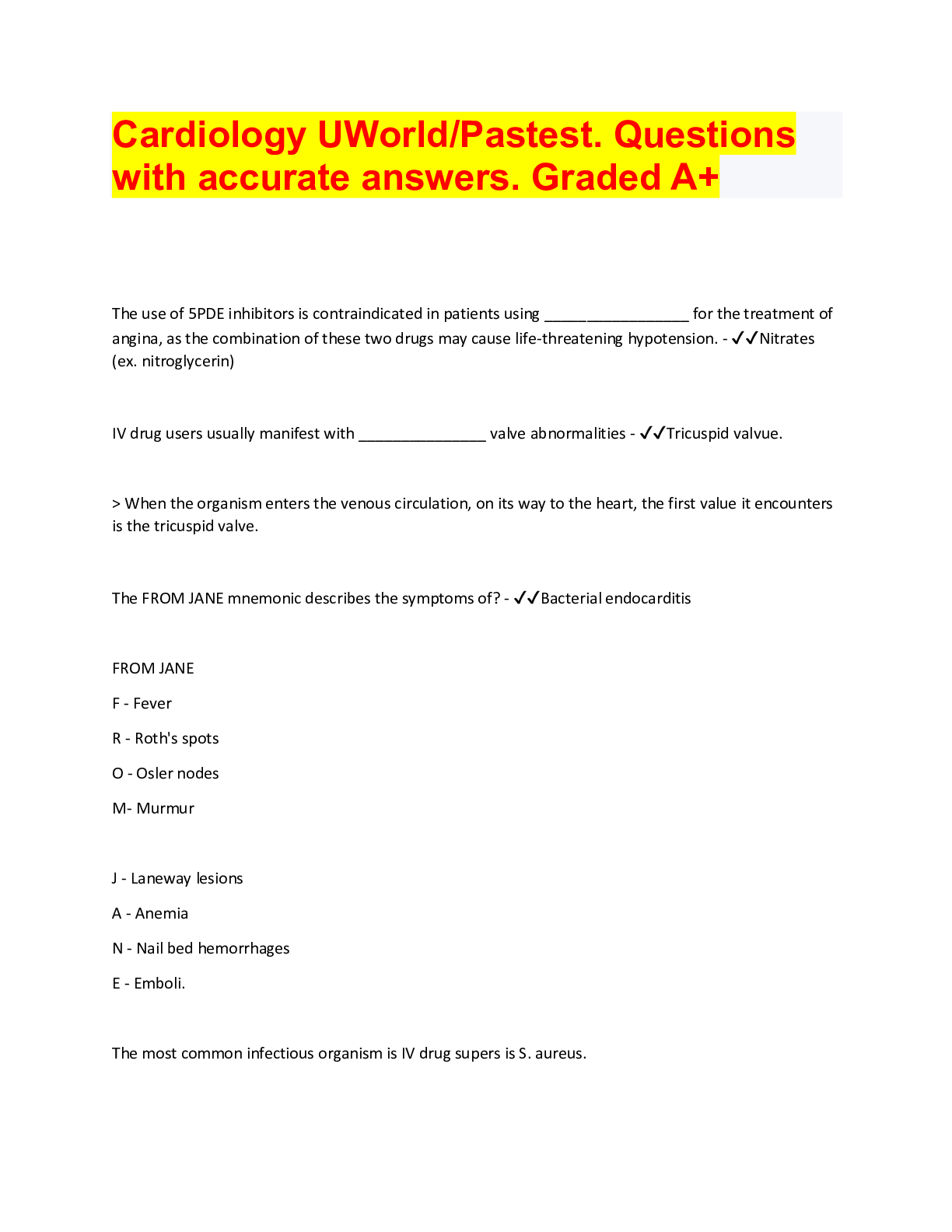
Cardiology BUNDLE, Questions and answers
All Cardiology exam papers questions with answers, download to score
By bundleHub Solution guider 1 year ago
$29
7
Reviews( 0 )
$12.00
Document information
Connected school, study & course
About the document
Uploaded On
Aug 21, 2022
Number of pages
100
Written in
Additional information
This document has been written for:
Uploaded
Aug 21, 2022
Downloads
0
Views
207

