Micro Biology > Experiment > DEPARTMENT OF MEDICAL MICROBIOLOGY : PRACTICAL TITLE: IDENTIFICATION OF PARASITES IN STOOL SAMPLE. (All)
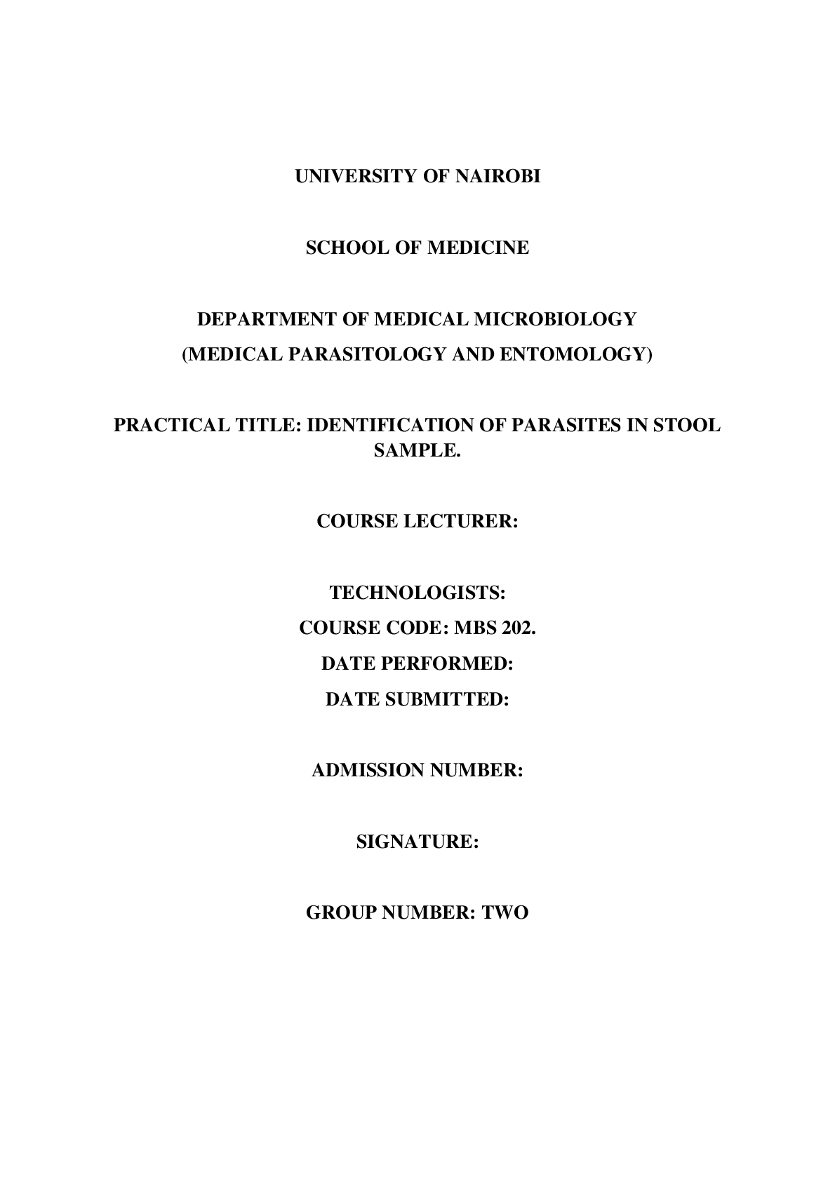
DEPARTMENT OF MEDICAL MICROBIOLOGY : PRACTICAL TITLE: IDENTIFICATION OF PARASITES IN STOOL SAMPLE.
Document Content and Description Below
TITLE IDENTIFICATION OF INTESTINAL PARASITE (Schistosoma mansoni) AIM • Examining faecal specimens for presence of intestinal trematodes • Identification of parasite’s eggs and larvae using... concentration technique also called Formalin-ether concentration technique. INTRODUCTION Faecal samples are examined for presence of parasite’s eggs, cysts, larvae from faecal material. Common stages of parasites found in stool are trophozoites and cysts. The main tasks involved in identification of intestinal parasites found in stool are collection of faecal samples, separation of eggs and larvae from faecal material and their concentration, microscopic examination of prepared specimens, preparation of faecal cultures, isolation and identification of larvae from cultures. Macroscopic and microscopic examination methods are extensively used in examination of intestinal parasites in stool. The adult worms and segments are visible to human eye. The eggs, larvae and cysts are seen through the microscope. Faecal material should be properly prepared and examined; care should be taken for accurate identification of parasite morphology. The morning stool is commonly used in which an applicator stick and container are used for the collection. Diet taken and drugs such as antibiotics can interfere with the examination. The stool is put in container immediately to prevent disintegration of parasites. When they disintegrate they become unrecognisable. In a scenario where the lab is far, the specimen is preserved .It should not go beyond twelve hours. The container should be labbled with name of patient and time of collection to prevent and avoid confusion. Urine and dust should not mix with the sample since urine destroys trophozoits and dusts interferes with examination .In lab the specimen should be placed in a cools and shaddy condition .Light can make the parasite to disintegrate. The technician performs macroscopic examination, checks moisture and water content and indicates whether it is loose, watery, formed and soft. If blood and mucus is noticed, it is indication of presence of trophozoites. Microscopic examination enables finer and further identification of the parasites. It involves the following types of wet mounts; direct saline wet mount which is employed to demonstrate worms, eggs, larvae, trophozoites and cysts. The other technique is iodine wet mount used for identification of cysts and buffered methylene blue wet mount used for identification of trophozoites in stool specimens. Other methods used are Concentration technique which is preferred when the level of infection is low and when a patient demonstrates clinical features and wet mounts are negative. Supplementary techniques include permanent staining technique and Kato Katz technique. [Show More]
Last updated: 1 year ago
Preview 1 out of 6 pages
Reviews( 0 )
Document information
Connected school, study & course
About the document
Uploaded On
Feb 04, 2021
Number of pages
6
Written in
Additional information
This document has been written for:
Uploaded
Feb 04, 2021
Downloads
0
Views
42








.png)

.png)


