Biology > EXAM REVIEW > Chamberlain College of Nursing BIOS 255 A&P 3 FINAL filled out/ Final exam review (All)
Chamberlain College of Nursing BIOS 255 A&P 3 FINAL filled out/ Final exam review
Document Content and Description Below
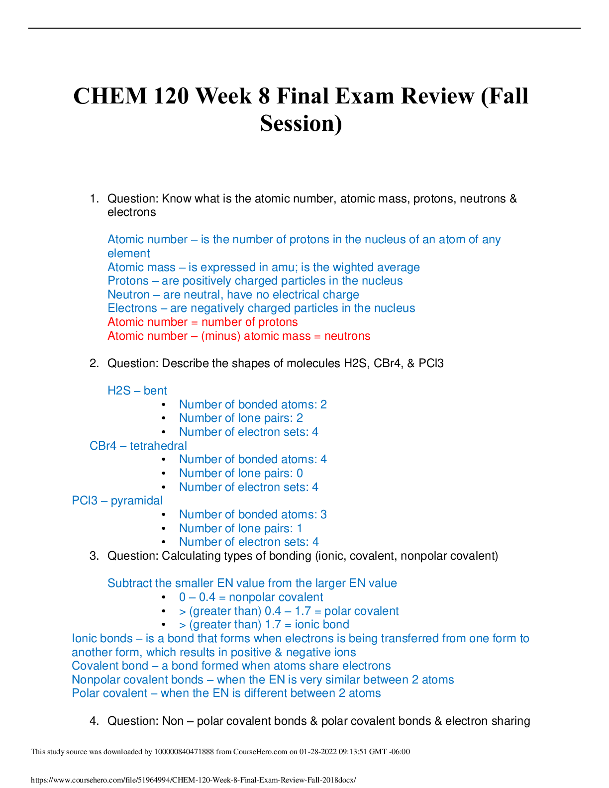
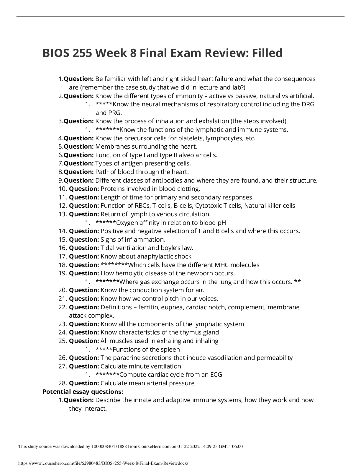
Bios 255 Final exam review Be familiar with left and right sided heart failure and what the consequences are (remember the case study that we did in lecture and lab?) (698) Left side: One cause of ... mitral insufficiency, in which there is backflow of blood from the left ventricle into the left atrium, is mitral valve prolapse (MVP). In MVP one or both cusps of the mitral valve protrude into the left atrium during ventricular contraction. Mitral valve prolapse is one of the most common valvular disorders, affecting as much as 30% of the population. It is more prevalent in women than in men, and does not always pose a serious threat. The left side of the heart receives blood rich in oxygen from the lungs and pumps it to the remainder of the body. As the ability to pump blood forward from the left side of the heart is decreased, the remainder of the body does not receive enough oxygen especially when exercising. This results in fatigue. In addition, the pressure in the veins of the lung increases, which may cause fluid accumulation in the lung. This results in shortness of breath and pulmonary edema. Right side: In aortic stenosis the aortic valve is narrowed, and in aortic insufficiency there is backflow of blood from the aorta into the left ventricle. In right-sided heart failure, the right ventricle loses its pumping function, and blood may back up into other areas of the body, producing congestion. Congestion affects the liver, the gastrointestinal tract, and the limbs. In addition, the right ventricle may be unable to pump blood efficiently to the lungs and to the left ventricle. Causes of right-sided heart failure include left-sided heart failure and lung diseases such as chronic bronchitis and emphysema. Other causes include congenital heart disease, clots in pulmonary arteries, pulmonary hypertension, and heart valve disease. Know the different types of immunity – active vs passive, natural vs artificial. First, let us define the following terms and then we can put them together. The term active generally means that your body is producing antibodies by exposure. Passive is a term where the antibodies are being transferred to you, i.e. breast milk IgA to infant. Natural terms the introduction of the antigen via exposure. Artificial is the introduction of antigens/antibodies via an altered state of the microbe/ antibody. Naturally Acquired Active Immunity- Following exposure to a microbe, antigen recognition by B cells and T cells and costimulation lead to formation of antibody-secreting plasma cells, cytotoxic T cells, and B and T memory cells. Naturally Acquired Passive Immunity- IgG antibodies are transferred from mother to fetus across placenta, or IgA antibodies are transferred from mother to baby in milk during breastfeeding. Artificially Acquired Active Immunity- Antigens introduced during vaccination stimulate cell-mediated and antibody-mediated immune responses, leading to production of memory cells. Antigens are pretreated to be immunogenic but not pathogenic (they will trigger an immune response but not cause significant illness.) Artificially Acquired Passive Immunity- Intravenous injection of immunoglobulins. *****Know the neural mechanisms of respiratory control including the DRG and PRG. (872) Respiratory center- Neurons in the pons and medulla oblongata of the brain stem that regulate breathing. It is divided into the medullary respiratory center and the pontine respiratory center. Within the medullary respiratory center, you find two respiratory groups, the ventral respiratory group (AKA expiratory area) and the dorsal respiratory group (AKA inspiratory area). The DRG generates impulses to the diaphragm via the phrenic nerves and the external intercostals via the intercostal nerves. These impulses trigger contraction of these muscles which in turn execute inhalation. When the nerves are not firing, this passive relaxation allows recoil of the lungs and thoracic wall, passive exhalation. The VRG is only activated during forceful inhalation and trigger the accessory muscles to work. An important part of the VRG is the Pre-Botzinger Complex which is believed to be important in the generation of the rhythm of breathing (Pacemaker cells) . PRG (Pontine Respiratory Group)- A collection of neurons in the pons that transmits nerve impulses to the dorsal respiratory group, and may modify the basic rhythm of breathing. (AKA pneumotaxic area) The PRG may play a role in both inhalation and exhalation by modifying the basic rhythm of breathing generated by the VRG, as when exercising, speaking, or sleeping. Know the process of inhalation and exhalation (the steps involved) (FIG. 24.13) During Inhalation, the diaphragm contracts and the external intercostals contract. The chest cavity expands, and the alveolar pressure drops below atmospheric pressure. Air flows into the lungs in response to the pressure gradient and the lung volume expands. During deep inhalation, the scalene and sternocleidomastoid muscles expand the chest further, thereby creating a greater drop in alveolar pressure. During exhalation, the diaphragm relaxes and the external intercostals relax. The chest and lungs recoil, the chest cavity retracts, and the alveolar pressure increases above atmospheric pressure. Air flows out of the lungs in response to the pressure gradient, and the lung volume decreases. During forced exhalations, the internal intercostals and the abdominal muscles contract, thereby reducing the size of the chest cavity further and creating a greater increase in alveolar pressure. Inhalation: • Diaphragm: contracts & moves down • Intercostal muscles: contract, move ribs out • Chest volume: increases • Pressure in lungs: decreases • Air Flow: higher percentage of oxygen Exhalation: • Diaphragm: relaxes & moves up • Intercostals muscles: relax, move ribs in. • Chest volume: lessens • Pressure in lungs: increases • Air flow: carbon dioxide out *******Know the functions of the lymphatic and immune systems. *****The primary functions of the lymphatic is system is to 1. Drain excess interstitial fluid, 2. Transport dietary lipids and lipid-soluble vitamins, and 3. Carry out immune response. Know the precursor cells for platelets, lymphocytes, etc. The precursor cells for platelets are megakaryocytes. Membranes surrounding the heart. (690) The pericardium surrounds and protects the heart. Covers the heart muscle. The pericardium consists of two main parts: (1) the fibrous pericardium (tough, inelastic, dense irregular connective tissue) -- prevents overstretching of the heart, provides protection, and anchors the heart in the mediastinum. and (2) the serous pericardium (double membrane of outer parietal layer and inner visceral layer, fluid-lubricating part between these layers, reduces friction as the heart moves.) (Walls of heart= 3= epicardium, myocardium, endocardium) Function of type I and type II alveolar cells. (854) The alveolar epithelium comprises two main cell types: the alveolar type I and alveolar type II cell. The type I cell: simple squamous epithelial cells that form a nearly continuous lining of the alveolar wall; main site of gas exchanges, The type II cell, also called septal cells, are fewer in number and are found between type I alveolar cells; rounded or cuboidal epithelial cells with free surfaces containing microvilli, secrete alveolar fluid, which keeps the surface between the cells and the air moist. Type II cells act as the "caretaker" of the alveolar compartment. It responds to damage of the vulnerable type I cell by dividing and acting as a progenitor cell for both type I and type II cells. In addition, it syntheses, stores and releases pulmonary surfactant into the alveolar hypophase, where it acts to optimize conditions for gas exchange. Types of antigen presenting cells. (Table 22.5) Macrophage- Processing and presentation of foreign antigens to T cells, secretion of interleukin-1, which stimulates secretion of interleukin-2 by helper T cells and induces proliferation of B cells; secretion of interferons that stimulate T cell growth. Dendritic Cells- Processes and presents antigens to T cells and B cells; Found in mucous membranes, skin, and lymph nodes. B cell- Processes and presents antigen to helper T cells. Path of blood through the heart. A drop of blood enters the heart via the Vena Cava (Superior or inferior) or coronary sinus into the Right Atrium. From the right atria, it flows into the Right ventricle. The right ventricle pumps the blood into the pulmonary trunk where is branches into the left and right pulmonary arteries. The blood then returns from the lungs via the pulmonary veins into the left atrium. It then leaves the le [Show More]
Last updated: 1 year ago
Preview 1 out of 32 pages
.png)
Reviews( 0 )
Document information
Connected school, study & course
About the document
Uploaded On
Aug 03, 2022
Number of pages
32
Written in

Additional information
This document has been written for:
Uploaded
Aug 03, 2022
Downloads
0
Views
187

.png)