Biology > QUESTIONS & ANSWERS > A 56-year-old woman was referred to a neurology practice specializing in movement disorders followin (All)
A 56-year-old woman was referred to a neurology practice specializing in movement disorders following a nine-year history of bradykinesia (slowness) and rigidity
Document Content and Description Below
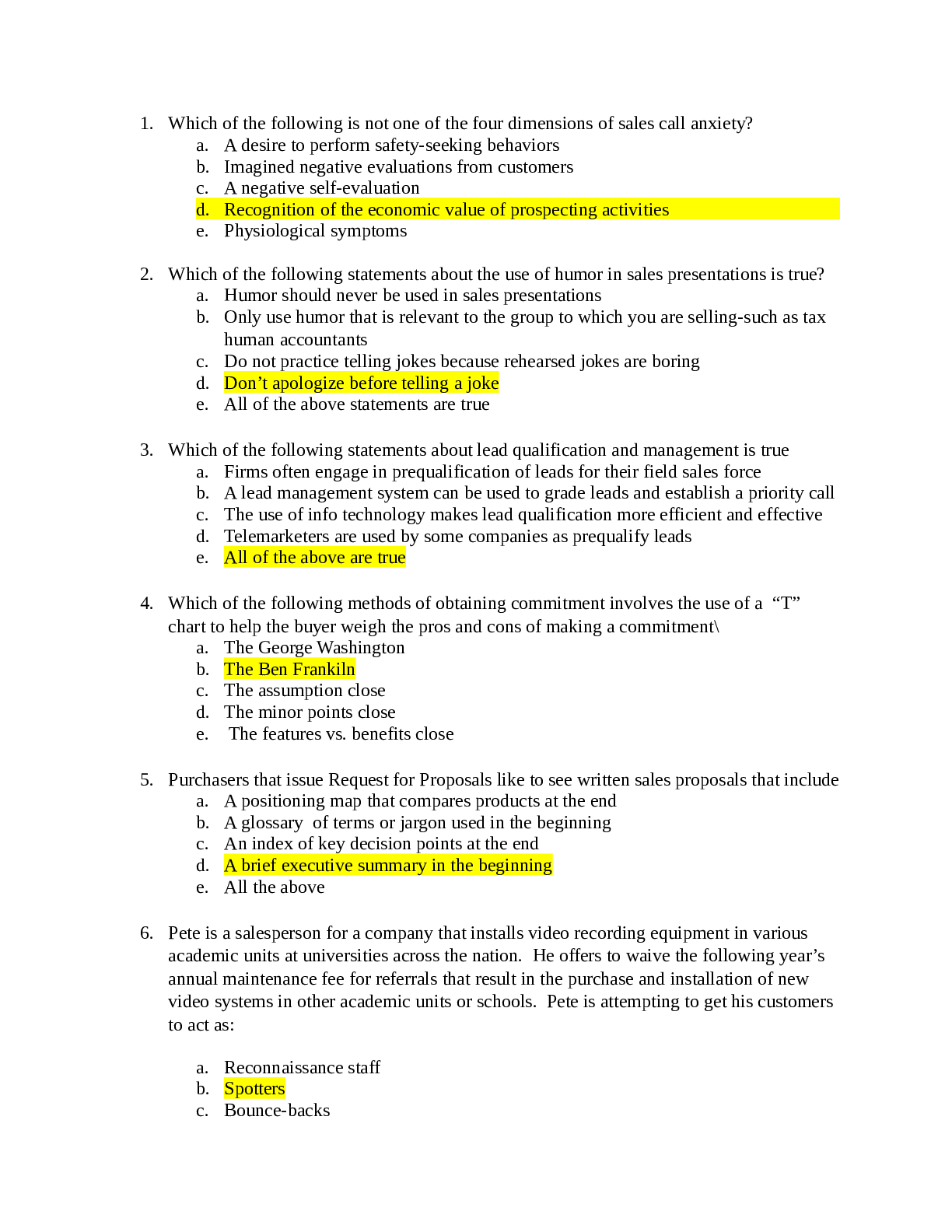
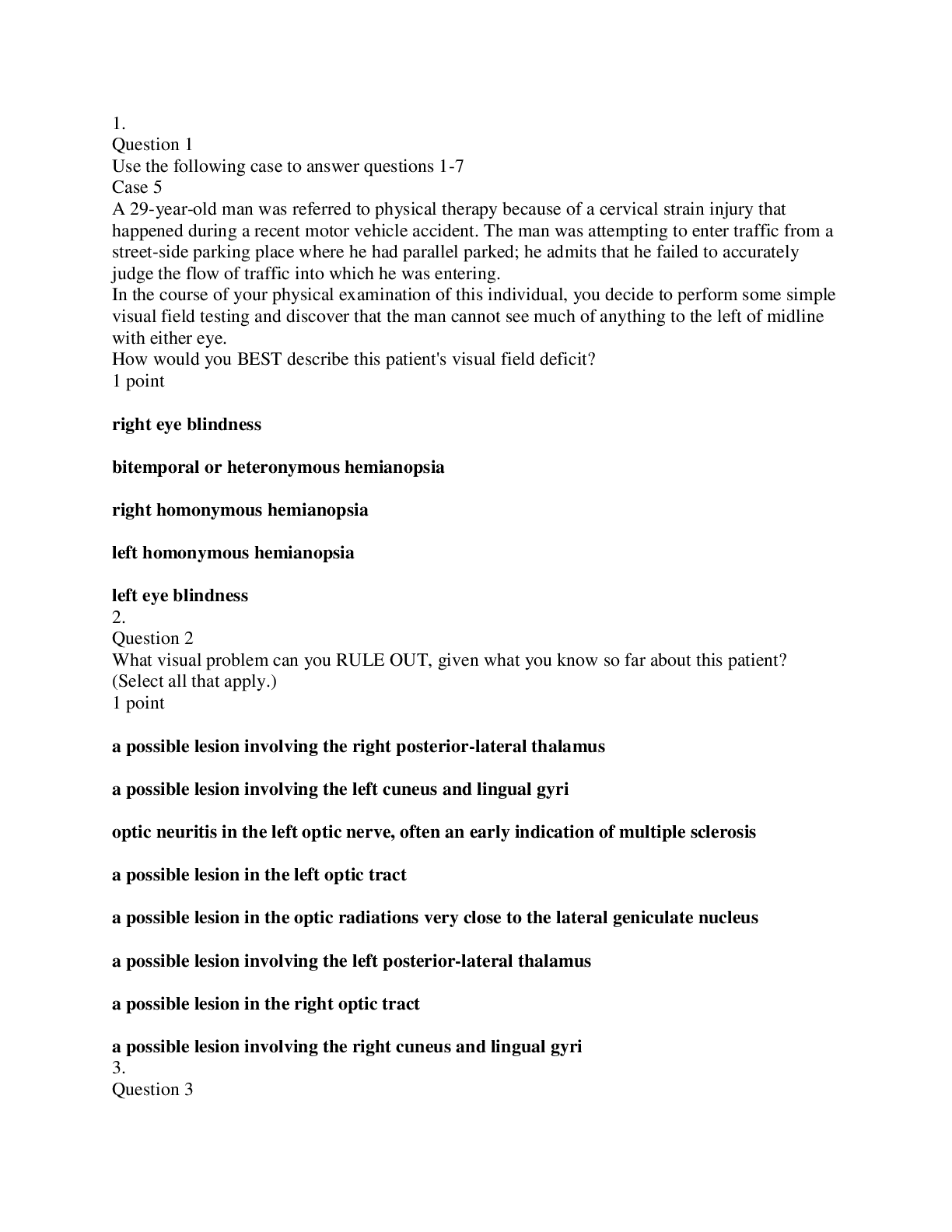
1. Question 1 Use the following case to answer questions 1-4 Case 7 A 56-year-old woman was referred to a neurology practice specializing in movement disorders following a nine-year histor... y of bradykinesia (slowness) and rigidity. In more recent years, she developed resting tremor in her hands and difficulties walking. She has been treated with levodopa (Sinemet), which helped for a few years, but then she developed severe ON-OFF fluctuations—alternating between incapacitating dyskinesias (involuntary, repetitive body movements) and total body freezing. Her sensory functions were intact, but bodily movements were increasingly difficult to govern. On the basis of the symptoms and signs described above, what is the most likely medical diagnosis? 1 point meningitis encephalitis amyotrophic lateral sclerosis Huntington's disease Parkinson's disease Alzheimer's disease 2. Question 2 Given the benefit of levodopa, at least for a few years, where in the nervous system of this patient is there likely to be histopathological degeneration of neuronal cell bodies? 1 point subthalamic nucleus ventral pallidum striatum putamen substantia nigra pars compacta substantia nigra pars reticulata nucleus accumbens globus pallidus 3. Question 3 How does this histopathology (if left untreated) affect the activity of neurons in the basal ganglia? 1 point activity decreases in the striatal neurons that contribute to the indirect pathway activity diminishes in the subthalamic nucleus activity increases in the internal segment of the globus pallidus activity diminishes in the internal segment of the globus pallidus activity increases in the striatal neurons that contribute to the direct pathway 4. Question 4 In the course of medical care, this patient was evaluated for neurosurgical placement of a deep brain stimulator for relief of her movement disorder. This procedure entailed MRI-based surgical planning using a stereotaxic system, intraoperative neurophysiological recording to identify target structures, and then final positioning of the bipolar electrode for deep brain stimulation. After a brief recovery period, the patient was then subject to a second minor surgical procedure that connected the device to a subcutaneous controller and power supply. The controller was then programmed and therapeutic stimulation parameters were determined empirically. Her bradykinesia and rigidity were greatly improved and there was a marked reduction of her ON-OFF fluctuations and dyskinesias. For this woman and many like her, deep brain stimulation for movement disorders targets the subthalamic nucleus. Although the mechanisms of therapeutic action remain uncertain, one rationale for targeting the subthalamic nucleus with deep brain stimulation is provided by which of the following statements? 1 point Deep brain stimulation of the subthalamic nucleus normalizes the balance of activity in the direct and indirect pathway, leading to more appropriate and functional disinhibition of the motor thalamus. Deep brain stimulation of the subthalamic nucleus is designed to increase the discharge rate of subthalamic neurons, which in turn disinhibits the motor thalamus. Deep brain stimulation of the subthalamic nucleus increases the steady-state output of the internal segment of the globus pallidus, which facilitates the initiation of movement. Deep brain stimulation of the subthalamic nucleus is safer than the alternative targets, since subthalamic nucleus neurons are GABAergic and stimulating them cannot induce excitotoxicity. 5. Question 5 Use the following case to answer questions 5-12 Case 8 A 50-year-old man went to bed feeling very dizzy, with complaints of an occipital headache. The next day, neurological examination revealed the following: The right eyelid was closed. The right globe was deviated laterally and could not be moved upward or inward. The right pupil was dilated and did not react to light directly or consensually, or during accommodation. Light shown into the right eye caused contraction of the left pupil. There was marked hemiparesis on the left, with moderately increased muscle tone and deep reflexes on the left. An extensor plantar response (positive Babinski sign) was elicited on the left. On the left side of the body, a constant rhythmical tremor at rest was noted that was worse on movement. Movements of the left limbs were ataxic. There was hemianesthesia to all sensory modalities over the left face and the left side of the body and limbs. If not described above, then all other aspects of the neurological exam and the integrity of all other body structures were found to be within normal limits. Beginning with the first cluster of signs listed above, which extra-ocular muscles are affected in this patient? (Select all that apply.) 1 point left inferior rectus right lateral rectus right medial rectus left medial rectus right inferior rectus left lateral rectus right superior rectus left superior rectus 6. Question 6 Damage to which pathways/structures provides for the BEST explanation of the pupillary signs in this case? 1 point damage to the right optic nerve damage to descending sympathetic fibers on the right damage to parasympathetic fibers coursing with the right oculomotor nerve damage to parasympathetic fibers coursing with the left oculomotor nerve damage to descending sympathetic fibers on the left damage to the posterior commissure damage to the left optic nerve 7. Question 7 Which nerve appears to be damaged in this man, and is the damage located within the brainstem or along the extramedullary (outside of the CNS) course of the nerve? 1 point long ciliary nerve (sympathetic fibers) within the left orbital fissure in the base of the skull left and right CNs II (optic nerve), within the optic chiasm right CN III (right oculomotor nerve), within the right cavernous sinus in the base of the skull right CN III (right oculomotor nerve), within the midbrain left CN III (left oculomotor nerve), within the midbrain right CN VII (right facial nerve), within the right stylomastoid foramen in the base of the skull long ciliary nerve (sympathetic fibers) within the right orbital fissure in the base of the skull left CN III (left oculomotor nerve), within the left cavernous sinus in the base of the skull left CN VII (left facial nerve), within the left stylomastoid foramen in the base of the skull 8. Question 8 Damage to which motor pathway (at the specified level or side) most likely accounts for the weakness as well as changes in muscle tone, reflexes and extensor plantar response? 1 point corticospinal fibers within the base of the left side of the pons (basis pontis) corticospinal pathway at the level of its decussation reticulospinal fibers on the left corticospinal fibers within the base of the left cerebral peduncle (basis pedunculi) lateral vestibulospinal pathway on the left lateral vestibulospinal pathway on the right reticulospinal fibers on the right corticospinal fibers within the base of the right cerebral peduncle (basis pedunculi) corticospinal fibers within the base of the right side of the pons (basis pontis) 9. Question 9 Damage to which neural structure(s) most likely accounts for the constant rhythmical tremor on the left side of the body at rest? 1 point substantia nigra, pars reticulata on the left subthalamic nucleus on the right subthalamic nucleus on the left substantia nigra, pars reticulata on the right globus pallidus, internal segment on the left substantia nigra, pars compacta on the right globus pallidus, internal segment on the right substantia nigra, pars compacta on the left 10. Question 10 Damage to which neural structure(s) most likely accounts for the uncoordinated (ataxic) behavior of the left limbs during movement in this patient? 1 point ventral lateral and ventral anterior nuclei of the thalamus (i.e., the motor thalamus) dentate-thalamic tract (projections from the cerebellar dentate nucleus to the contralateral motor thalamus) on the right side of the brainstem rostral to the decussation of the superior cerebellar peduncles dentate nucleus in the right cerebellar hemisphere dentate-thalamic tract (projections from the cerebellar dentate nucleus to the contralateral motor thalamus) within the decussation of the superior cerebellar peduncles in the rostral pons dentate nucleus in the left cerebellar hemisphere superior cerebellar peduncle on the right superior cerebellar peduncle on the left 11. Question 11 Based on your analysis of the case, where is the most likely site of the lesion? 1 point caudal midbrain (level of inferior colliculus), paramedian tegmental area caudal pons, right lateral tegmental area right central cortex, involving the precentral and postcentral gyri right rostral midbrain (level of superior colliculus) involving the cerebral peduncle and extensive portions of the tegmentum, with the midline being the medial margin of the damage rostral pons, paramedian tegmental area left central cortex, involving the precentral and postcentral gyri caudal pons, left lateral tegmental area left rostral midbrain (level of superior colliculus) involving the cerebral peduncle and extensive portions of the tegmentum, with the midline being the medial margin of the damage 12. Question 12 The localized and relatively rapid onset of symptoms in this gentleman suggests a pathological process involving the blood supply or drainage of the affected region. Which vessel is likely affected in this case? 1 point penetrating lateral branches of the left posterior inferior cerebellar artery right middle cerebral artery penetrating medial branches of the right posterior cerebral artery penetrating lateral branches of the right anterior inferior cerebellar artery penetrating lateral branches of the right posterior inferior cerebellar artery penetrating medial branches of the left posterior cerebral artery left middle cerebral artery penetrating medial branches from the pontine segment of the basilar artery penetrating lateral branches of the right vertebral artery penetrating lateral branches of the left vertebral artery penetrating lateral branches of the left anterior inferior cerebellar artery [Show More]
Last updated: 1 year ago
Preview 1 out of 10 pages
Reviews( 0 )
Document information
Connected school, study & course
About the document
Uploaded On
Sep 20, 2021
Number of pages
10
Written in

Additional information
This document has been written for:
Uploaded
Sep 20, 2021
Downloads
0
Views
86


.png)
.png)

.png)
.png)
.png)