Pathophysiology > QUESTIONS & ANSWERS > NURS 5315 PATHO Module 7 N5315 Advanced Pathophysiology Neurologic System Core Knowledge Objectives (All)
NURS 5315 PATHO Module 7 N5315 Advanced Pathophysiology Neurologic System Core Knowledge Objectives with Advanced Organizers
Document Content and Description Below
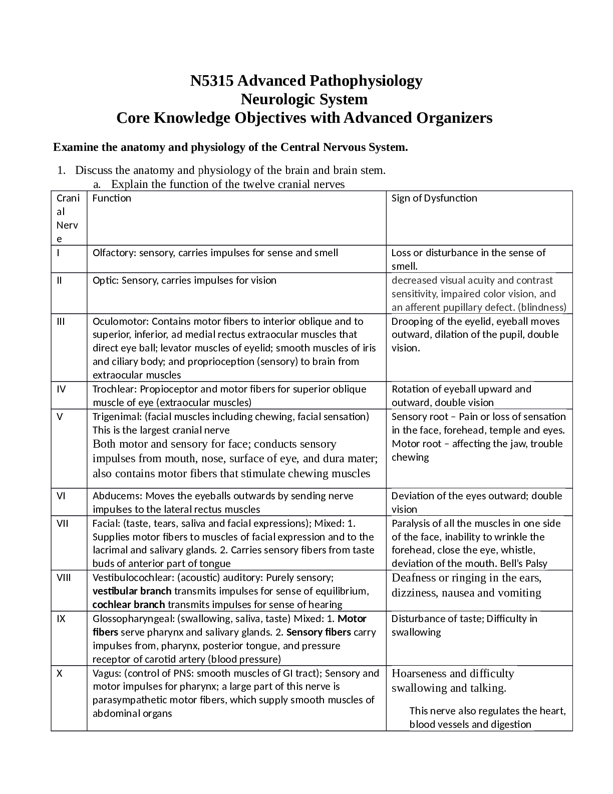
N5315 Advanced Pathophysiology Neurologic System Core Knowledge Objectives with Advanced Organizers Examine the anatomy and physiology of the Central Nervous System. 1. Discuss the anatomy and phy... siology of the brain and brain stem. a. Explain the function of the twelve cranial nerves Crani al Nerv e Function Sign of Dysfunction I Olfactory: sensory, carries impulses for sense and smell Loss or disturbance in the sense of smell. II Optic: Sensory, carries impulses for vision decreased visual acuity and contrast sensitivity, impaired color vision, and an afferent pupillary defect. (blindness) III Oculomotor: Contains motor fibers to interior oblique and to superior, inferior, ad medial rectus extraocular muscles that direct eye ball; levator muscles of eyelid; smooth muscles of iris and ciliary body; and proprioception (sensory) to brain from extraocular muscles Drooping of the eyelid, eyeball moves outward, dilation of the pupil, double vision. IV Trochlear: Propioceptor and motor fibers for superior oblique muscle of eye (extraocular muscles) Rotation of eyeball upward and outward, double vision V Trigenimal: (facial muscles including chewing, facial sensation) This is the largest cranial nerve Both motor and sensory for face; conducts sensory impulses from mouth, nose, surface of eye, and dura mater; also contains motor fibers that stimulate chewing muscles Sensory root – Pain or loss of sensation in the face, forehead, temple and eyes. Motor root – affecting the jaw, trouble chewing VI Abducems: Moves the eyeballs outwards by sending nerve impulses to the lateral rectus muscles Deviation of the eyes outward; double vision VII Facial: (taste, tears, saliva and facial expressions); Mixed: 1. Supplies motor fibers to muscles of facial expression and to the lacrimal and salivary glands. 2. Carries sensory fibers from taste buds of anterior part of tongue Paralysis of all the muscles in one side of the face, inability to wrinkle the forehead, close the eye, whistle, deviation of the mouth. Bell’s Palsy VIII Vestibulocochlear: (acoustic) auditory: Purely sensory; vestibular branch transmits impulses for sense of equilibrium, cochlear branch transmits impulses for sense of hearing Deafness or ringing in the ears, dizziness, nausea and vomiting IX Glossopharyngeal: (swallowing, saliva, taste) Mixed: 1. Motor fibers serve pharynx and salivary glands. 2. Sensory fibers carry impulses from, pharynx, posterior tongue, and pressure receptor of carotid artery (blood pressure) Disturbance of taste; Difficulty in swallowing X Vagus: (control of PNS: smooth muscles of GI tract); Sensory and motor impulses for pharynx; a large part of this nerve is parasympathetic motor fibers, which supply smooth muscles of abdominal organs Hoarseness and difficulty swallowing and talking. This nerve also regulates the heart, blood vessels and digestion resulting in irregular heartbeat and lowered blood pressure. It regulates the stomach telling it to move food through the digestive system, thus damage can result in decreased digestion and thus nausea, bloating and vomiting. XI Spinal Accessory: (Moving head & shoulders, swallowing); provides sensory and motor fibers for sternocleidomastoid and trapezius muscles (movement of head and shoulders) and muscles of soft palate, pharynx, and larynx (swallowing) Dropping of the shoulder; inability to rotate the head away from affected area. XII Hypoglossal: (tounge muscles, speech, swallowing);Carries motor fibers to muscles of tongue and sensory impulses from tongue to brain. Paralysis of one side of the tongue; deviation of tongue toward paralyzed side; thick speech. b. Explain the function of the cerebrum, cerebellum, parietal lobe, frontal lobe, occipital lobe, temporal lobe, brain stem, reticular formation reticular activating system, and limbic system. Lobe Location Function Sign of Injury/Lesion / Disorder Cerebrum forebrain Largest part of the bain. Derived from the telencephalon, characterized by numerous convolutions called gyri. Comprised of two hemispheres Contain- cerebral cortex, basal ganglia, epithalamus, thalamus, hypothalamus, subthalamus. Prefrontal lobe- goal-oriented behavior/concentration, shortterm memory, elaboration of thought and inhibition on the limbic areas of the CNS. Prefrontal (Brodmann 6) programming motor movements. Basal ganglia systemextrapyramidal system- efferent pathways outside the pyramids of the medulla. Frontal eye fields (lower portion of Brodmann 8) controlling eye movement in the [Show More]
Last updated: 1 year ago
Preview 1 out of 28 pages
Reviews( 0 )
Document information
Connected school, study & course
About the document
Uploaded On
Mar 09, 2022
Number of pages
28
Written in

Additional information
This document has been written for:
Uploaded
Mar 09, 2022
Downloads
1
Views
114
.png)
.png)
.png)
.png)
.png)
.png)
.png)
.png)
.png)
.png)
.png)

.png)
.png)
.png)